Tag Archive for: deep brain stimulation

Drs. Oluigbo and Myseros

Spotlight on Children’s National Hospital Neurosurgery

Drs. Oluigbo and Myseros

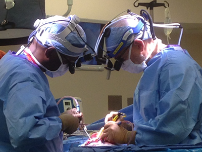
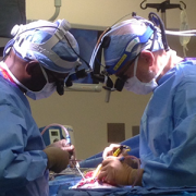
Our neurosurgery team is among the most experienced in the nation. We have performed thousands of surgeries and are dedicated to giving the best possible care. The Children’s National Hospital Division of Neurosurgery consistently ranks among the country’s top programs according to U.S. News & World Report.

Patients travel to us from all over the world because we have the resources and expertise necessary to care for their neurological conditions through multidisciplinary programs such as:

  • Spine Disorders
  • Deep Brain Stimulation Program
  • Neuro Intensive Care Unit (Neuro ICU)
  • Neuro-ophthalmology
  • Spina Bifida Program
  • Brain and Spinal Cord Tumors
  • Craniofacial Disorders
  • Chiari Malformations
  • Epilepsy
  • Brachial Plexus Injury
  • Spasticity Program
  • Neurovascular diseases such as AVM’s and Moyamoya

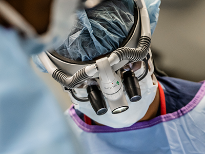
Minimally invasive surgery

The Children’s National Hospital Division of Neurosurgery is among the first in the country to develop new techniques and adopt the latest technologies that make minimally invasive neurosurgery possible by utilizing state of the art equipment and developing new techniques, including:

  • ROSA surgical robot / SEEG placement
  • Surgical Theater with virtual reality visualization
  • Visualase® magnetic resonance imaging (MRI)-guided laser ablation
  • 5T intra-operative MRI (iMRI)
  • Deep brain stimulation
  • Neuropace epilepsy control

Advanced treatment and cutting edge research

Children’s National is involved in cutting edge scientific research offering new hope for our patients and new methods of treatment. Our doctors have developed some of the most advanced treatments and clinics for our patients including:

  • Multidisciplinary skull base neurosurgery program
  • Participating in the 1st generation of genetic modulation trials
  • CAR T-Cell Therapy research
  • Ehlers-Danlos syndrome (EDS) /Hypermobility Program
  • Pseudotumor Cerebri Multidisciplinary panel
  • Leader in open and endoscopic craniosynostosis surgery

Ranked No. 5 in the nation

U.S. News & World Report ranks our neurosurgery program number five in the nation, reflecting our commitment to excellence in care for our patients and families.

Level 1 surgery verification

Children’s National is one of only 12 children’s hospitals in the country to attain Level 1 Surgery Verification from the American College of Surgeons.

doctor performing neurosurgery

Successful outcomes

Children with rare and medically complex conditions, such as brain tumors, craniofacial disorders, Chiari malformations, vascular disorders and brachial plexus palsy, to name a few, achieve exceptional outcomes at Children’s National. Our patients experience fewer complications, go home sooner and maintain long-term symptom relief.

Specialized expertise

Our entire team is dedicated to meeting your child’s unique needs. Our Neuro-Intensive Care Unit nurses recognize signs of pain and complications your child may not be able to explain.

Pioneering new treatments

Children’s National is at the forefront of new device-based treatments that not only fix neurologic problems, but also restore brain function. We are one of the few pediatric programs in the country offering dedicated pediatric deep brain stimulation, which uses a pacemaker-like device to significantly reduce the burden of movement disorders and difficult-to-control epilepsy, as well as Neuropace implantation to help with seizures in eloquent areas of the brain.

Training the next generation of top neurosurgeons

We are proudly training the next generation of pediatric neurosurgeons through residency programs and fellowships in conjunction with several area medical schools.

illustration of brain showing cerebellum

Focusing on the “little brain” to rescue cognition

illustration of brain showing cerebellum

Research faculty at Children’s National in Washington, D.C., with colleagues recently published a review article in Nature Reviews Neuroscience that covers the latest research about how abnormal development of the cerebellum leads to a variety of neurodevelopmental disorders.

Cerebellum translates as “little brain” in Latin. This piece of anatomy – that appears almost separate from the rest of the brain, tucked under the two cerebral hemispheres – long has been known to play a pivotal role in voluntary motor functions, such as walking or reaching for objects, as well as involuntary ones, such as maintaining posture.

But more recently, says Aaron Sathyanesan, Ph.D., a postdoctoral research fellow at the Children’s Research Institute, the research arm of Children’s National  in Washington, D.C., researchers have discovered that the cerebellum is also critically important for a variety of non-motor functions, including cognition and emotion.

Sathyanesan, who studies this brain region in the laboratory of Vittorio Gallo, Ph.D., Chief Research Officer at Children’s National and scientific director of the Children’s Research Institute, recently published a review article with colleagues in Nature Reviews Neuroscience covering the latest research about how altered development of the cerebellum contributes to a variety of neurodevelopmental disorders.

These disorders, he explains, are marked by problems in the nervous system that arise while it’s maturing, leading to effects on emotion, learning ability, self-control, or memory, or any combination of these. They include diagnoses as diverse as intellectual disability, autism spectrum disorder (ASD), attention-deficit/hyperactivity disorder and Down syndrome.

“One reason why the cerebellum might be critically involved in each of these disorders,” Sathyanesan says, “is because its developmental trajectory takes so long.”

Unlike other brain structures, which have relatively short windows of development spanning weeks or months, the principal cells of the cerebellum – known as Purkinje cells – start to differentiate from stem cell precursors at the beginning of the seventh gestational week, with new cells continuing to appear until babies are nearly one year old.  In contrast, cells in the neocortex, a part of the brain involved in higher-order brain functions such as cognition, sensory perception and language is mostly finished forming while fetuses are still gestating in the womb.

This long window for maturation allows the cerebellum to make connections with other regions throughout the brain, such as extensive connections with the cerebral cortex, the outer layer of the cerebrum that plays a key role in perception, attention, awareness, thought, memory, language and consciousness. It also allows ample time for things to go wrong.

“Together,” Sathyanesan says, “these two characteristics are at the root of the cerebellum’s involvement in a host of neurodevelopmental disorders.”

For example, the review article notes, researchers have discovered both structural and functional abnormalities in the cerebellums of patients with ASD. Functional magnetic resonance imaging (MRI), an imaging technique that measures activity in different parts of the brain, suggests that significant differences exist between connectivity between the cerebellum and cortex in people with ASD compared with neurotypical individuals. Differences in cerebellar connectivity are also evident in resting-state functional connectivity MRI, an imaging technique that measures brain activity in subjects when they are not performing a specific task. Some of these differences appear to involve patterns of overconnectivity to different brain regions, explains Sathyanesan; other differences suggest that the cerebellums of patients with ASD don’t have enough connections to other brain regions.

These findings could clarify research from Children’s National and elsewhere that has shown that babies born prematurely often sustain cerebellar injuries due to multiple hits, including a lack of oxygen supplied by infants’ immature lungs, he adds. Besides having a sibling with ASD, premature birth is the most prevalent risk factor for an ASD diagnosis.

The review also notes that researchers have discovered structural changes in the cerebellums of patients with Down syndrome, who tend to have smaller cerebellar volumes than neurotypical individuals. Experimental models of this trisomy recapitulate this difference, along with abnormal connectivity to the cerebral cortex and other brain regions.

Although the cerebellum is a pivotal contributor toward these conditions, Sathyanesan says, learning more about this brain region helps make it an important target for treating these neurodevelopmental disorders. For example, he says, researchers are investigating whether problems with the cerebellum and abnormal connectivity could be lessened through a non-invasive form of brain stimulation called transcranial direct current stimulation or an invasive one known as deep brain stimulation. Similarly, a variety of existing pharmaceuticals or new ones in development could modify the cerebellum’s biochemistry and, consequently, its function.

“If we can rescue the cerebellum’s normal activity in these disorders, we may be able to alleviate the problems with cognition that pervade them all,” he says.

In addition to Sathyanesan and Senior Author Gallo, Children’s National study co-authors include Joseph Scafidi, D.O., neonatal neurologist; Joy Zhou and Roy V. Sillitoe, Baylor College of Medicine; and Detlef H. Heck, of University of Tennessee Health Science Center.

Financial support for research described in this post was provided by the National Institute of Neurological Disorders and Stroke under grant numbers 5R01NS099461, R01NS089664, R01NS100874, R01NS105138 and R37NS109478; the Hamill Foundation; the Baylor College of Medicine Intellectual and Developmental Disabilities Research Center under grant number U54HD083092; the University of Tennessee Health Science Center (UTHSC) Neuroscience Institute; the UTHSC Cornet Award; the National Institute of Mental Health under grant number R01MH112143; and the District of Columbia Intellectual and Developmental Disabilities Research Center under grant number U54 HD090257.

Chima Oluigbo

The benefits of deep brain stimulation for pediatric patients

Chima Oluigbo

There was no effective treatment for uncontrolled, difficult, and sometime painful movements associated with movement disorders. That is, before the development of deep brain stimulation (DBS) techniques.

Children’s National Health System is one of only two children’s hospitals with fully integrated DBS programs. Chima Oluigbo, M.D., who leads the pioneering Deep Brain Stimulation Program within Children’s Division of Neurosurgery, is one of few pediatric deep brain stimulation experts in North America and cross-trained in pediatric and functional neurosurgery.

Dr. Oluigbo says the effects of DBS are often dramatic: 90 percent of children with primary dystonia show up to 90 percent symptom improvement.

A 6-year-old boy with dystonia so severe that his body curved like a “C” was one of the first patients to undergo the procedure at Children’s National. Six weeks later, he gained the ability to sit straight and to control his hands and legs. He also was able to smile, an improvement that brought particular joy to his parents.

Inside the brain with movement disorders

Patients with movement disorders experience difficulties due to neurological dysfunction that impact the speed, fluency, quality, and ease in which they move. In these cases, neurons in the brain’s motor circuits misfire. Through the use of DBS, neurosurgeons can synchronize neuronal firing and accomplish the previously impossible: restoring muscle control to patients with these disorders.

Movement disorders are common in children. “It’s not just numbers, it’s also about impact. Think about the potential of a child who is very intelligent and can contribute to society. When that child is not able to contribute because he or she is disabled by a movement disorder, the lost potential is very significant. It has an impact,” Dr. Oluigbo says.

What is deep brain stimulation?

DBS uses an implantable device to send continuous, low-level electrical impulses to areas deep within the brain. The impulses prevent the brain from firing abnormal signals that are linked to movement disorders and seizures. When a child is considered to be a candidate for the technique, here’s what happens next:

  1. Imaging: Magnetic resonance imaging (MRI) helps pinpoint the area of brain tissue responsible for movement disorders and informs the treatment plans.
  2. Neurotransmitter implant procedure: Using minimally invasive neurosurgery techniques, doctors access the brain through a tiny incision in the child’s skull and place thin, insulated wires (leads) in the area of brain tissue responsible for the condition.
  3. Pulse generator implant procedure: The pulse generator (neurostimulator) is a battery-operated device that sends low-level electrical impulses to the leads. During a separate procedure, the pulse generator is implanted near the child’s collarbone. Leads are threaded under the child’s skin to connect with the pulse generator.
  4. Stimulation treatments: Once the leads and pulse generator are connected, the child receives a continuous stream of electrical impulses. Impulses are generated by the neurostimulator, travel through the leads, and end up in the deep tissue of the brain. Here, they block abnormal signals that are linked to the child’s movement disorder.
  5. Follow-up care: The child will likely need deep brain stimulation throughout his or her lifetime to make sure the device is working correctly and to adjust the neurotransmitter settings to meet his or her changing needs.

Deep brain stimulation at Children’s National

Children’s National is currently conducting clinical trials seeking to expand the use of this procedure to patients with cerebral palsy, one of the most common dystonias. The effective use of deep brain stimulation requires ongoing attention from a multidisciplinary team (from neuropsychology to rehabilitation medicine), giving seamless care under one roof.

There is evidence to suggest that this technique could be used to aid people with memory disorders, patients in minimally conscious states, and patients with incurable epilepsies.