Tag Archive for: Congenital Zika Virus Program

Sarah Mulkey

MRI finds novel brain defects in Zika-exposed newborns

Sarah Mulkey

“Imaging is constantly helping us make new discoveries with this virus, and in these two cases we found things that had not been previously described,” says Sarah Mulkey, M.D., Ph.D.

Magnetic resonance imaging (MRI) has identified two brain abnormalities never before reported in newborns with prenatal exposure to the Zika virus. Children’s National Health System researchers reported these findings from a study of more than 70 fetuses or newborns with Zika exposure in utero. The study was published in the January 2018 edition of Pediatric Neurology.

The two novel defects – cranial nerve enhancement and cerebral infarction – may join the growing list of neurological findings associated with congenital Zika infection.

“Imaging is constantly helping us make new discoveries with this virus, and in these two cases we found things that had not been previously described,” says Sarah Mulkey, M.D., Ph.D., the study’s lead author and a fetal-neonatal neurologist at Children’s National. Dr. Mulkey works in Children’s Congenital Zika Virus Program, one of the nation’s first comprehensive, dedicated Zika programs.

The research team recommends that postnatal brain MRI be considered in addition to ultrasound for newborns exposed to Zika in utero. “Brain MRI can be performed in the newborn often without sedation and provides an opportunity to look for brain abnormalities we might not catch otherwise – or might not detect until much later,” says Dr. Mulkey.

Birth defects are seen in 6 to 11 percent of pregnancies affected by Zika, and some of the neurological complications in infants are not apparent until well after birth.

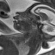
Of the two infants in which the new abnormalities were observed, both had normal head size at birth. Neither had smaller-than-normal head size (microcephaly), one of the more severe effects associated with congenital Zika syndrome.

One infant had a normal neurological evaluation at 2 days of age. However, a brain MRI conducted the following day, using gadolinium contrast due to concern of infection, showed enhancement of multiple cranial nerves. “Nerve root enhancement is very rare in a newborn and had not been described with Zika before,” Dr. Mulkey says. “Yet, there was no neurological deficit that we could identify by physical exam.”

The research team acknowledges that the clinical significance of this finding is not yet known.

In the second patient, brain MRI conducted without contrast at 16 days of age revealed a small area consistent with chronic infarction (ischemic stroke) that likely occurred during the third trimester.

“We followed the mother throughout her pregnancy, and both MRI and ultrasound imaging were normal at 28 weeks gestation,” Dr. Mulkey says. “A postnatal ultrasound was also normal, but the postnatal MRI showed a stroke that had occurred at least one month prior to the MRI and after the last fetal study.”

She adds: “This is the first published report of fetal stroke associated with Zika infection, and it may add to our knowledge of what can occur with congenital Zika infection.”

Unlike most congenital infections, Zika virus does not appear to cause viral-induced placental inflammation, which can lead to fetal stroke. So, the authors say they cannot be sure that congenital Zika contributed to the infarct in this case. However, they write, “Given the relatively low incidence of perinatal ischemic infarct and the lack of other maternal- or birth-related risk factors for this patient, Zika infection is considered a possible etiology.”

In both patients, neonatal brain MRI identified subclinical findings that had not previously been described as part of congenital Zika syndrome. As the body of evidence about the Zika virus has grown, the spectrum of associated brain abnormalities has expanded to include considerably more findings than isolated microcephaly.

Data gathered in 2017 from the Centers for Disease Control and Prevention’s Zika pregnancy and infant registry indicates that 25 percent of eligible U.S. infants receive recommended postnatal imaging. Dr. Mulkey said this represents many possible missed opportunities for earlier identification of brain abnormalities.

“Brain MRI should be considered in all newborns exposed to Zika virus in utero, even in the presence of normal birth head circumference, normal cranial ultrasound and normal fetal imaging,” she says. “In both of these patients, the changes we observed were not evident on cranial ultrasound or on fetal MRI and fetal ultrasound.”

In addition to Dr. Mulkey, Children’s co-authors include L. Gilbert Vezina, M.D., Neuroradiology Program director; Dorothy I. Bulas, M.D., chief of Diagnostic Imaging and Radiology; Zarir Khademian, M.D., radiologist; Anna Blask, M.D., radiologist; Youssef A. Kousa, M.S., D.O., Ph.D., child neurology fellow; Lindsay Pesacreta, FNP; Adré  J. du Plessis, M.B.Ch.B., M.P.H., Fetal Medicine Institute director; and Roberta L. DeBiasi, M.D., M.S., senior author and Pediatric Infectious Disease division chief; and Caitlin Cristante, B.S.

Financial support for this research was provided by the Thrasher Research Fund.

Neonatal baby

Multidisciplinary experts help CDC’s Zika research

“We are very excited about this next phase in our Zika research,” says Roberta L. DeBiasi, M.D., M.S. “It is a natural extension of our earlier participation as subject matter experts assisting as the CDC developed and published guidelines to inform the care of Zika-exposed and Zika-infected infants across the nation and U.S. territories.”

The Centers for Disease Control and Prevention (CDC) is funding three multidisciplinary experts from the Congenital Zika Virus Program at Children’s National Health System to collaborate on two of the CDC’s longitudinal Zika research projects in Colombia, South America.

“Zika en embarazadas y niños en Colombia” (ZEN) is a research study jointly designed by Colombia’s Instituto Nacional de Salud (INS) and the CDC to evaluate the association between Zika virus infection and adverse maternal, fetal and infant health outcomes. The study is following a large cohort of Colombian women from the first trimester of pregnancy, their male partners and their infants.

Under the six-month contract, Roberta L. DeBiasi, M.D., M.S., Sarah B. Mulkey, M.D., Ph.D., and Cara Biddle, M.D., M.P.H., will serve as consultants for the ZEN study providing expertise in pediatric infectious diseases, neurology, neurodevelopment and coordination of the complex care needs of Zika-affected infants.

The federal funding will underwrite the consultants’ work effort, as well as travel to the CDC’s headquarters in Atlanta and to research sites in Colombia. To that end, Drs. DeBiasi, Mulkey and Biddle participated in a December 2017 kickoff meeting, joining ZEN team leaders based in the U.S. at the CDC, as well as the INS in Colombia, with whom they will conduct research and collaborate academically.

Cara-Biddle-and-Sarah-Mulkey

Cara Biddle, M.D., M.P.H., and Sarah B. Mulkey, M.D., Ph.D., also will serve as consultants for the ZEN study.

“We are very excited about this next phase in our Zika research,” says Dr. DeBiasi, chief of the Division of Pediatric Infectious Diseases and co-director of the Children’s Zika program. “It is a natural extension of our earlier participation as subject matter experts assisting as the CDC developed and published guidelines to inform the care of Zika-exposed and Zika-infected infants across the nation and U.S. territories.”

Children’s National is leading its own longitudinal studies in Colombia that explore such questions as whether Zika-exposed infants whose neuroimaging appears normal when they are born experience any longer-term neurological issues and the role of genetics in neurologic injury following congenital Zika virus exposure and infection.

Roberta DeBiasi and Sarah Mulkey

Children’s National experts contribute to new Zika guidelines

Roberta DeBiasi and Sarah Mulkey

Roberta DeBiasi, M.D., M. S., and Sarah B. Mulkey, M.D., Ph.D., members of Children’s multidisciplinary Congenital Zika Virus Program, were among the experts invited to participate in a forum held in Atlanta at CDC headquarters in late August to formulate new Zika recommendations.

The Centers for Disease Control and Prevention (CDC) on Oct. 19, 2017 updated guidelines for evaluation of women, fetuses and infants exposed to the Zika virus during pregnancy. Although only women with symptoms will now be routinely tested, asymptomatic and symptomatic infants born to these women will still be tested for the Zika virus using blood and urine tests.

Infants who appear normal, whose mothers either had negative Zika results or who had not undergone testing, will not undergo Zika testing. These infants still will undergo a standard evaluation, including a detailed physical exam, hearing screen and routine developmental assessments. The revised Zika guidance includes input from practitioners on the front lines of the Zika epidemic, including Children’s National Health System clinicians.

“These changes in the recommendations for Zika testing should not be interpreted as Zika infection risks subsiding for pregnant women and their infants in the United States. It’s simply an acknowledgement of the limitations of current testing methods – which must occur within a narrow window after Zika exposure – and the poor predictive value of Zika testing right now,” says Roberta L. DeBiasi, M.D., M.S., chief of Children’s Division of Pediatric Infectious Diseases. Dr. DeBiasi and Sarah B. Mulkey, M.D., Ph.D., members of Children’s multidisciplinary Congenital Zika Virus Program, were among the experts invited to participate in the Zika forum held in Atlanta at CDC headquarters in late August to formulate the recommendations.

While all infants will receive a standard evaluation, expanded evaluations that include an ophthalmologic assessment, more detailed hearing evaluation and ultrasound of the newborn’s head will be reserved for infants born to mothers confirmed to be Zika positive or Zika probable, or for infants born with abnormalities potentially consistent with congenital Zika syndrome, regardless of maternal status.

The majority of U.S. infants who have been exposed to Zika in the womb appeared normal at birth, according to CDC registries. Now, the next wave of these normal-appearing babies will receive standard evaluations when they are born, including a newborn hearing screening. At each well-child visit, these infants will receive:

  • A comprehensive physical examination
  • An age-appropriate vision screening
  • Developmental monitoring and screening using validated tools

“This is a natural evolution in the diagnosis and screening strategy now that the peak of the first wave of Zika transmission appears to be over,” Dr. DeBiasi says. “While we continue to evaluate new possible cases of Zika infection among pregnant women in our practice, a sizable proportion of Children’s cases are Zika-exposed infants whose physical exam and neuroimaging appeared normal at birth. Through ongoing monitoring, we hope to learn more about these children’s long-term neurodevelopment outcomes.”

What Children’s has learned about congenital Zika infection

Roberta DeBiasi

Roberta DeBiasi, M.D., M.S., outlined lessons learned during a pediatric virology workshop at IDWeek2017, one of three such Zika presentations led by Children’s National research-clinicians during this year’s meeting of pediatric infectious disease specialists.

The Congenital Zika Virus Program at Children’s National Health System provides a range of advanced testing and services for exposed and infected fetuses and newborns. Data that the program has gathered in evaluating and managing Zika-affected pregnancies and births may offer instructive insights to other centers developing similar programs.

The program evaluated 36 pregnant women and their fetuses from January 2016 through May 2017. Another 14 women and their infants were referred to the Zika program for postnatal consultations during that time.

“As the days grow shorter and temperatures drop, we continue to receive referrals to our Zika program, and this is a testament to the critical need it fulfills in the greater metropolitan D.C. region,” says Roberta L. DeBiasi, M.D., M.S., chief of the Division of Pediatric Infectious Diseases and co-leader of the program. “Our multidisciplinary team now has consulted on 90 dyads (mothers and their Zika-affected fetuses/infants). The lessons we learned about when and how these women were infected and how their offspring were affected by Zika may be instructive to institutions considering launching their own programs.”

Dr. DeBiasi outlined lessons learned during a pediatric virology workshop at IDWeek2017, one of three such Zika presentations led by Children’s National research-clinicians during this year’s meeting of pediatric infectious disease specialists.

“The Zika virus continues to circulate in dozens of nations, from Angola to the U.S. Virgin Islands. Clinicians considering a strategic approach to managing pregnancies complicated by Zika may consider enlisting an array of specialists to attend to infants’ complex care needs, including experts in fetal imaging, pediatric infectious disease, physical therapists, audiologists, ophthalmologists and radiologists skilled at reading serial magnetic resonance images as well as ultrasounds,” Dr. DeBiasi says. “At Children’s we have a devoted Zika hotline to triage patient and family concerns. We provide detailed instructions for referring institutions explaining protocols before and after childbirth, and we provide continuing education for health care professionals.”

Of the 36 pregnant women possibly exposed to Zika during pregnancy seen in the program’s first year, 32 lived in the United States and traveled to countries where Zika virus was circulating. Two women had partners who traveled to Zika hot zones. And two moved to the Washington region from places where Zika is endemic. Including the postnatal cases, 89 percent of patients had been bitten by Zika-tainted mosquitoes, while 48 percent of women could have been exposed to Zika via sex with an infected partner.

Twenty percent of the women were exposed before conception; 46 percent were exposed to Zika in the first trimester of pregnancy; 26 percent were exposed in the second trimester; and 8 percent were exposed in the final trimester. In only six of 50 cases (12 percent) did the Zika-infected individual experience symptoms.

Zika infection can be confirmed by detecting viral fragments but only if the test occurs shortly after infection. Twenty-four of the 50 women (nearly 50 percent) arrived for a Zika consultation outside that 12-week testing window. Eleven women (22 percent) had confirmed Zika infection and another 28 percent tested positive for the broader family of flavivirus infections that includes Zika. Another detection method picks up antibodies that the body produces to neutralize Zika virus. For seven women (14 percent), Zika infection was ruled out by either testing method.

“Tragically, four fetuses had severe Zika-related birth defects,” Dr. DeBiasi says. “Due to the gravity of those abnormalities, two pregnancies were not carried to term. The third pregnancy was carried to term, but the infant died immediately after birth. The fourth pregnancy was carried to term, but that infant survived less than one year.”

Roberta DeBiasi

Panel: Significant Zika risks linger for pregnant women and developing fetuses in US

Roberta DeBiasi

The threat from Zika “is not over. It is just beginning for the families who are affected by this,” says Roberta L. DeBiasi, M.D., M.S., chief of the Division of Pediatric Infectious Diseases and co-director of the Congenital Zika Virus Program at Children’s National Health System.

The Zika virus epidemic may have fallen off the radar for many media outlets, but significant risks continue to linger for pregnant women and developing fetuses, a panel of experts told staff working for U.S. Congressional leaders.

“The threat of this virus is real, and the threat continues,” Margaret Honein, Ph.D., M.P.H., of the Centers for Disease Control and Prevention’s (CDC) pregnancy and birth defects task force, said during the July 13 briefing held in the Russell Senate Office Building.

Dr. Honein told about 100 attendees that more than 200 Zika-affected babies have been born in the United States suffering from serious birth defects, such as rigid joints, inconsolable distress that causes them to cry continuously and difficulties swallowing. Some of these infants experience seizures that cause further brain damage.

Predicting what Zika will do next in the United States is very difficult, Dr. Honein said, adding that local outbreaks could occur “at any time.” A map she displayed showed Zika’s impact in shades of blue, with Zika infections documented in nearly every state and the highest number of infections – and deepest shade of blue­ – for California, Florida and Texas.

The threat from Zika “is not over. It is just beginning for the families who are affected by this,” agreed Roberta L. DeBiasi, M.D., M.S., chief of the Division of Pediatric Infectious Diseases and co-director of the Congenital Zika Virus Program at Children’s National Health System.

Since Children’s National launched its Zika program in May 2016, the multidisciplinary team has consulted on 65 mother-fetus/infant pairs, Dr. DeBiasi said. Because in utero Zika infection can result in a wide range of side effects, the Children’s team includes pediatric infectious diseases experts, fetal/neonatal neurologists to consult on seizures, audiologists to assess hearing, physical therapists and orthopaedists to contend with limb contractures, pulmonologists to relieve breathing problems and ophthalmologists to diagnose and treat vision disorders – among other specialists.

“You really need a program that has all of these areas of expertise available for a family,” Dr. DeBiasi told attendees. “It is not possible for a family to organize 27 different appointments if you have a child with these needs.”

Children’s Zika experts also collaborate with researchers in Colombia to gauge the ability of magnetic resonance imaging to produce earlier Zika diagnoses, to assess the role of viral load as biomarkers and to document Zika’s long-term impact on children’s neurodevelopment. The Colombia study has enrolled an additional 85 women/infant pairs.

In one presentation slide, Dr. DeBiasi showed sharp magnetic resonance imaging scans from their research study of a fetal brain at 18 and 22 weeks gestation that indicated clear abnormalities, including abnormal cortical folding. Ultrasound images taken at the exact same time points did not detect these abnormalities, she said.

Asked for advice by an attendee whose clinic treats women who regularly travel between California and Mexico, Dr. DeBiasi underscored the fact that Zika infection poses a risk to developing fetuses even if the pregnant woman has no symptoms of infection. “Whether or not they’re symptomatic, the risk is the same. It’s hard for people to understand that. That is No. 1,” she said.

Another challenge is for women who scrupulously follow the CDC’s guidance on lowering their infection risk while traveling. Upon return, those women may be unaware that they could still be exposed to Zika through unprotected sex with their partner who also has travelled, for as long as six months after travel.

Drs. DeBiasi and du Plessis

Zika virus, one year later

Drs. DeBiasi and du Plessis

A multidisciplinary team at Children’s National has consulted on 66 Zika-affected pregnancies and births since May 2016.

The first pregnant patient with worries about a possible Zika virus infection arrived at the Children’s National Health System Fetal Medicine Institute on Jan. 26, 2016, shortly after returning from international travel.

Sixteen months ago, the world was just beginning to learn how devastating the mosquito-borne illness could be to fetuses developing in utero. As the epidemic spread, a growing number of sun-splashed regions that harbor mosquitoes that efficiently spread the virus experienced a ballooning number of Zika-affected pregnancies and began to record sobering birth defects.

The Washington, D.C. patient’s concerns were well-founded. Exposure to Zika virus early in her pregnancy led to significant fetal brain abnormalities, and Zika virus lingered in the woman’s bloodstream months after the initial exposure — longer than the Centers for Disease Control and Prevention (CDC) then thought was possible.

The research paper describing the woman’s lengthy Zika infection, published by The New England Journal of Medicine, was selected as one of the most impactful research papers written by Children’s National authors in 2016.

In the intervening months, a multidisciplinary team at Children National has consulted on 66 pregnancies and infants with confirmed or suspected Zika exposure. Thirty-five of the Zika-related evaluations were prenatal, and 31 postnatal evaluations assessed the impact of in utero Zika exposure after the babies were born.

The continuum of Zika-related injuries includes tragedies, such as a 28-year-old pregnant woman who was referred to Children’s National after imaging hinted at microcephaly. Follow-up with sharper magnetic resonance imaging (MRI) identified severe diffuse thinning of the cerebral cortical mantle, evidence of parenchymal cysts in the white matter and multiple contractures of upper and lower extremities with muscular atrophy.

According to a registry of Zika-affected pregnancies maintained by the CDC, one in 10 pregnancies across the United States with laboratory-confirmed Zika virus infection has resulted in birth defects in the fetus or infant.

“More surprising than that percentage is the fact that just 25 percent of infants underwent neuroimaging after birth – despite the CDC’s recommendation that all Zika-exposed infants undergo postnatal imaging,” says Roberta L. DeBiasi, M.D., M.S., chief of the Division of Pediatric Infectious Diseases and co-director of the Congenital Zika Virus Program at Children’s National. “Clinicians should follow the CDC’s guidance to the letter, asking women about possible exposure to Zika and providing multidisciplinary care to babies after birth. Imaging is an essential tool to accurately monitor the growing baby’s brain development.”

Adré du Plessis, M.B.Ch.B., M.P.H., director of the Fetal Medicine Institute and Congenital Zika Virus Program co-leader, explains the challenges: ”When it comes to understanding the long-term consequences for fetuses exposed to the Zika virus, we are still on the steepest part of the learning curve. Identifying those children at risk for adverse outcomes will require a sustained and concerted multidisciplinary effort from conception well beyond childhood.”

In addition to counseling families in the greater Washington, D.C. region, the Children’s research team is collaborating with international colleagues to conduct a clinical trial that has been recruiting Zika-infected women and their babies in Colombia. Pediatric Resident Youssef A. Kousa, D.O., Ph.D., M.S., and Neurologist Sarah B. Mulkey, M.D., Ph.D., will present preliminary findings during Research and Education Week 2017.

In Colombia as well as the District of Columbia, a growing challenge continues to be assessing Zika’s more subtle effects on pregnancies, developing fetuses and infants, says Radiologist Dorothy Bulas, M.D., another member of Children’s multidisciplinary Congenital Zika Virus Program.

The most severe cases from Brazil were characterized by interrupted fetal brain development, smaller-than-normal infant head circumference, brain calcifications, enlarged ventricles, seizures and limbs folded at odd angles. In the United States and many other Zika-affected regions, Zika-affected cases with such severe birth defects are outnumbered by infants who were exposed to Zika in utero but have imaging that appears normal.

In a darkened room, Dr. Bulas pores over magnified images of the brains of Zika-infected babies, looking for subtle differences in structure that may portend future problems.

“There are some questions we have answered in the past year, but a number of questions remain unanswered,” Dr. Bulas says. “For neonates, that whole area needs assessment. As the fetal brain is developing, the Zika virus seems to affect the progenitor cells. They’re getting hit quite early on. While we may not detect brain damage during the prenatal period, it may appear in postnatal images. And mild side effects that may not be as obvious early on still have the potential to be devastating.”

Drs. DeBiasi and du Plessis

Suspected domestic zika virus infection in Florida underscores the importance of ongoing vigilance

Drs. DeBiasi and du Plessis

Federal health officials continue to investigate the first possible cases of domestic Zika virus transmission in Florida. In light of the growing number of Zika infections, the vast majority of which have been associated with foreign travel, vigilance for additional cases is warranted – particularly as summer heat intensifies and mosquito populations grow. The Centers for Disease Control and Prevention (CDC) now advises that all pregnant women in the continental United States and U.S. territories be evaluated for Zika infection at each prenatal care visit. The CDC also recognizes that Zika-exposed infants will require long-term, multidisciplinary care.

In mid-May, Children’s National Health System Fetal Medicine Institute and Division of Pediatric Infectious Disease announced the formation of a Congenital Zika Virus Program to serve as a dedicated resource for referring clinicians and for pregnant women to receive counseling and science-driven answers about the impact of the Zika virus on pregnancies and newborns. Children’s clinicians have consulted on 30 pregnancies or births with potential Zika virus exposure and/or infection. As of Aug. 31, eight were Zika-positive or probable. One of the pregnancies was the subject of an article published by The New England Journal of Medicine.

”While we’re hopeful there are few local cases, the Congenital Zika Virus Program has been developing emergency response plans in collaboration with local departments of health to prepare for any eventuality,” says Roberta DeBiasi, MD, MS, Chief of the Division of Infectious Disease and Congenital Zika Virus Program co-leader.

Over the years, Children’s National has invested in equipment and highly trained personnel, building world-class expertise in infectious diseases, pediatric neurology, pediatric cardiology, genetics, neurodevelopment, and other specialties. Children’s clinicians are recognized leaders in next-generation imaging techniques, such as fetal MRI, which detects more subtle and earlier indications of impaired brain growth. A variety of divisions work together to offer multidisciplinary support and coordinated care to infants born with special needs. As the nation braces for the possible expansion of Zika virus infection to other states, Children’s National is facilitating the multi-step process of testing blood, urine, and tissue with state health departments, helping to ensure timely and precise information. Children’s National specialists guide Zika-affected pregnancies through the fetal period and are able to oversee and coordinate the care of Zika-affected infants after delivery. Care and clinical support is provided by a multidisciplinary team of pediatric neurologists, ophthalmologists, audiologists, physical and occupational therapists, infectious disease experts, and neurodevelopmental physicians.

The Children’s National multidisciplinary team includes:

  • Adre du Plessis, M.B.Ch.B., Director of the Fetal Medicine Institute, Chief of the Fetal and Transitional Medicine Division, and Congenital Zika Virus Program co-leader;
  • Roberta DeBiasi, M.D., M.S., Chief of the Division of Infectious Disease and Congenital Zika Virus Program co-leader;
  • Cara Biddle, M.D., M.P.H., Medical Director, Children’s Health Center, and a bilingual expert on complex care;
  • Dorothy Bulas, M.D., Radiologist in the Division of Diagnostic Imaging and Radiology;
  • Taeun Chang, M.D., Director, Neonatal Neurology Program in the Division of Neurophysiology, Epilepsy and Critical Care Neurology;
  • Sarah Mulkey, M.D., Ph.D., Fetal-Neonatal Neurologist, Fetal Medicine Institute;
  • Lindsay Pesacreta, M.S., F.N.P.-B.C., Board-Certified Family Nurse Practitioner; and
  • Gilbert Vezina, M.D., attending Radiologist in the Division of Diagnostic Imaging and Radiology and Director of the Neuroradiology Program.

[Updated Sept. 13, 2016]

New program provides science-driven answers about zika virus’s impact on pregnancies

Drs. DeBiasi and du Plessis

Each week, as temperatures rise, the likelihood increases that the United States will experience domestic Zika virus transmission. Indeed, such domestic Zika transmission already is occurring in Puerto Rico and the U.S. Virgin Islands. The Children’s National Health System Fetal Medicine Institute and Division of Pediatric Infectious Disease announced the formation of a Congenital Zika Virus Program to serve as a dedicated resource for referring clinicians and for pregnant women to receive counseling and science-driven answers about the impact of the Zika virus on their pregnancies.

Over years, Children’s National has invested in equipment and highly trained personnel, building expertise in infectious diseases, pediatric neurology, pediatric cardiology, genetics, neurodevelopment, and other specialties. Children’s clinicians are recognized as national leaders in next-generation imaging techniques, such as fetal MRI, and a variety of divisions work together to offer multidisciplinary support and coordinated care to infants born with special needs. As the nation prepares for the Zika virus, Children’s National is facilitating the multi-step process of blood testing, helping to ensure timely and precise information. Children’s National specialists are able to guide Zika-affected pregnancies through the fetal period and can oversee the care of Zika-affected infants after delivery. Care and clinical support is provided by a multidisciplinary team of pediatric neurologists, physical therapists, infectious disease experts, and neurodevelopmental physicians.