Winners announced in pediatric medical device competition focused on cardiology
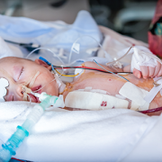
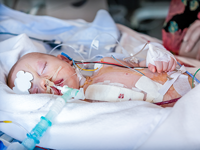
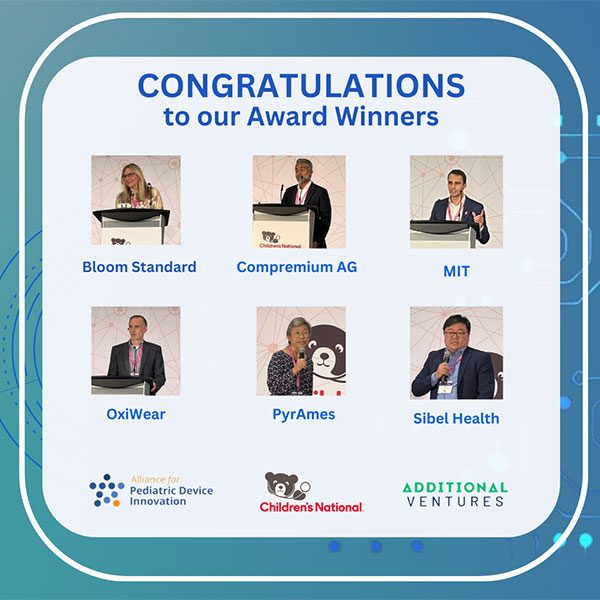 Six medical technology innovators focused on pediatric cardiology were selected to receive grants of $50,000 each in the “Make Your Medical Device Pitch for Kids!TM” competition in Toronto. The funds will help awardees bring their devices to the market and improve care for children with heart conditions.
The awardees, selected from a highly competitive field of ten finalists, are:
- Bloom Standard, Minneapolis – Autonomous, hands-free ultrasound
- Compremium AG, Bern, Switzerland – Noninvasive central venous pressure estimation for pediatric patients
- Massachusetts Institute of Technology, Cambridge, Mass. – Polymeric auxetic stent to treat pediatric aortic coarctation
- OxiWear, Arlington, Va. – Home measurement of oxygen levels in pediatric congenital heart disease
- PyrAmes Inc., Cupertino, Calif. – Improved, wearable, noninvasive pediatric blood pressure monitor
- Sibel Health, Chicago – Hospital-to-home monitoring for pediatric heart conditions
The competition is presented by the Alliance for Pediatric Device Innovation (APDI), a nonprofit consortium led by Children’s National Hospital and funded through the Food and Drug Administration (FDA), and Additional Ventures, a nonprofit focused on accelerating research progress and improving clinical care for individuals born with single ventricle heart defects. Along with grant funding, awardees gain access to support services and technical expertise provided by APDI and Additional Ventures in areas that include engineering, regulatory, reimbursement, clinical trials study design and data science services.
According to the Centers for Disease Control and Prevention, about 40,000 children are born annually with a congenital heart defect. Children with heart conditions need medical devices tailored to their specific physiological needs. There is a significant unmet need for pediatric devices designed to monitor and treat young patients effectively in cardiology, interventional cardiology, cardiac surgery and electrophysiology. This competitive grant program is designed to identify and support the development and commercialization of devices addressing these needs.
“Congratulations to our awardees, whose innovative technologies show great promise in advancing care for pediatric heart patients,” said Kolaleh Eskandanian, Ph.D., M.B.A., vice president and chief innovation officer at Children’s National and APDI program director and principal investigator. “We are thrilled to welcome this new cohort into our pediatric device accelerator, where they will have the opportunity to collaborate with clinician-scientists at Children’s National and connect to Additional Ventures’ network. Along with these collaborations, the awardees will benefit from a full range of APDI wraparound services designed to support the development of devices specifically for pediatric patients, helping them navigate the complex path to market.”
The competition was held in conjunction with the 12th Annual Symposium on Pediatric Device Innovation, presented by Children’s National and co-located with The MedTech Conference powered by AdvaMed. Focused on transforming pediatric care with exclusive innovations for children, this year’s symposium featured panel discussions and keynote presentations with leading experts in pediatrics and medical technology to exchange information and ideas on critical issues in pediatric device development and pediatric healthcare innovation gaps.
“Additional Ventures is thrilled to support this new class of innovators whose products will make a profound impact in the management and care of pediatric heart patients,” said Additional Ventures CEO Kristie Keller, Ph.D. “We welcome them to our growing community of inventors, researchers and clinicians, and we look forward to working together with our awarded teams and ADPI to bring these products to market. We hope that this competition both inspires and activates the community and brings much-needed new entrants and new ideas to pediatric-first device development.”
APDI is one of five nonprofit consortia in the FDA’s Pediatric Device Consortia grant program. It receives funding to provide a platform of services, expertise and grants that support pediatric innovators in bringing medical devices to the market that specifically address the unmet needs of children. Led by Children’s National, APDI partners include Johns Hopkins University, CIMIT at Mass General Brigham, Tufts Medical Center, MedStar Health Research Institute, MedTech Color and OrthoPediatrics Corp.