Tag Archive for: cognitive development

Newborn baby in a crib

Pioneering research center aims to revolutionize prenatal and neonatal health

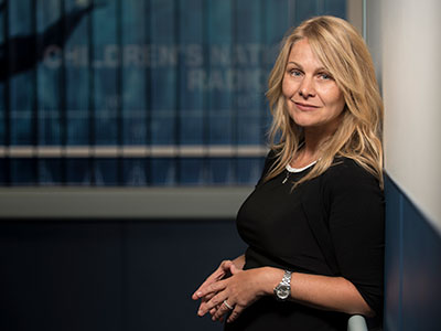
Catherine Limperopoulos, Ph.D., was drawn to understanding the developing brain, examining how early adverse environments for a mother can impact the baby at birth and extend throughout its entire lifetime. She has widened her lens – and expanded her team – to create the new Center for Prenatal, Neonatal & Maternal Health Research at Children’s National Hospital.

“Despite the obvious connection between mothers and babies, we know that conventional medicine often addresses the two beings separately. We want to change that,” said Dr. Limperopoulos, who also directs the Developing Brain Institute. “Given the current trajectory of medicine toward precision care and advanced imaging, we thought this was the right moment to channel our talent and resources into understanding this delicate and highly dynamic relationship.”

Moving the field forward

Since its establishment in July 2023, the new research center has gained recognition through high-impact scientific publications, featuring noteworthy studies exploring the early phases of human development.

Dr. Limperopoulos has been at the forefront of groundbreaking research, directing attention to the consequences of maternal stress on the unborn baby and the placenta. In addition, under the guidance of Kevin Cook, Ph.D., investigators published a pivotal study on the correlation between pain experienced by premature infants in the Neonatal Intensive Care Unit and the associated risks of autism and developmental delays.

Another area of research has focused on understanding the impact of congenital heart disease (CHD) on prenatal brain development, given the altered blood flow to the brain caused by these conditions during this period of rapid development. Led by Josepheen De Asis-Cruz, M.D., Ph.D., a research team uncovered variations in the functional connectivity of the brains of infants with CHD. In parallel, Nickie Andescavage, M.D., and her team employed advanced imaging techniques to identify potential biomarkers in infants with CHD, holding promise for guiding improved diagnostics and postnatal care. Separately, she is investigating the impact of COVID-19 on fetal brain development.

In the months ahead, the team plans to concentrate its efforts on these areas and several others, including the impact of infectious disease, social determinants of health and protecting developing brains from the negative impacts of maternal stress, pre-eclampsia and other conditions prevalent among expectant mothers.

Assembling a team

Given its robust research plan and opportunities for collaboration, the center pulled together expertise from across the hospital’s faculty and has attracted new talent from across the country, including several prominent faculty members:

  • Daniel Licht, M.D., has joined Children’s National to build a noninvasive optical device research group to better care for children with CHD. Dr. Licht brings decades of experience in pediatric neurology, psychiatry and critical care and is recognized internationally for his expertise in neurodevelopmental outcomes in babies with CHD.
  • Katherine L. Wisner, M.S., M.D., has accumulated extensive knowledge on the impact of maternal stress on babies throughout her career, and her deep background in psychiatry made her a natural addition to the center. While Dr. Wisner conducts research into the urgent need to prioritize maternal mental health, she will also be treating mothers as part of the DC Mother-Baby Wellness Initiative — a novel program based at Children’s National that allows mothers to more seamlessly get care for themselves and participate in mother-infant play groups timed to align with their clinical appointments.
  • Catherine J. Stoodley, B.S., M.S., D.Phil., brings extensive research into the role of the cerebellum in cognitive development. Dr. Stoodley uses clinical studies, neuroimaging, neuromodulation and behavioral testing to investigate the functional anatomy of the part of the brain responsible for cognition.
  • Katherine M. Ottolini, M.D., attending neonatologist, is developing NICU THRIVE – a research program studying the effects of tailored nutrition on the developing newborn brain, including the impact of fortifying human milk with protein, fat and carbohydrates. With a grant from the Gerber Foundation, Dr. Ottolini is working to understand how personalized fortification for high-risk babies could help them grow.

Early accolades

The new center brings together award-winning talent. This includes Yao Wu, Ph.D., who recently earned the American Heart Association’s Outstanding Research in Pediatric Cardiology award for her groundbreaking work in CHD, particularly for her research on the role of altered placental function and neurodevelopmental outcomes in toddlers with CHD. Dr. Wu became the third Children’s National faculty member to earn the distinction, joining an honor roll that includes Dr. Limperopoulos and David Wessel, M.D., executive vice president and chief medical officer.

Interim Chief Academic Officer Catherine Bollard, M.D., M.B.Ch.B., said the cross-disciplinary collaboration now underway at the new center has the potential to make a dramatic impact on the field of neonatology and early child development. “This group epitomizes the Team Science approach that we work tirelessly to foster at Children’s National,” Dr. Bollard said. “Given their energetic start, we know these scientists and physicians are poised to tackle some of the toughest questions in maternal-fetal medicine and beyond, which will improve outcomes for our most fragile patients.”

A blue background showcases a brain made of gears, symbolizing the intricate workings of the mind.A blue background showcases a brain made of gears, symbolizing the intricate workings of the mind.

Measuring preschoolers’ cognitive development in Latin America

A blue background showcases a brain made of gears, symbolizing the intricate workings of the mind.A blue background showcases a brain made of gears, symbolizing the intricate workings of the mind.

Neurodevelopment occurs at a rapid pace during the preschool years, research shows.

Measuring and tracking development in early childhood is important for understanding how to best support long-term positive health outcomes for children. However, the art of selecting measurement tools that are appropriate for a given study sample is complex; variations in language, cultural relevance and adaptations, availability of normative data, and more can impact the administration and interpretation of study findings.

In a recent study published in the Journal of Pediatric Psychology, the authors systematically review the standardized neurodevelopmental assessments used to study preschool-aged children’s cognitive development in Spanish-speaking Latin America.

“Thinking critically about the ways we measure these outcomes is an important first step in conducting ethical and accurate global research,” said Meagan Williams, M.S.P.H., C.C.R.C., senior clinical research coordinator for the division of Infectious Diseases and the Zickler Family Prenatal Pediatrics Institute at Children’s National Hospital and corresponding author of the study.

The hold-up in the field

The field of child development research is complicated by the presence and use of many different neurodevelopmental assessment tools, only some of which are standardized and use norms to allow researchers to compare their cohorts’ performance with population-level data.

“Many cognitive assessments, including some of the most commonly used assessments identified in our review of research conducted in Spanish-speaking Latin America, have only been officially normed in the United States,” Williams added. “Others have been translated into Spanish and normed in large countries such as Mexico or Spain, but Latin America is a large and culturally diverse region, and it is unclear whether these normative samples are generalizable and culturally relevant to other regions of Spanish-speaking Latin America.”

This systematic review outlined 41 different neurodevelopmental assessments used in 97 studies, including information about each study’s sample, use of each measurement tool and findings related to child cognitive development. The review goes into detail about the most widely used assessments, including background information of each assessment and trends in how authors of the studies in this review reported their methods and results.

Moving the field forward

When researchers use similar methods and valid outcome measures across studies, it becomes possible to combine data between cohorts and draw stronger conclusions about child outcomes. For example, The Wechsler Preschool and Primary Scale of Intelligence (WPPSI) was identified as the most used tool to measure intelligence of young children ages 2-6 in the systematic review. The WPPSI is also one of many assessments that Williams’s research team, led by Sarah Mulkey, M.D., is using to measure neurodevelopment among children exposed to Zika virus in utero.

“If researchers studying longitudinal outcomes after Zika virus exposure also use the WPPSI for their study samples – and if they openly report on their methods, adaptations, and sample characteristics – then it may be possible to harmonize data from multiple cohorts and draw stronger conclusions across studies,” Williams said.

But even the most popular and widespread assessments such as the WPPSI are not without their limitations. While norms exist for regions such as the United States and Mexico, additional work needs to be done to appropriately adapt and validate these tools for use in other populations.

Benefiting patients

Neurodevelopment occurs at a rapid pace during the preschool years. The earlier neurodevelopmental delays can be identified, the earlier experts can administer interventions to respond to these delays and help children get back on track with their peers.

This is particularly salient for research conducted in low- and middle-income countries, where socioeconomic, environmental and other exposures that are known to impact child development differ from those in higher-resource settings. Internationally, there appears to be significant variability in not only the assessment tools which are selected for use, but also in the methods reported across studies to conduct and interpret these assessments.

“By coming together as a field and prioritizing the use and validation of common and culturally appropriate assessments, we can better understand child outcome data on both an individual study level as well as on a broader population level, which will lead to a better understanding of the unique needs and strengths of the children we serve,” Williams said.

The team at Children’s National has been studying the neurodevelopment of children exposed to the Zika virus in Colombia since the beginning of the Zika epidemic.

“As our study on Zika virus has overlapped with the COVID-19 pandemic, we have had the opportunity to collect child outcome data at a very interesting time in history. We are excited that this review revealed that we chose a popular and well-studied assessment for our data collection in this region and we look forward to publishing our 5-year outcome data (which will include WPPSI data) in the coming year,” Williams and Mulkey concluded.

Dr. Mulkey is committed to studying the long-term neurodevelopmental impacts that viruses like Zika and SARS-CoV-2 have on infants born to mothers who were infected during pregnancy through research with the Congenital Infection Program at Children’s National and in collaboration with colleagues in Colombia.

Additional Children’s National authors include Madison Berl, Ph.D.

pregnant woman by window

Stress during pregnancy may hinder cognitive development

pregnant woman by window

This is the first study to shed light on an important link between altered in-utero fetal brain development and the long-term cognitive development consequences for fetuses exposed to high levels of toxic stress during pregnancy.

Women’s elevated anxiety, depression and stress during pregnancy altered key features of the fetal brain, which subsequently decreased their offspring’s cognitive development at 18 months. These changes also increased internalizing and dysregulation behaviors, according to a new study by Children’s National Hospital published in JAMA Network Open. Researchers followed a cohort of 97 pregnant women and their babies. The findings further suggest that persistent psychological distress after the baby is born may influence the parent-child interaction and infant self-regulation.

This is the first study to shed light on an important link between altered in-utero fetal brain development and the long-term cognitive development consequences for fetuses exposed to high levels of toxic stress during pregnancy. While in the womb, the researchers observed changes in the sulcal depth and left hippocampal volume, which could explain the neurodevelopment issues seen after birth. Once they grow into toddlers, these children may experience persistent social-emotional problems and have difficulty establishing positive relationships with others, including their mothers. To further confirm this, future studies with a larger sample size that reflect more regions and populations are needed.

“By identifying the pregnant women with elevated levels of psychological distress, clinicians could recognize those babies who are at risk for later neurodevelopmental impairment and might benefit from early, targeted interventions,” said Catherine Limperopoulos, Ph.D., chief and director of the Developing Brain Institute at Children’s National and senior author of the study.

Catherine Limperopoulos

“By identifying the pregnant women with elevated levels of psychological distress, clinicians could recognize those babies who are at risk for later neurodevelopmental impairment and might benefit from early, targeted interventions,” said Catherine Limperopoulos, Ph.D., chief and director of the Developing Brain Institute at Children’s National and senior author of the study.

Regardless of their socioeconomic status, about one of every four pregnant women suffers from stress-related symptoms, the most common pregnancy complication. The relationship between altered fetal brain development, prenatal maternal psychological distress and long-term neurodevelopmental outcomes remain unknown. Studying in utero fetal brain development poses challenges due to fetal and maternal movements, imaging technology, signal-to-noise ratio issues and changes in brain growth.

All pregnant participants were healthy, most had some level of education and were employed. To quantify prenatal maternal stress, anxiety and depression, the researchers used validated self-reported questionnaires. Fetal brain volumes and cortical folding were measured from three-dimensional reconstructed images derived from MRI scans. Fetal brain creatine and choline were quantified using proton magnetic resonance spectroscopy. The 18-month child neurodevelopment was measured using validated scales and assessments.

This study builds upon previous work from the Developing Brain Institute led by Limperopoulos, which discovered that anxiety in pregnant women appears to affect the brain development of their babies. Her team also found that maternal mental health, even for women with high socioeconomic status, alters the structure and biochemistry of the developing fetal brain. The growing evidence underscores the importance of mental health support for pregnant women.

“We’re looking at shifting the health care paradigm and adopting these changes more broadly to better support moms,” said Limperopoulos. “What’s clear is early interventions could help moms reduce their stress, which can positively impact their symptoms and thereby their baby long after birth.”

Pregnant-Mom

Safeguarding fetal brain health in pregnancies complicated by CHD

Pregnant-Mom

During the last few weeks of pregnancy, certain regions of the fetal brain experience exponential growth but also are more vulnerable to injury during that high-growth period.

Yao Wu, Ph.D., a research postdoctoral fellow in the Developing Brain Research Laboratory at Children’s National Health System, has received a Thrasher Research Fund early career award to expand knowledge about regions of the fetal brain that are vulnerable to injury from congenital heart disease (CHD) during pregnancy.

CHD, the most common birth defect, can have lasting effects, including overall health issues; difficulty achieving milestones such as crawling, walking or running; and missed days at daycare or school, according to the Centers for Disease Control and Prevention. Brain injury is a major complication for infants born with CHD. Catherine Limperopoulos, Ph.D., director of Children’s brain imaging lab, was the first to provide in vivo evidence that fetal brain growth and metabolism in the third trimester of pregnancy is impaired within the womb.

“It remains unclear which specific regions of the fetal brain are more vulnerable to these insults in utero,” Limperopoulos says. “We first need to identify early brain abnormalities attributed to CHD and understand their impact on infants’ later behavioral and cognitive development in order to better counsel parents and effectively intervene during the prenatal period to safeguard brain health.”

During the last few weeks of pregnancy, certain regions of the fetal brain experience exponential growth but also are more vulnerable to injury during that high-growth period. The grant, $26,749 over two years, will underwrite “Brain Development in Fetuses With Congenital Heart Disease,” research that enables Wu to utilize quantitative, non-invasive magnetic resonance imaging (MRI) to compare fetal brain development in pregnancies complicated by CHD with brain development in healthy fetuses of the same gestational age.Wu will leverage quantitative, in vivo 3-D volumetric MRI to compare overall fetal and neonatal brain growth as well as growth in key regions including cortical grey matter, white matter, deep grey matter, lateral ventricles, external cerebrospinal fluid, cerebellum, brain stem, amygdala and the hippocampus.

The research is an offshoot of a prospective study funded by the National Institutes of Health that uses advanced imaging techniques to record brain growth in 50 fetuses in pregnancies complicated by CHD who need open heart surgery and 50 healthy fetuses. MRI studies are conducted during the second trimester (24 to 28 weeks gestational age), third trimester (33 to 37 weeks gestational age) and shortly after birth but before surgery. In addition, fetal and neonatal MRI measurements will be correlated with validated scales that measure infants’ and toddlers’ overall development, behavior and social/emotional maturity.

“I am humbled to be selected for this prestigious award,” Wu says. “The findings from our ongoing work could be instrumental in identifying strategies for clinicians and care teams managing high-risk pregnancies to optimize fetal brain development and infants’ overall quality of life.”