Tag Archive for: cerebral cortex

structure of EGFR

Study suggests EGFR inhibition reverses alterations induced by hypoxia

structure of EGFR

The study suggests that specific molecular responses modulated by EGFR (seen here) may be targeted as a therapeutic strategy for HX injury in the neonatal brain.

Hypoxic (HX) encephalopathy is a major cause of death and neurodevelopmental disability in newborns. While it is known that decreased oxygen and energy failure in the brain lead to neuronal cell death, the cellular and molecular mechanisms of HX-induced neuronal and glial cell damage are still largely undefined.

Panagiotis Kratimenos, M.D., and colleagues from the Center for Neuroscience Research at the Children’s National Research Institute, discovered increased expression of activated-epidermal growth factor receptor (EGFR) in affected cortical areas of neonates with HX and decided to further investigate the functional role of EGFR-related signaling pathways in the cellular and molecular changes induced by HX in the cerebral cortex.

The researchers found that HX-induced activation of EGFR and Ca2+/calmodulin kinase IV (CaMKIV) caused cell death and pathological alterations in neurons and glia. EGFR blockade inhibited CaMKIV activation, attenuated neuronal loss, increased oligodendrocyte proliferation and reversed HX-induced astrogliosis.

The researchers also performed, for the first time, high-throughput transcriptomic analysis of the cortex to define molecular responses to HX and to uncover genes specifically involved in EGFR signaling in brain injury. Their results indicate that specific molecular responses modulated by EGFR may be targeted as a therapeutic strategy for HX injury in the neonatal brain.

This study defines many new exciting avenues of scientific exploration to further elucidate the beneficial impact of EGFR blockade on perinatal brain injury at the cellular and molecular levels. This analysis could potentially result in the identification of new therapeutic targets associated with EGFR signaling in the developing mammalian brain that are linked with specific long-term abnormalities caused by perinatal brain injury.

Children’s National researchers who contributed to this study include Panagiotis Kratimenos, M.D., Ioannis Koutroulis, M.D., Ph.D., M.B.A., Susan Knoblach, Ph.D., Payal Banerjee, Surajit Bhattacharya, Ph.D., Maria Almira-Suarez, M.D., and Vittorio Gallo, Ph.D.

Read the full article in iScience.

illustration of the brain

New research provides glimpse into landscape of the developing brain

illustration of the brain

Stem and progenitor cells exhibit diversity in early brain development that likely contributes to later neural complexity in the adult cerebral cortex, this according to a new study in Science Advances. This research expands on existing ideas about brain development, and could significantly impact the clinical care of neurodevelopmental diseases in the future.

Stem and progenitor cells exhibit diversity in early brain development that likely contributes to later neural complexity in the adult cerebral cortex, this according to a study published Nov. 6, 2020, in Science Advances. Researchers from the Center for Neuroscience Research (CNR) at Children’s National Hospital say this research expands on existing ideas about brain development, and could significantly impact the clinical care of neurodevelopmental diseases in the future. The study was done in collaboration with a research team at Yale University led by Nenad Sestan, M.D, Ph.D.

“Our study provides a new glimpse into the landscape of the developing brain. What we are seeing are new complex families of cells very early in development,” says Tarik Haydar, Ph.D., director of CNR at Children’s National, who led this study. “Understanding the role of these cells in forming the cerebral cortex is now possible in a way that wasn’t possible before.”

The cerebral cortex emerges early in development and is the seat of higher-order cognition, social behavior and motor control. While the rich neural diversity of the cerebral cortex and the brain in general is well-documented, how this variation arises is relatively poorly understood.

“We’ve shown in our previous work that neurons generated from different classes of cortical stem and progenitor cells have different functional properties,” says William Tyler, Ph.D., CNR research faculty member and co-first author of the study. “Part of the reason for doing this study was to go back and try to classify all the different progenitors that exist so that eventually we can figure out how each contributes to the diversity of neurons in the adult brain.”

Using a preclinical model, the researchers were able to identify numerous groups of cortical stem and precursor cells with distinct gene expression profiles. The team also found that these cells showed early signs of lineage diversification likely driven by transcriptional priming, a process by which a mother cell produces RNA for the sole purpose of passing it on to its daughter cells for later protein production.

Tarik Haydar

“Our study provides a new glimpse into the landscape of the developing brain. What we are seeing are new complex families of cells very early in development,” says Tarik Haydar, Ph.D., director of CNR at Children’s National, who led this study. “Understanding the role of these cells in forming the cerebral cortex is now possible in a way that wasn’t possible before.”

Using novel trajectory reconstruction methods, the team observed distinct developmental streams linking precursor cell types to particular excitatory neurons. After comparing the dataset of the preclinical model to a human cell database, notable similarities were found, such as the surprising cross-species presence of basal radial glial cells (bRGCs), an important type of progenitor cell previously thought to be found mainly in the primate brain.

“At a very high level, the study is important because we are directly testing a fundamental theory of brain development,” says Zhen Li, Ph.D., CNR research postdoctoral fellow and co-first author of the study. The results add support to the protomap theory, which posits that early stem and progenitor diversity paves the way for later neuronal diversity and cortical complexity. Furthermore, the results also hold exciting translational potential.

“There is evidence showing that neurodevelopmental diseases affect different populations of the neural stem cells differently,” says Dr. Li. “If we can have a better understanding of the complexity of these neural stem cells there is huge implication of disease prevention and treatment in the future.”

“If we can understand how this early landscape is affected in disorders, we can predict the resulting changes to the cortical architecture and then very narrowly define ways that groups of cells behave in these disorders,” adds Dr. Haydar. “If we can understand how the cortex normally achieves its complex architecture, then we have key entry points into improving the clinical course of a given disorder and improving quality of life.”

Future topics the researchers hope to study include the effects of developmental changes on brain function, the origin and operational importance of bRGCs, and the activity, connections and cognitive features enabled by different families of neurons.

illustration of brain showing cerebellum

Focusing on the “little brain” to rescue cognition

illustration of brain showing cerebellum

Research faculty at Children’s National in Washington, D.C., with colleagues recently published a review article in Nature Reviews Neuroscience that covers the latest research about how abnormal development of the cerebellum leads to a variety of neurodevelopmental disorders.

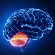
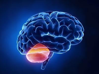
Cerebellum translates as “little brain” in Latin. This piece of anatomy – that appears almost separate from the rest of the brain, tucked under the two cerebral hemispheres – long has been known to play a pivotal role in voluntary motor functions, such as walking or reaching for objects, as well as involuntary ones, such as maintaining posture.

But more recently, says Aaron Sathyanesan, Ph.D., a postdoctoral research fellow at the Children’s Research Institute, the research arm of Children’s National  in Washington, D.C., researchers have discovered that the cerebellum is also critically important for a variety of non-motor functions, including cognition and emotion.

Sathyanesan, who studies this brain region in the laboratory of Vittorio Gallo, Ph.D., Chief Research Officer at Children’s National and scientific director of the Children’s Research Institute, recently published a review article with colleagues in Nature Reviews Neuroscience covering the latest research about how altered development of the cerebellum contributes to a variety of neurodevelopmental disorders.

These disorders, he explains, are marked by problems in the nervous system that arise while it’s maturing, leading to effects on emotion, learning ability, self-control, or memory, or any combination of these. They include diagnoses as diverse as intellectual disability, autism spectrum disorder (ASD), attention-deficit/hyperactivity disorder and Down syndrome.

“One reason why the cerebellum might be critically involved in each of these disorders,” Sathyanesan says, “is because its developmental trajectory takes so long.”

Unlike other brain structures, which have relatively short windows of development spanning weeks or months, the principal cells of the cerebellum – known as Purkinje cells – start to differentiate from stem cell precursors at the beginning of the seventh gestational week, with new cells continuing to appear until babies are nearly one year old.  In contrast, cells in the neocortex, a part of the brain involved in higher-order brain functions such as cognition, sensory perception and language is mostly finished forming while fetuses are still gestating in the womb.

This long window for maturation allows the cerebellum to make connections with other regions throughout the brain, such as extensive connections with the cerebral cortex, the outer layer of the cerebrum that plays a key role in perception, attention, awareness, thought, memory, language and consciousness. It also allows ample time for things to go wrong.

“Together,” Sathyanesan says, “these two characteristics are at the root of the cerebellum’s involvement in a host of neurodevelopmental disorders.”

For example, the review article notes, researchers have discovered both structural and functional abnormalities in the cerebellums of patients with ASD. Functional magnetic resonance imaging (MRI), an imaging technique that measures activity in different parts of the brain, suggests that significant differences exist between connectivity between the cerebellum and cortex in people with ASD compared with neurotypical individuals. Differences in cerebellar connectivity are also evident in resting-state functional connectivity MRI, an imaging technique that measures brain activity in subjects when they are not performing a specific task. Some of these differences appear to involve patterns of overconnectivity to different brain regions, explains Sathyanesan; other differences suggest that the cerebellums of patients with ASD don’t have enough connections to other brain regions.

These findings could clarify research from Children’s National and elsewhere that has shown that babies born prematurely often sustain cerebellar injuries due to multiple hits, including a lack of oxygen supplied by infants’ immature lungs, he adds. Besides having a sibling with ASD, premature birth is the most prevalent risk factor for an ASD diagnosis.

The review also notes that researchers have discovered structural changes in the cerebellums of patients with Down syndrome, who tend to have smaller cerebellar volumes than neurotypical individuals. Experimental models of this trisomy recapitulate this difference, along with abnormal connectivity to the cerebral cortex and other brain regions.

Although the cerebellum is a pivotal contributor toward these conditions, Sathyanesan says, learning more about this brain region helps make it an important target for treating these neurodevelopmental disorders. For example, he says, researchers are investigating whether problems with the cerebellum and abnormal connectivity could be lessened through a non-invasive form of brain stimulation called transcranial direct current stimulation or an invasive one known as deep brain stimulation. Similarly, a variety of existing pharmaceuticals or new ones in development could modify the cerebellum’s biochemistry and, consequently, its function.

“If we can rescue the cerebellum’s normal activity in these disorders, we may be able to alleviate the problems with cognition that pervade them all,” he says.

In addition to Sathyanesan and Senior Author Gallo, Children’s National study co-authors include Joseph Scafidi, D.O., neonatal neurologist; Joy Zhou and Roy V. Sillitoe, Baylor College of Medicine; and Detlef H. Heck, of University of Tennessee Health Science Center.

Financial support for research described in this post was provided by the National Institute of Neurological Disorders and Stroke under grant numbers 5R01NS099461, R01NS089664, R01NS100874, R01NS105138 and R37NS109478; the Hamill Foundation; the Baylor College of Medicine Intellectual and Developmental Disabilities Research Center under grant number U54HD083092; the University of Tennessee Health Science Center (UTHSC) Neuroscience Institute; the UTHSC Cornet Award; the National Institute of Mental Health under grant number R01MH112143; and the District of Columbia Intellectual and Developmental Disabilities Research Center under grant number U54 HD090257.