Tag Archive for: cerebellum

Purkinje cell

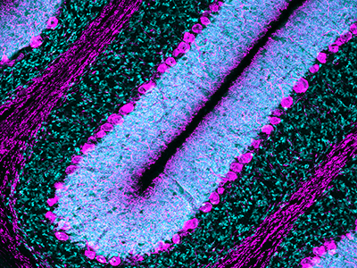
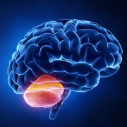
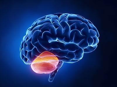
Premature birth disrupts Purkinje cell function, resulting in locomotor learning deficits

Purkinje cell

Children’s National Hospital researchers explored how preterm birth disrupts Purkinje cell function, resulting in locomotor learning deficits.

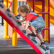
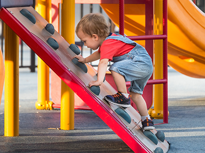
As the care of preterm babies continues to improve, neonatologists face new challenges to ensure babies are protected from injury during critical development of the cerebellum during birth and immediately after birth. How does this early injury affect locomotor function, and to what extent are clinicians able to protect the brain of preterm babies?
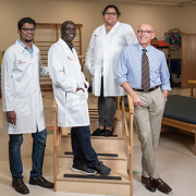
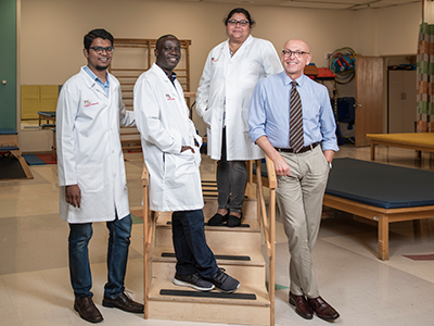
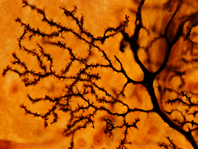
A new peer-reviewed study by Aaron Sathyanesan, Ph.D., Panagiotis Kratimenos, M.D., Ph.D., and Vittorio Gallo, Ph.D., published in the Proceedings of the National Academy of Sciences of the United States of America (PNAS), explores exactly what neural circuitry of the cerebellum is affected due to complications that occur around the time of birth causing these learning deficits, and finds a specific type of neurons — Purkinje cells — to play a central role.

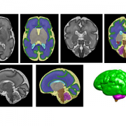
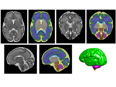
Up until now, there has been a sparsity of techniques available to measure neuronal activity during locomotor learning tasks that engage the cerebellum. To surmount this challenge, Children’s National used a multidisciplinary approach, bringing together a team of neuroscientists with neonatologists who leveraged their joint expertise to devise a novel and unique way to measure real-time Purkinje cell activity in a pre-clinical model with clinical relevance to humans.

Researchers measured neural circuit function by pairing GCaMP6f fiber photometry, used to measure neuronal activity in the brain of a free moving subject, with an ErasmusLadder, in which it needs to travel from point A to point B on a horizontal ladder with touch-sensitive rungs that register the type and length of steps. By introducing a sudden obstacle to movement, researchers observed how the subject coped and learned accordingly to avoid this obstacle. By playing a high-pitch tone just before the obstacle was introduced, researchers were able to measure how quickly the subjects were able to anticipate the obstacle and adjust their steps accordingly. Subjects with neonatal brain injury and normal models were run through a series of learning trials while simultaneously monitoring brain activity. In this way, the team was able to quantify cerebellum-dependent locomotor learning and adaptive behavior, unlocking a functional and mechanistic understanding of behavioral pathology that was previously unseen in this field.

In addition to showing that normal Purkinje cells are highly active during movement on the ErasmusLadder, the team explored the question of whether Purkinje cells of injured pre-clinical models were generally non-responsive to any kind of stimuli. They found that while Purkinje cells in injured subjects responded to puffs of air, which generally cue the subject to start moving on the ErasmusLadder, dysfunction in these cells was specific to the period of adaptive learning. Lastly, through chemogenetic inhibition, which specifically silences neonatal Purkinje cell activity, the team was able to mimic the effects of perinatal cerebellar injury, further solidifying the role of these cells in learning deficits.

The study results have implications for clinical practice. As the care of premature babies continues to improve, neonatologists face new challenges to ensure that babies not only survive but thrive. They need to find ways to prevent against the lifelong impacts that preterm birth would otherwise have on the cerebellum and developing brain.

Read the full press release here.

Read the full journal article here.

Vittorio Gallo

Special issue of “Neurochemical Research” honors Vittorio Gallo, Ph.D.

Vittorio Gallo

Investigators from around the world penned manuscripts that were assembled in a special issue of “Neurochemical Research” that honors Vittorio Gallo, Ph.D., for his leadership in the field of neural development and regeneration.

At a pivotal moment early in his career, Vittorio Gallo, Ph.D., was accepted to work with Professor Giulio Levi at the Institute for Cell Biology in Rome, a position that leveraged courses Gallo had taken in neurobiology and neurochemistry, and allowed him to work in the top research institute in Italy directed by the Nobel laureate, Professor Rita Levi-Montalcini.

For four years as a student and later as Levi’s collaborator, Gallo focused on amino acid neurotransmitters in the brain and mechanisms of glutamate and GABA release from nerve terminals. Those early years cemented a research focus on glutamate neurotransmission that would lead to a number of pivotal publications and research collaborations that have spanned decades.

Now, investigators from around the world who have worked most closely with Gallo penned tributes in the form of manuscripts that were assembled in a special issue of “Neurochemical Research” that honors Gallo “for his contributions to our understanding of glutamatergic and GABAergic transmission during brain development and to his leadership in the field of neural development and regeneration,” writes guest editor Arne Schousboe, of the University of Copenhagen in Denmark.

Dr. Gallo as a grad student

Vittorio Gallo, Ph.D. as a 21-year-old mustachioed graduate student.

“In spite of news headlines about competition in research and many of the negative things we hear about the research world, this shows that research is also able to create a community around us,” says Gallo, chief research officer at Children’s National Hospital and scientific director for the Children’s National Research Institute.

As just one example, he first met Schousboe 44 years ago when Gallo was a 21-year-old mustachioed graduate student.

“Research can really create a sense of community that we carry on from the time we are in training, nurture as we meet our colleagues at periodic conferences, and continue up to the present. Creating community is bi-directional: influencing people and being influenced by people. People were willing to contribute these 17 articles because they value me,” Gallo says. “This is a lot of work for the editor and the people who prepared papers for this special issue.”

In addition to Gallo publishing more than 140 peer-reviewed papers, 30 review articles and book chapters, Schousboe notes a number of Gallo’s accomplishments, including:

  • He helped to develop the cerebellar granule cell cultures as a model system to study how electrical activity and voltage-dependent calcium channels modulate granule neuron development and glutamate release.
  • He developed a biochemical/neuropharmacological assay to monitor the effects of GABA receptor modulators on the activity of GABA chloride channels in living neurons.
  • He and Maria Usowicz used patch-clamp recording and single channel analysis to demonstrate for the first time that astrocytes express glutamate-activated channels that display functional properties similar to neuronal counterparts.
  • He characterized one of the spliced isoforms of the AMPA receptor subunit gene Gria4 and demonstrated that this isoform was highly expressed in the cerebellum.
  • He and his Children’s National colleagues demonstrated that glutamate and GABA regulate oligodendrocyte progenitor cell proliferation and differentiation.
Purkinje cells

Purkinje cells are large neurons located in the cerebellum that are elaborately branched like interlocking tree limbs and represent the only source of output for the entire cerebellar cortex.

Even the image selected to grace the special issue’s cover continues the theme of continuity and leaving behind a legacy. That image of Purkinje cells was created by a young scientist who works in Gallo’s lab, Aaron Sathyanesan, Ph.D. Gallo began his career working on the cerebellum – a region of the brain important for motor control – and now studies with a team of scientists and clinician-scientists Purkinje cells’ role in locomotor adaptive behavior and how that is disrupted after neonatal brain injury.

“These cells are the main players in cerebellar circuitry,” Gallo says. “It’s a meaningful image because goes back to my roots as a graduate student and is also an image that someone produced in my lab early in his career. It’s very meaningful to me that Aaron agreed to provide this image for the cover of the special issue.”

T2-Weighted Magnetic Resonance (MR) Imaging Brain Segmentation

Maternal mental health alters structure and biochemistry of developing fetal brain

Even when pregnant women have uncomplicated pregnancies and high socioeconomic status, when they experience elevated anxiety, stress or depression these prenatal stressors can alter the structure of the developing fetal brain and disrupt its biochemistry, according to Children’s National Hospital research published online Jan. 29, 2020, in JAMA Network Open.

The Children’s National research findings “have enormous scientific, clinical and public health implications,” Charles A. Nelson III, Ph.D.,  Boston Children’s Hospital, writes in a companion editorial.

“Previously we found that 65% of pregnant women who received a diagnosis of fetal congenital heart disease had elevated levels of stress. It’s concerning but not surprising that pregnant women who wonder if their baby will need open heart surgery would feel stress,” says Catherine Limperopoulos, Ph.D., director of the Center for the Developing Brain at Children’s National and the study’s senior author. “In this latest study, we ran the same panel of questionnaires and were surprised to find a high proportion of otherwise healthy pregnant women whose unborn babies are doing well also report high levels of stress.”

Anxiety and depression are the most common mental health problems during pregnancy. To learn more about the implications for the developing fetal brain, the Children’s National research team recruited 119 healthy volunteers with low-risk pregnancies from obstetric clinics in Washington, D.C., from Jan. 1, 2016, to April 17, 2019. The women’s mean age was 34.4 years old. All were high school graduates, 83% were college graduates, and 84% reported professional employment.

T2-Weighted Magnetic Resonance (MR) Imaging Brain Segmentation.

T2-Weighted Magnetic Resonance (MR) Imaging Brain Segmentation. Segmentation results of total brain (orange), cortical gray matter (green), white matter (blue), deep gray matter (brown), brainstem (yellow), cerebellum (light blue), left hippocampus (purple) and right hippocampus (red) on a 3-Dimensional reconstructed T2-weighted MR image of a fetus at 26.4 gestational weeks. The hippocampus plays a central role in memory and behavioral inhibition and contains high concentrations of corticosteroid receptors and, thus, this brain region is sensitive to stress. Credit: JAMA Network Open.

The team performed 193 fetal brain magnetic resonance imaging (MRI) sessions between 24-40 weeks gestation and measured the volume of the total fetal brain as well as the cortical gray matter, white matter, deep gray matter, cerebellum, brainstem and hippocampus volumes. On the same day as their MRI visit, the pregnant women completed validated questionnaires to measure maternal stress, anxiety and depression, answering questions such as “how do you feel right now,” “how do you generally feel” as well as the degree of stressful feelings they experienced the month prior.

Of the pregnant women in the study:

  • 27% tested positive for stress
  • 26% tested positive for anxiety
  • 11% tested positive for depression
  • Maternal anxiety and stress were associated with increased fetal cortical gyrification
  • Elevated maternal depression was associated with decreased creatine and choline levels in the fetal brain
  • Maternal stress scores decreased with increasing gestational age, while anxiety and depression did not

“We report for the first time that maternal psychological distress may be associated with increased fetal local gyrification index in the frontal and temporal lobes,” says Yao Wu, Ph.D., a research associate working with Limperopoulos at Children’s National and the study’s lead author. “We also found an association with left fetal hippocampal volume, with maternal psychological distress selectively stunting the left hippocampal volumetric growth more than the right. And elevated maternal depression was associated with decreased creatine and choline levels in the fetal brain,” Wu adds.

Late in pregnancy – at the time these women were recruited into the cohort study – the fetal brain grows exponentially and key metabolite levels also rise. Creatine facilitates recycling of adenosine triphosphate, the cell’s energy currency. Typically, levels of this metabolite rise, denoting rapid changes and higher cellular maturation; creatine also is known to support cognitive function. Choline levels also typically rise, marking cell membrane turnover as new cells are generated and support memory, mental focus and concentration.

“These women were healthy, and of high socioeconomic status and educational level, leading us to conclude that the prevalence of prenatal maternal psychological distress may be underestimated,” Limperopoulos adds. “While stress is an everyday reality for most of us, this is different because elevated stress during pregnancy can alter fetal brain programming. Our findings underscore the critical need to universally screen all pregnant women for prenatal psychological distress, even young mothers whose pregnancies wouldn’t otherwise raise red flags.”

In addition to Limperopoulos and Wu, Children’s National study co-authors include Yuan-Chiao Lu, Ph.D., research associate; Marni Jacobs, Ph.D., biostatistician; Subechhya Pradhan, Ph.D., research faculty; Kushal Kapse, MS, staff engineer; Li Zhao, Ph.D., research faculty; Nickie Niforatos-Andescavage, M.D., neonatologist; Gilbert Vezina, M.D., director of the neuroradiology program; and Adré  J. du Plessis, M.B.Ch.B., director, Fetal Medicine Institute. Research coordinators Catherine Lopez, MS, Kathryn Lee Bannantine, BSN, and Jessica Lynn Quistorff, MPH, assisted with subject recruitment.

Financial support for the research described in this post was provided by the National Institutes of Health under grant No. RO1 HL116585-01 and the Thrasher Research Fund under Early Career award No. 14764.

Journal Reference:
Yao Wu, Yuan-Chiao Lu, Marni Jacobs, Subechhya Pradhan, Kushal Kapse, Li Zhao, Nickie Niforatos-Andescavage, Gilbert Vezina, Adré J. du Plessis, Catherine Limperopoulos. “Association of prenatal maternal psychological distress with fetal brain growth, metabolism and cortical maturation,” JAMA Network Open, 3(1): e1919940, 2020

Catherine Limperopoulos

Stressful pregnancies can leave fingerprint on fetal brain

Catherine Limperopoulos

“We were alarmed by the high percentage of pregnant women with a diagnosis of a major fetal heart problem who tested positive for stress, anxiety and depression,” says Catherine Limperopoulos, Ph.D., director of the Center for the Developing Brain at Children’s National and the study’s corresponding author.

When a diagnosis of fetal congenital heart disease causes pregnant moms to test positive for stress, anxiety and depression, powerful imaging can detect impaired development in key fetal brain regions, according to Children’s National Hospital research published online Jan. 13, 2020, in JAMA Pediatrics.

While additional research is needed, the Children’s National study authors say their unprecedented findings underscore the need for universal screening for psychological distress as a routine part of prenatal care and taking other steps to support stressed-out pregnant women and safeguard their newborns’ developing brains.

“We were alarmed by the high percentage of pregnant women with a diagnosis of a major fetal heart problem who tested positive for stress, anxiety and depression,” says Catherine Limperopoulos, Ph.D., director of the Center for the Developing Brain at Children’s National and the study’s corresponding author. “Equally concerning is how prevalent psychological distress is among pregnant women generally. We report for the first time that this challenging prenatal environment impairs regions of the fetal brain that play a major role in learning, memory, coordination, and social and behavioral development, making it all the more important for us to identify these women early during pregnancy to intervene,” Limperopoulos adds.

Congenital heart disease (CHD), structural problems with the heart, is the most common birth defect. Still, it remains unclear how exposure to maternal stress impacts brain development in fetuses with CHD.

The multidisciplinary study team enrolled 48 women whose unborn fetuses had been diagnosed with CHD and 92 healthy women with uncomplicated pregnancies. Using validated screening tools, they found:

  • 65% of pregnant women expecting a baby with CHD tested positive for stress
  • 27% of women with uncomplicated pregnancies tested positive for stress
  • 44% of pregnant women expecting a baby with CHD tested positive for anxiety
  • 26% of women with uncomplicated pregnancies tested positive for anxiety
  • 29% of pregnant women expecting a baby with CHD tested positive for depression and
  • 9% women with uncomplicated pregnancies tested positive for depression

All told, they performed 223 fetal magnetic resonance imaging sessions for these 140 fetuses between 21 and 40 weeks of gestation. They measured brain volume in cubic centimeters for the total brain as well as volumetric measurements for key regions such as the cerebrum, cerebellum, brainstem, and left and right hippocampus.

Maternal stress and anxiety in the second trimester were associated with smaller left hippocampi and smaller cerebellums only in pregnancies affected by fetal CHD. What’s more, specific regions — the hippocampus head and body and the left cerebellar lobe – were more susceptible to stunted growth. The hippocampus is key to memory and learning, while the cerebellum controls motor coordination and plays a role in social and behavioral development.

The hippocampus is a brain structure that is known to be very sensitive to stress. The timing of the CHD diagnosis may have occurred at a particularly vulnerable time for the developing fetal cerebellum, which grows faster than any other brain structure in the second half of gestation, particularly in the third trimester.

“None of these women had been screened for prenatal depression or anxiety. None of them were taking medications. And none of them had received mental health interventions. In the group of women contending with fetal CHD, 81% had attended college and 75% had professional educations, so this does not appear to be an issue of insufficient resources,” Limperopoulos adds. “It’s critical that we routinely to do these screenings and provide pregnant women with access to interventions to lower their stress levels. Working with our community partners, Children’s National is doing just that to help reduce toxic prenatal stress for both the health of the mother and for the future newborns. We hope this becomes standard practice elsewhere.”

Adds Yao Wu, Ph.D., a research associate working with Limperopoulos at Children’s National and the study’s lead author: “Our next goal is exploring effective prenatal cognitive behavioral interventions to reduce psychological distress felt by pregnant women and improve neurodevelopment in babies with CHD.”

In addition to Limperopoulos and Wu , Children’s National study co-authors include Kushal Kapse, MS, staff engineer; Marni Jacobs, Ph.D., biostatistician; Nickie Niforatos-Andescavage, M.D., neonatologist; Mary T. Donofrio, M.D., director, Fetal Heart Program; Anita Krishnan, M.D., associate director, echocardiography; Gilbert Vezina, M.D., director, Neuroradiology Program; David Wessel, M.D., Executive Vice President and Chief Medical Officer; and Adré  J. du Plessis, M.B.Ch.B., director, Fetal Medicine Institute. Jessica Lynn Quistorff, MPH, Catherine Lopez, MS, and Kathryn Lee Bannantine, BSN, assisted with subject recruitment and study coordination.

Financial support for the research described in this post was provided by the National Institutes of Health under grant No. R01 HL116585-01 and the Thrasher Research Fund under Early Career award No. 14764.

allopregnanolone molecule

Autism spectrum disorder risk linked to insufficient placental steroid

allopregnanolone molecule

A study led by Children’s National Hospital and presented during Neuroscience 2019 finds that loss of allopregnanolone, a key hormone supplied by the placenta, leads to long-term structural alterations of the cerebellum – a brain region essential for smooth motor coordination, balance and social cognition – and increases the risk of developing autism.

An experimental model study suggests that allopregnanolone, one of many hormones produced by the placenta during pregnancy, is so essential to normal fetal brain development that when provision of that hormone decreases – as occurs with premature birth – offspring are more likely to develop autism-like behaviors, a Children’s National Hospital research team reports at the Neuroscience 2019 annual meeting.

“To our knowledge, no other research team has studied how placental allopregnanolone (ALLO) contributes to brain development and long-term behaviors,” says Claire-Marie Vacher, Ph.D., lead author. “Our study finds that targeted loss of ALLO in the womb leads to long-term structural alterations of the cerebellum – a brain region that is essential for motor coordination, balance and social cognition ­– and increases the risk of developing autism,” Vacher says.

According to the Centers for Disease Control and Prevention, about 1 in 10 infants is born preterm, before 37 weeks gestation; and 1 in 59 children has autism spectrum disorder.

In addition to presenting the abstract, on Monday, Oct. 21, Anna Penn, M.D., Ph.D., the abstract’s senior author, will discuss the research with reporters during a Neuroscience 2019 news conference. This Children’s National abstract is among 14,000 abstracts submitted for the meeting, the world’s largest source of emerging news about brain science and health.

ALLO production by the placenta rises in the second trimester of pregnancy, and levels of the neurosteroid peak as fetuses approach full term.

To investigate what happens when ALLO supplies are disrupted, a research team led by Children’s National created a novel transgenic preclinical model in which they deleted a gene essential in ALLO synthesis. When production of ALLO in the placentas of these experimental models declines, offspring had permanent neurodevelopmental changes in a sex- and region-specific manner.

“From a structural perspective, the most pronounced cerebellar abnormalities appeared in the cerebellum’s white matter,” Vacher adds. “We found increased thickness of the myelin, a lipid-rich insulating layer that protects nerve fibers. From a behavioral perspective, male offspring whose ALLO supply was abruptly reduced exhibited increased repetitive behavior and sociability deficits – two hallmarks in humans of autism spectrum disorder.”

On a positive note, providing a single ALLO injection during pregnancy was enough to avert both the cerebellar abnormalities and the aberrant social behaviors.

The research team is now launching a new area of research focus they call “neuroplacentology” to better understand the role of placenta function on fetal and newborn brain development.

“Our team’s data provide exciting new evidence that underscores the importance of placental hormones on shaping and programming the developing fetal brain,” Vacher notes.

  • Neuroscience 2019 presentation
    Sunday, Oct. 20, 9:30 a.m. (CDT)
    “Preterm ASD risk linked to cerebellar white matter changes”
    Claire-Marie Vacher, lead author; Sonia Sebaoui, co-author; Helene Lacaille, co-author; Jackie Salzbank, co-author; Jiaqi O’Reilly, co-author; Diana Bakalar, co-author; Panagiotis Kratimenos, M.D., neonatologist and co-author; and Anna Penn, M.D., clinical neonatologist and developmental neuroscientist and senior author.
illustration of brain showing cerebellum

Focusing on the “little brain” to rescue cognition

illustration of brain showing cerebellum

Research faculty at Children’s National in Washington, D.C., with colleagues recently published a review article in Nature Reviews Neuroscience that covers the latest research about how abnormal development of the cerebellum leads to a variety of neurodevelopmental disorders.

Cerebellum translates as “little brain” in Latin. This piece of anatomy – that appears almost separate from the rest of the brain, tucked under the two cerebral hemispheres – long has been known to play a pivotal role in voluntary motor functions, such as walking or reaching for objects, as well as involuntary ones, such as maintaining posture.

But more recently, says Aaron Sathyanesan, Ph.D., a postdoctoral research fellow at the Children’s Research Institute, the research arm of Children’s National  in Washington, D.C., researchers have discovered that the cerebellum is also critically important for a variety of non-motor functions, including cognition and emotion.

Sathyanesan, who studies this brain region in the laboratory of Vittorio Gallo, Ph.D., Chief Research Officer at Children’s National and scientific director of the Children’s Research Institute, recently published a review article with colleagues in Nature Reviews Neuroscience covering the latest research about how altered development of the cerebellum contributes to a variety of neurodevelopmental disorders.

These disorders, he explains, are marked by problems in the nervous system that arise while it’s maturing, leading to effects on emotion, learning ability, self-control, or memory, or any combination of these. They include diagnoses as diverse as intellectual disability, autism spectrum disorder (ASD), attention-deficit/hyperactivity disorder and Down syndrome.

“One reason why the cerebellum might be critically involved in each of these disorders,” Sathyanesan says, “is because its developmental trajectory takes so long.”

Unlike other brain structures, which have relatively short windows of development spanning weeks or months, the principal cells of the cerebellum – known as Purkinje cells – start to differentiate from stem cell precursors at the beginning of the seventh gestational week, with new cells continuing to appear until babies are nearly one year old.  In contrast, cells in the neocortex, a part of the brain involved in higher-order brain functions such as cognition, sensory perception and language is mostly finished forming while fetuses are still gestating in the womb.

This long window for maturation allows the cerebellum to make connections with other regions throughout the brain, such as extensive connections with the cerebral cortex, the outer layer of the cerebrum that plays a key role in perception, attention, awareness, thought, memory, language and consciousness. It also allows ample time for things to go wrong.

“Together,” Sathyanesan says, “these two characteristics are at the root of the cerebellum’s involvement in a host of neurodevelopmental disorders.”

For example, the review article notes, researchers have discovered both structural and functional abnormalities in the cerebellums of patients with ASD. Functional magnetic resonance imaging (MRI), an imaging technique that measures activity in different parts of the brain, suggests that significant differences exist between connectivity between the cerebellum and cortex in people with ASD compared with neurotypical individuals. Differences in cerebellar connectivity are also evident in resting-state functional connectivity MRI, an imaging technique that measures brain activity in subjects when they are not performing a specific task. Some of these differences appear to involve patterns of overconnectivity to different brain regions, explains Sathyanesan; other differences suggest that the cerebellums of patients with ASD don’t have enough connections to other brain regions.

These findings could clarify research from Children’s National and elsewhere that has shown that babies born prematurely often sustain cerebellar injuries due to multiple hits, including a lack of oxygen supplied by infants’ immature lungs, he adds. Besides having a sibling with ASD, premature birth is the most prevalent risk factor for an ASD diagnosis.

The review also notes that researchers have discovered structural changes in the cerebellums of patients with Down syndrome, who tend to have smaller cerebellar volumes than neurotypical individuals. Experimental models of this trisomy recapitulate this difference, along with abnormal connectivity to the cerebral cortex and other brain regions.

Although the cerebellum is a pivotal contributor toward these conditions, Sathyanesan says, learning more about this brain region helps make it an important target for treating these neurodevelopmental disorders. For example, he says, researchers are investigating whether problems with the cerebellum and abnormal connectivity could be lessened through a non-invasive form of brain stimulation called transcranial direct current stimulation or an invasive one known as deep brain stimulation. Similarly, a variety of existing pharmaceuticals or new ones in development could modify the cerebellum’s biochemistry and, consequently, its function.

“If we can rescue the cerebellum’s normal activity in these disorders, we may be able to alleviate the problems with cognition that pervade them all,” he says.

In addition to Sathyanesan and Senior Author Gallo, Children’s National study co-authors include Joseph Scafidi, D.O., neonatal neurologist; Joy Zhou and Roy V. Sillitoe, Baylor College of Medicine; and Detlef H. Heck, of University of Tennessee Health Science Center.

Financial support for research described in this post was provided by the National Institute of Neurological Disorders and Stroke under grant numbers 5R01NS099461, R01NS089664, R01NS100874, R01NS105138 and R37NS109478; the Hamill Foundation; the Baylor College of Medicine Intellectual and Developmental Disabilities Research Center under grant number U54HD083092; the University of Tennessee Health Science Center (UTHSC) Neuroscience Institute; the UTHSC Cornet Award; the National Institute of Mental Health under grant number R01MH112143; and the District of Columbia Intellectual and Developmental Disabilities Research Center under grant number U54 HD090257.

preterm brain scans

Early lipids in micropreemies’ diets can boost brain growth

preterm brain scans

Segmentation of a preterm brain T2-weighted MRI image at 30 gestational weeks [green=cortical grey matter; blue=white matter; grey=deep grey matter; cyan=lateral ventricle; purple=cerebellum; orange=brainstem; red=hippocampus; yellow=cerebrospinal fluid].

Dietary lipids, already an important source of energy for tiny preemies, also provide a much-needed brain boost by significantly increasing global brain volume as well as increasing volume in regions involved in motor activities and memory, according to research presented during the Pediatric Academic Societies 2019 Annual Meeting.

“Compared with macronutrients like carbohydrates and proteins, lipid intake during the first month of life is associated with increased overall and regional brain volume for micro-preemies,” says Catherine Limperopoulos, Ph.D., director of MRI Research of the Developing Brain at Children’s National and senior author. “Using non-invasive magnetic resonance imaging, we see increased volume in the cerebellum by 2 weeks of age. And at four weeks of life, lipids increase total brain volume and boost regional brain volume in the cerebellum, amygdala-hippocampus and brainstem.”

The cerebellum is involved in virtually all physical movement and enables coordination and balance. The amygdala processes and stores short-term memories. The hippocampus manages emotion and mood. And the brainstem acts like a router, passing messages from the brain to the rest of the body, as well as enabling essential functions like breathing, a steady heart rate and swallowing.

According to the Centers for Disease Control and Prevention, about 1 in 10 U.S. babies is born preterm, or before 37 weeks gestation. Regions of the brain that play vital roles in complex cognitive and motor activities experience exponential growth late in pregnancy, making the developing brains of preterm infants particularly vulnerable to injury and impaired growth.

Children’s research faculty examined the impact of lipid intake in the first month of life on brain volumes for very low birth weight infants, who weighed 1,500 grams or less at birth. These micro-preemies are especially vulnerable to growth failure and neurocognitive impairment after birth.

The team enrolled 68 micro-preemies who were 32 weeks gestational age and younger when they were admitted to Children’s neonatal intensive care unit during their first week of life. They measured cumulative macronutrients – carbohydrates, proteins, lipids and calories – consumed by these newborns at 2 and 4 weeks of life. Over years, Limperopoulos’ lab has amassed a large database of babies who were born full-term; this data provides unprecedented insights into normal brain development and will help to advance understanding of brain development in high-risk preterm infants.

“Even after controlling for average weight gain and other health conditions, lipid intake was positively associated with cerebellar and brainstem volumes in very low birthweight preterm infants,” adds Katherine M. Ottolini, the study’s lead author.

According to Limperopoulos, Children’s future research will examine the optimal timing and volume of lipids to boost neurodevelopment for micro-preemies.

Pediatric Academic Societies 2019 Annual Meeting presentation

  • “Early lipid intake improves brain growth in premature infants.”
    • Saturday, April 27, 2019, 1:15-2:30 p.m. (EST)

Katherine M. Ottolini, lead author; Nickie Andescavage, M.D., Attending, Neonatal-Perinatal Medicine and co-author; Kushal Kapse, research and development staff engineer and co-author; and Catherine Limperopoulos, Ph.D., director of MRI Research of the Developing Brain and senior author, all of Children’s National.

Claire Marie Vacher

Placental function linked to brain injuries associated with autism

Claire Marie Vacher

“We saw long-term cerebellar white matter alterations in male experimental models, and behavioral testing revealed social impairments and increased repetitive behaviors, two hallmark features of ASD,” says Claire-Marie Vacher, Ph.D., lead study author.

Allopregnanolone (ALLO), a hormone made by the placenta late in pregnancy, is such a potent neurosteroid that disrupting its steady supply to the developing fetus can leave it vulnerable to brain injuries associated with autism spectrum disorder (ASD), according to Children’s research presented during the Pediatric Academic Societies 2019 Annual Meeting.

In order to more effectively treat vulnerable babies, the Children’s research team first had to tease out what goes wrong in the careful choreography that is pregnancy. According to the Centers for Disease Control and Prevention, about 1 in 10 babies is born preterm, before 37 weeks of gestation. Premature birth is a major risk factor for ASD.

The placenta is an essential and understudied organ that is shared by the developing fetus and the pregnant mother, delivering oxygen, glucose and nutrients and ferrying out waste products. The placenta also delivers ALLO, a progesterone derivative, needed to ready the developing fetal brain for life outside the womb.

ALLO ramps up late in gestation. When babies are born prematurely, their supply of ALLO stops abruptly. That occurs at the same time the cerebellum – a brain region essential for motor coordination, posture, balance and social cognition– typically undergoes a dramatic growth spurt.

“Our experimental model demonstrates that losing placental ALLO alters cerebellar development, including white matter development,” says Anna Penn, M.D., Ph.D., a neonatologist in the divisions of Neonatology and Fetal Medicine, and a developmental neuroscientist at Children’s National. “Cerebellar white matter development occurs primarily after babies are born, so connecting a change in placental function during pregnancy with lingering impacts on later brain development is a particularly striking result.”

The research team created a novel experimental model in which the gene encoding the enzyme responsible for producing ALLO is deleted in the placenta. They compared these preclinical models with a control group and performed whole brain imaging and RNAseq gene expression analyses for both groups.

“We saw long-term cerebellar white matter alterations in male experimental models, and behavioral testing revealed social impairments and increased repetitive behaviors, two hallmark features of ASD,” says Claire-Marie Vacher, Ph.D., lead study author. “These male-specific outcomes parallel the increased risk of brain injury and ASD we see in human babies born prematurely.”

ALLO binds to specific GABA receptors, which control most inhibitory signaling in the nervous system.

“Our findings provide a new way to frame poor placental function: Subtle but significant changes in utero may set in motion neurodevelopmental disorders that children experience later in life,” adds Dr. Penn, the study’s senior author. “Future directions for our research could include identifying new targets in the placenta or brain that could be amenable to hormone supplementation, opening the potential for earlier treatment for high-risk fetuses.”

Pediatric Academic Societies 2019 Annual Meeting presentation

  • “Placental allopregnanolone loss alters postnatal cerebellar development and function.”
    • Sunday, April 28, 2019, 5:15 p.m. to 5:30 p.m. (EST)

Claire-Marie Vacher, Ph.D., lead author; Jackie Salzbank, co-author; Helene Lacaille, co-author; Dana Bakalar, co-author; Jiaqi O’Reilly, co-author; and Anna Penn, M.D., Ph.D., a neonatologist in the divisions of Neonatology and Fetal Medicine, developmental neuroscientist and senior study author.

Catherine Limperopoulos

Breastfeeding boosts metabolites important for brain growth

Catherine Limperopoulos

“Proton magnetic resonance spectroscopy, a non-invasive imaging technique that describes the chemical composition of specific brain structures, enables us to measure metabolites that may play a critical role for growth and explain what makes breastfeeding beneficial for newborns’ developing brains,” says Catherine Limperopoulos, Ph.D.

Micro-preemies who primarily consume breast milk have significantly higher levels of metabolites important for brain growth and development, according to sophisticated imaging conducted by an interdisciplinary research team at Children’s National.

“Our previous research established that vulnerable preterm infants who are fed breast milk early in life have improved brain growth and neurodevelopmental outcomes. It was unclear what makes breastfeeding so beneficial for newborns’ developing brains,” says Catherine Limperopoulos, Ph.D., director of MRI Research of the Developing Brain at Children’s National. “Proton magnetic resonance spectroscopy, a non-invasive imaging technique that describes the chemical composition of specific brain structures, enables us to measure metabolites essential for growth and answer that lingering question.”

According to the Centers for Disease Control and Prevention, about 1 in 10 U.S. infants is born preterm. The Children’s research team presented their findings during the Pediatric Academic Societies 2019 Annual Meeting.

The research-clinicians enrolled babies who were very low birthweight (less than 1,500 grams) and 32 weeks gestational age or younger at birth when they were admitted to Children’s neonatal intensive care unit in the first week of life. The team gathered data from the right frontal white matter and the cerebellum – a brain region that enables people to maintain balance and proper muscle coordination and that supports high-order cognitive functions.

Each chemical has its own a unique spectral fingerprint. The team generated light signatures for key metabolites and calculated the quantity of each metabolite. Of note:

  • Cerebral white matter spectra showed significantly greater levels of inositol (a molecule similar to glucose) for babies fed breast milk, compared with babies fed formula.
  • Cerebellar spectra had significantly greater creatine levels for breastfed babies compared with infants fed formula.
  • And the percentage of days infants were fed breast milk was associated with significantly greater levels of both creatine and choline, a water soluble nutrient.

“Key metabolite levels ramp up during the times babies’ brains experience exponential growth,” says Katherine M. Ottolini, the study’s lead author. “Creatine facilitates recycling of ATP, the cell’s energy currency. Seeing greater quantities of this metabolite denotes more rapid changes and higher cellular maturation. Choline is a marker of cell membrane turnover; when new cells are generated, we see choline levels rise.”

Already, Children’s National leverages an array of imaging options that describe normal brain growth, which makes it easier to spot when fetal or neonatal brain development goes awry, enabling earlier intervention and more effective treatment. “Proton magnetic resonance spectroscopy may serve as an important additional tool to advance our understanding of how breastfeeding boosts neurodevelopment for preterm infants,” Limperopoulos adds.

Pediatric Academic Societies 2019 Annual Meeting presentation

  • “Improved cerebral and cerebellar metabolism in breast milk-fed VLBW infants.”
    • Monday, April 29, 2019, 3:30–3:45 p.m. (EST)

Katherine M. Ottolini, lead author; Nickie Andescavage, M.D., Attending, Neonatal-Perinatal Medicine and co-author; Kushal Kapse, research and development staff engineer and co-author; Sudeepta Basu, M.D., neonatologist and co-author; and Catherine Limperopoulos, Ph.D., director of MRI Research of the Developing Brain and senior author, all of Children’s National.

Study authors Aaron Sathyanesan, Ph.D., Joseph Abbah, B.Pharm., Ph.D., Srikanya Kundu, Ph.D. and Vittorio Gallo, Ph.D.

Children’s perinatal hypoxia research lauded

Study authors Aaron Sathyanesan, Ph.D., Joseph Abbah, B.Pharm., Ph.D., Srikanya Kundu, Ph.D. and Vittorio Gallo, Ph.D.

Study authors Aaron Sathyanesan, Ph.D., Joseph Abbah, B.Pharm., Ph.D., Srikanya Kundu, Ph.D. and Vittorio Gallo, Ph.D.

Chronic sublethal hypoxia is associated with locomotor miscoordination and long-term cerebellar learning deficits in a clinically relevant model of neonatal brain injury, according to a study led by Children’s National Health System researchers published by Nature Communications. Using high-tech optical and physiological methods that allow researchers to turn neurons on and off and an advanced behavioral tool, the research team found that Purkinje cells fire significantly less often after injury due to perinatal hypoxia.

The research team leveraged a fully automated, computerized apparatus – an Erasmus Ladder – to test experimental models’ adaptive cerebellar locomotor learning skills, tracking their missteps as well as how long it took the models to learn the course.

The research project, led by Aaron Sathyanesan, Ph.D., a Children’s postdoctoral research fellow, was honored with a F1000 prime “very good rating.” The Children’s research team used both quantitative behavior tests and electrophysiological assays, “a valuable and objective platform for functional assessment of targeted therapeutics in neurological disorders,” according to the recommendation on a digital forum in which the world’s leading scientists and clinicians highlight the best articles published in the field.

Calling the Erasmus Ladder an “elegant” behavioral system, Richard Lu, Ph.D., and Kalen Berry write that the Children’s National Health System research team “revealed locomotor behavior and cerebellar learning deficits, and further utilized multielectrode recording/optogenetics approaches to define critical pathophysiological features, such as defects in Purkinje cell firing after neonatal brain injury.”

Lu, Beatrice C. Lampkin Endowed Chair in Cancer Epigenetics, and Berry, an associate faculty member in the Cancer and Blood Diseases Institute, both at Cincinnati Children’s, note that the Children’s results “suggest that GABA signaling may represent a potential therapeutic target for hypoxia-related neonatal brain injury that, if provided at the correct time during development post-injury, could offer lifelong improvements.”

In addition to Sathyanesan, Children’s co-authors include Co-Lead Author, Srikanya Kundu, Ph.D., and Joseph Abbah, both of Children’s Center for Neuroscience Research, and Vittorio Gallo, Ph.D., Children’s Chief Research Officer and the study’s senior author.

Research covered in this story was supported by the Intellectual and Developmental Disability Research Center under award number U54HD090257.

DNA moleucle

PAC1R mutation may be linked to severity of social deficits in autism

DNA moleucle

A mutation of the gene PAC1R may be linked to the severity of social deficits experienced by kids with autism spectrum disorder (ASD), finds a study from a multi-institutional research team led by Children’s National faculty. If the pilot findings are corroborated in larger, multi-center studies, the research published online Dec. 17, 2018, in Autism Research represents the first step toward identifying a potential novel biomarker to guide interventions and better predict outcomes for children with autism.

As many as 1 in 40 children are affected by ASD. Symptoms of the disorder – such as not making eye contact, not responding to one’s name when called, an inability to follow a conversation of more than one speaker or incessantly repeating certain words or phrases – usually crop up by the time a child turns 3.

The developmental disorder is believed to be linked, in part, to disrupted circuitry within the amygdala, a brain structure integral for processing social-emotional information. This study reveals that PAC1R is expressed during key periods of brain development when the amygdala – an almond-shaped cluster of neurons – develops and matures. A properly functioning amygdala, along with brain structures like the prefrontal cortex and cerebellum, are crucial to neurotypical social-emotional processing.

“Our study suggests that an individual with autism who is carrying a mutation in PAC1R may have a greater chance of more severe social problems and disrupted functional brain connectivity with the amygdala,” says Joshua G. Corbin, Ph.D., interim director of the Center for Neuroscience Research at Children’s National Health System and the study’s co-senior author. “Our study is one important step along the pathway to developing new biomarkers for autism spectrum disorder and, hopefully, predicting patients’ outcomes.”

The research team’s insights came through investigating multiple lines of evidence:

  • They looked at gene expression in the brains of an experimental model at days 13.5 and 18.5 of fetal development and day 7 of life, dates that correspond with early, mid and late amygdala development. They confirmed that Pac1r is expressed in the experimental model at a critical time frame for brain development that coincides with the timing for altered brain trajectories with ASD.
  • They looked at gene expression in the human brain by mining publicly available genome-wide transcriptome data, plotting median PAC1R expression values for key brain regions. They found high levels of PAC1R expression at multiple ages with higher PAC1R expression in male brains during the fetal period and higher PAC1R expression in female brains during childhood and early adulthood.
  • One hundred twenty-nine patients with ASD aged 6 to 14 were recruited for behavioral assessment. Of the 48 patients who also participated in neuroimaging, 20 were able to stay awake for five minutes without too much movement as the resting state functional magnetic resonance images were captured. Children who were carriers of the high-risk genotype had higher resting-state connectivity between the amygdala and right posterior temporal gyrus. Connectivity alterations in a region of the brain involved in processing visual motion may influence how kids with ASD perceive socially meaningful information, the authors write.
  • Each child also submitted a saliva sample for DNA genotyping. Previously published research finds that a G to C single nucleotide polymorphism, a single swap in the nucleotides that make up DNA, in PAC1R is associated with higher risk for post traumatic stress disorder in girls. In this behavioral assessment, the research team found children with autism who carried the homozygous CC genotype had higher scores as measured through a validated tool, meaning they had greater social deficits than kids with the heterozygous genotype.

All told, the project is the fruit of six years of painstaking research and data collection, say the researchers. That includes banking patients’ saliva samples collected during clinical visits for future retrospective analyses to determine which genetic mutations were correlated with behavioral and functional brain deficits, Corbin adds.

Lauren Kenworthy, who directs our Center for Autism Spectrum Disorders, and I have been talking over the years about how we could bring our programs together. We homed in on this project to look at about a dozen genes to assess correlations and brought in experts from genetics and genomics at Children’s National to sequence genes of interest,” he adds. “Linking the bench to bedside is especially difficult in neuroscience. It takes a huge amount of effort and dozens of discussions, and it’s very rare. It’s an exemplar of what we strive for.”

In addition to Corbin, study co-authors include Lead Author Meredith Goodrich and Maria Jesus Herrero, post-doctoral fellow, Children’s Center for Neuroscience Research; Anna Chelsea Armour and co-Senior Author Lauren Kenworthy, Ph.D., Children’s Center for Autism Spectrum Disorders; Karuna Panchapakesan, Joseph Devaney and Susan Knoblach, Ph.D., Children’s Center for Genetic Medicine Research; Xiaozhen You and Chandan J. Vaidya, Georgetown University; and Catherine A.W. Sullivan and Abha R. Gupta, Yale School of Medicine.

Financial support for the research described in this report was provided by DC-IDDRC under awards HD040677-07 and 1U54HD090257, the Clinical and Translational Science Institute at Children’s National, The Isidore and Bertha Gudelsky Family Foundation and the National Institutes of Health under awards MH083053-01A2 and MH084961.

Vittorio Gallo

Vittorio Gallo, Ph.D., honored with Senator Jacob Javits Award in the Neurosciences

Vittorio Gallo

Vittorio Gallo, Ph.D., Children’s Chief Research Officer, has been awarded a prestigious Senator Jacob Javits Award in the Neurosciences, which extends federal funding for Gallo’s lab for at least seven years. The long-term support is offered to “investigators with a history of exceptional talent, imagination and preeminent scientific achievement.”

Only National Institute of Neurological Disorders and Stroke (NINDS) staff members or NINDS Council members may nominate researchers for the coveted awards, named in honor of Sen. Jacob Javits, (R-New York). Before his death, Sen. Javits advocated for additional research in a wide variety of disorders of the brain and nervous system.

“It’s a great recognition from the neuroscience community and from NINDS for contributions to neuroscience and outstanding service to the neuroscience community,” Gallo says. “It’s also very exciting because it gives additional national visibility to our Center for Neuroscience Research and to Children’s National Health System, as one of the nation’s leading research institutions.”

Through the award, Gallo’s successful five-year Research Project Grant from the National Institutes of Health will be converted to a seven-year award. In the fourth year of federal funding, he can apply for a budgetary increase.

“Thanks to this funding, I predict we will be able to identify cellular and molecular mechanisms that underlie developmental delays in children who experienced neonatal brain injury,” Gallo says.

“We are really starting to understand this very complex problem: How does neonatal brain injury lead to developmental delays later in a child’s life? What are the mechanisms? We know there are cognitive and behavioral abnormalities that are common to children who have experienced hypoxia as newborns. But we don’t really know how these behavioral abnormalities arise at the physiological, cellular and molecular levels.”

Gallo says identifying these cellular targets will make it possible to tailor interventions that target distinct cell types at different times in the child’s life.

Recent work by Gallo’s lab includes a research paper published online Aug. 13, 2018, by Nature Communications that found chronic sublethal hypoxia is associated with locomotor miscoordination and long-term cerebellar learning deficits in a clinically relevant model of neonatal brain injury.

toddler on a playground

Perinatal hypoxia associated with long-term cerebellar learning deficits and Purkinje cell misfiring

toddler on a playground

The type of hypoxia that occurs with preterm birth is associated with locomotor miscoordination and long-term cerebellar learning deficits but can be partially alleviated with an off-the-shelf medicine, according to a study using a preclinical model.

Oxygen deprivation associated with preterm birth leaves telltale signs on the brains of newborns in the form of alterations to cerebellar white matter at the cellular and the physiological levels. Now, an experimental model of this chronic hypoxia reveals that those cellular alterations have behavioral consequences.

Chronic sublethal hypoxia is associated with locomotor miscoordination and long-term cerebellar learning deficits in a clinically relevant model of neonatal brain injury, according to a study led by Children’s National Health System researchers published online Aug. 13, 2018, by Nature Communications. Using high-tech optical and physiological methods that allow researchers to turn neurons on and off and an advanced behavioral tool, the research team finds that Purkinje cells fire significantly less often after injury due to perinatal hypoxia. However, an off-the-shelf medicine now used to treat epilepsy enables those specialized brain cells to regain their ability to fire, improving locomotor performance.

Step out of the car onto the pavement, hop up to the level of the curb, stride to the entrance, and climb a flight of stairs. Or, play a round of tennis. The cerebellum coordinates such locomotor performance and muscle memory, guiding people of all ages as they adapt to a changing environment.

“Most of us successfully coordinate our movements to navigate the three-dimensional spaces we encounter daily,” says Vittorio Gallo, Ph.D., Children’s Chief Research Officer and the study’s senior author. “After children start walking, they also have to learn how to navigate the environment and the spaces around them.”

These essential tasks, Gallo says, are coordinated by Purkinje cells, large neurons located in the cerebellum that are elaborately branched like interlocking tree limbs and represent the only source of output for the entire cerebellar cortex. The rate of development of the fetal cerebellum dramatically increases at a time during pregnancy that often coincides with preterm birth, which can delay or disrupt normal brain development.

“It’s almost like a short circuit. Purkinje cells play a very crucial role, and when the frequency of their firing is diminished by injury the whole output of this brain region is impaired,” Gallo says. “For a family of a child who has this type of impaired neural development, if we understand the nature of this disrupted circuitry and can better quantify it, in terms of locomotor performance, then we can develop new therapeutic approaches.”

Study authors Aaron Sathyanesan, Ph.D., Joseph Abbah, B.Pharm., Ph.D., Srikanya Kundu, Ph.D. and Vittorio Gallo, Ph.D.

The research team leveraged a fully automated, computerized apparatus that looks like a ladder placed on a flat surface, encased in glass, with a darkened box at either end. Both the hypoxic and control groups had training sessions during which they learned how to traverse the horizontal ladder, coaxed out of the darkened room by a gentle puff of air and a light cue. Challenge sessions tested their adaptive cerebellar locomotor learning skills. The pads they strode across were pressure-sensitive and analyzed individual stepping patterns to predict how long it should take each to complete the course.

During challenge sessions, obstacles were presented in the course, announced by an audible tone. If learning was normal, then the response to the tone paired with the obstacle would be a quick adjustment of movement, without breaking stride, says Aaron Sathyanesan, Ph.D., co-lead author. Experimental models exposed to perinatal hypoxia showed significant deficits in associating that tone with the obstacle.

“With the control group, we saw fewer missteps during any given trial,” Sathyanesan says. “And, when they got really comfortable, they took longer steps. With the hypoxic group, it took them longer to learn the course. They made a significantly higher number of missteps from day one. By the end of the training period, they could walk along all of the default rungs, but it took them longer to learn how to do so.”

Purkinje cells fire two different kinds of spikes. Simple spikes are a form of constant activity as rhythmic and automatic as a heartbeat. Complex spikes, by contrast, occur less frequently. Sathyanesan and co-authors say that some of the deficits that they observed were due to a reduction in the frequency of simple spiking.

Two weeks after experiencing hypoxia, the hypoxic group’s locomotor performance remained significantly worse than the control group, and delays in learning could still be seen five weeks after hypoxia.

Gamma-aminobutyric acid (GABA), a neurotransmitter, excites immature neurons before and shortly after birth but soon afterward switches to having an inhibitory effect within in the cerebellum, Sathyanesan says. The research team hypothesizes that reduced levels of excitatory GABA during early development leads to long-term motor problems. Using an off-the-shelf drug to increase GABA levels immediately after hypoxia dramatically improved locomotor performance.

“Treating experimental models with tiagabine after hypoxic injury elevates GABA levels, partially restoring Purkinje cells’ ability to fire,” Gallo says. “We now know that restoring GABA levels during this specific window of time has a beneficial effect. However, our approach was not specifically targeted to Purkinje cells. We elevated GABA everywhere in the brain. With more targeted and selective administration to Purkinje cells, we want to gauge whether tiagabine has a more powerful effect on normalizing firing frequency.”

In addition to Gallo and Sathyanesan, Children’s co-authors include Co-Lead Author, Srikanya Kundu, Ph.D., and Joseph Abbah, B.Pharm., Ph.D., both of Children’s Center for Neuroscience Research.

Research covered in this story was supported by the Intellectual and Developmental Disability Research Center under award number U54HD090257.

LCModel output from 32 GA baby

Understanding the long-term consequences of prematurity

LCModel output from 32 GA baby

Children’s National Health System researchers processed H1-MRS data using LCModel software to calculate absolute metabolite concentrations for N-acetyl-aspartate (NAA), choline (Cho) and creatine (Cr). Preterm infants had significantly lower cerebellar NAA (p=<0.025) and higher Cho (p=<0.001) when compared with healthy term-equivalent infants. The area of the brain within the red box is the cerebellum, the region of interest for this study.

Premature birth, a condition that affects approximately 10 percent of births in the United States, often is accompanied by health problems ranging from difficulties breathing and eating to long-term neurocognitive delays and disabilities. However, the reasons for these problems have been unclear.

In a study published online Aug. 15, 2017 in Scientific Reports, a team of Children’s National Health System clinician-researchers reports that prematurity is associated with altered metabolite profiles in the infants’ cerebellum, the part of the brain that controls coordination and balance. Pre-term infants in the study had significantly lower levels of a chemical marker of nerve cell integrity and significantly higher concentrations of a chemical marker of cellular membrane turnover.

“These data suggest that interrupting the developing fetal brain’s usual growth plan during gestation – which can occur through early birth, infection or experiencing brain damage – might trigger a compensatory mechanism. The infant’s brain tries to make up for lost time or heal injured tissue by producing a certain type of cells more quickly than it normally would,” says Catherine Limperopoulos, Ph.D., director of the Developing Brain Research Laboratory at Children’s National and senior study author. “The more sensitive imaging technique that we used also revealed nerve cell damage from brain injuries extends beyond the site of injury, a finding that contrasts with what is found through conventional magnetic resonance imaging (MRI).”

It has long been clear that prematurity – birth before 37 weeks gestation – is accompanied by a number of immediate and long-term complications, from potential problems breathing and feeding at birth to impairments in hearing and sight that can last throughout an individual’s life.

Neurocognitive developmental delays often accompany pre-term birth, many of which can have long-lasting consequences. Studies have shown that children born prematurely are more likely to struggle in school, have documented learning disabilities and experience significant delays in developing gross and fine motor skills compared with children born at full-term.

Several studies have investigated the root cause of these issues in the cerebrum, the structure that takes up the majority of the brain and is responsible for functions including learning and memory, language and communication, sensory processing and movement. However, the cerebellum – a part of the brain that plays an important role in motor control – has not received as much research attention.

In the new study, Limperopoulos and colleagues used a specialized MRI technique that allowed them to parse out differences in which molecules are present in the cerebellum of full-term infants compared with premature infants. Their findings show a variety of differences that could offer clues to explain developmental differences between these two populations – and potentially identify ways to intervene to improve outcomes.

The researchers recruited 59 premature infants, born at 32 or fewer weeks’ gestation, and 61 healthy, full-term infants. Each baby received a special type of MRI known as proton magnetic resonance spectroscopy, or H1-MRS, that measures the concentrations of particular molecules in the brain. The full-term infants had these MRIs shortly after birth; the pre-term infants had them at 39 to 41 weeks gestational age, or around the time that they would have been born had the pregnancy continued to term.

Looking specifically at the cerebellum, the researchers found that the pre-term infants overall had significantly lower concentrations of N-acetyl-aspartate (NAA), a marker of the integrity of nerve cells. They also had significantly higher concentrations of choline, a marker of cell membrane integrity and membrane turnover.

Concentrations of creatine, a marker of stores of cellular energy, were about the same overall between the two groups. However, the researchers found that brain injuries, which affected 35 of the pre-term infants but none of the full-term infants, were associated with significantly lower concentrations of NAA, choline and creatine. Having a neonatal infection, which affected 21 of the pre-term infants but none of the full-term ones, was associated with lower NAA and creatine.

The findings could offer insight into exactly what’s happening in the brain when infants are born pre-term and when these vulnerable babies develop infections or their brains become injured – conditions that convey dramatically higher risks for babies born too early, Limperopoulos says. The differences between the full-term babies and the pre-term ones reflect disturbances these cells are experiencing at a biochemical level, she explains.

Limperopoulos and colleagues note that more research will be necessary to connect these findings to what is already known about developmental problems in pre-term infants. Eventually, she says, scientists might be able to use this knowledge to develop treatments that might be able to change the course of brain development in babies born too early, getting them on track with infants born at term.

“We know that the bodies of pre-term infants demonstrate a remarkable ability to catch up with peers who were born at full-term, in terms of weight and height. Our challenge is to ensure that preemies’ brains also have an opportunity to develop as normally as possible to ensure optimal long-term outcomes,” Limperopoulos says.