Tag Archive for: cardiac surgery

illustration of brain with stem cells

Innovative phase 1 trial to protect brains of infants with CHD during and after surgery

A novel phase 1 trial looking at how best to optimize brain development of babies with congenital heart disease (CHD) is currently underway at Children’s National Hospital.

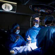
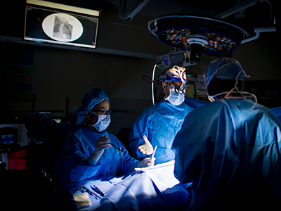
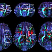
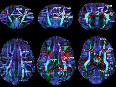
Children with CHD sometimes demonstrate delay in the development of cognitive and motor skills. This can be a result of multiple factors including altered prenatal oxygen delivery, brain blood flow and genetic factors associated with surgery including exposure to cardiopulmonary bypass, also known as the heart lung machine.

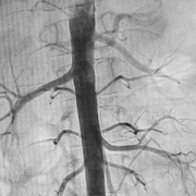
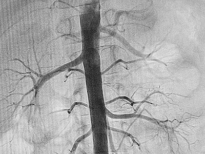
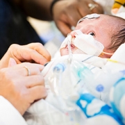
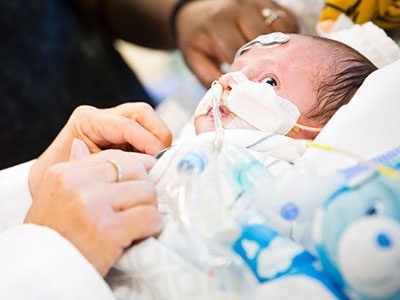
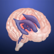
This phase 1 trial is the first to deliver mesenchymal stromal cells from bone marrow manufactured in a lab (BM-MSC) into infants already undergoing cardiac surgery via cardiopulmonary bypass. The hypothesis is that by directly infusing the MSCs into the blood flow to the brain, more MSCs quickly and efficiently reach the subventricular zone and other areas of the brain that are prone to inflammation. The trial is open to eligible patients ages newborn to six months of age.


Learn more in this overview video.

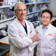
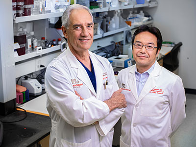
The trial is part of a $2.5 million, three-year grant from the National Institutes of Health (NIH) led by Richard Jonas, M.D.Catherine Bollard, M.B.Ch.B., M.D., and Nobuyuki Ishibashi, M.D.. The project involves collaboration between the Prenatal Cardiology program of Children’s National Heart Institute, the Center for Cancer and Immunology Research, the Center for Neuroscience Research and the Sheikh Zayed Institute for Pediatric Surgical Innovation.

“NIH supported studies in our laboratory have shown that MSC therapy may be extremely helpful in improving brain development in animal models after cardiac surgery,” says Dr. Ishibashi. “MSC infusion can help reduce inflammation including prolonged microglia activation that can occur during surgery that involves the heart lung machine.”

Staff from the Cellular Therapy Laboratory, led by director Patrick Hanley, Ph.D., manufactured the BM-MSC at the Center for Cancer and Immunology Research, led by Dr. Bollard.

The phase 1 safety study will set the stage for a phase 2 effectiveness trial of this highly innovative MSC treatment aimed at reducing brain damage, minimizing neurodevelopmental disabilities and improving the postoperative course in children with CHD. The resulting improvement in developmental outcome and lessened behavioral impairment will be of enormous benefit to individuals with CHD.

For more information about this new treatment, contact the clinical research team: Gil Wernovsky, M.D., Shriprasad Deshpande, M.D., Maria Fortiz.

Mended Little Hearts’ Volunteer of the Year, Maryann Mayhood, and her son Joseph delivered the Hospital of the Year award to Dr. Donofrio in November 2020.

Mended Little Hearts names Children’s National Hospital as ‘Hospital of the Year’

Mended Little Hearts’ Volunteer of the Year, Maryann Mayhood, and her son Joseph delivered the Hospital of the Year award to Dr. Donofrio in November 2020.

Mended Little Hearts’ Volunteer of the Year, Maryann Mayhood, and her son Joseph delivered the Hospital of the Year award to Dr. Donofrio in November 2020.

Children’s National Hospital was named Hospital of the Year by Mended Little Hearts, one of the top organizations in the U.S. for patients with congenital heart disease and their families. Children’s National was selected as the Hospital of the Year across all divisions of the Mended Little Hearts national network and the Washington, D.C. region. The hospital is recognized with the award for its efforts to empower Mended Little Hearts volunteers and make it possible for the group to provide peer support and education to children and adults with congenital heart disease, their families and the surrounding communities.

“It’s an honor to be recognized as a champion by a group like Mended Little Hearts that truly represents the voices and needs of patients and their families. We embrace and encourage their work because we know that providing the best care for children and their families goes beyond simply outstanding clinical service,” says Charles Berul, M.D., chief of Cardiology and co-director of the Children’s National Heart Institute. “We are privileged to have a group of dedicated volunteers from Mended Little Hearts who are willing to work side-by-side with our team to share peer support, education and guidance for our families at Children’s National.”

Though many in-person activities are currently on hold or held virtually for the health and safety of everyone during the COVID-19 public health emergency, Children’s National and Mended Little Hearts continue to coordinate closely together to support families as much as possible by making virtual connections and via the Mended Little Hearts “Bravery Bags,” which are given to every family and include personal essentials for a hospital stay as well as important guidance such as questions to ask care providers and how to seek more information about the care plan.

For the last few years, the hospital has also provided space within the hospital for the group to host family breakfasts and other events, making sure families have access to the information and support items they need during a hospital stay. They are also welcomed to many of the hospital’s annual events for adults and children with congenital heart disease and their families, to connect and share experiences.

“We are honored to recognize Children’s National Hospital for the outstanding work they have done to support heart patients and their families,” said Mended Hearts Inc. President Ron Manriquez. “That they have won this award is proof of the deep commitment they have to their members, families and the community at large. We are grateful for the work they do to support the Mended Little Hearts mission.”

Mended Little Hearts and its parent group, Mended Hearts, are organizations that inspire hope and seek to improve the quality of life for heart patients and their families through ongoing peer-to-peer support.

Yves d'Udekem

Yves d’Udekem, M.D., Ph.D., joins Children’s National as Cardiac Surgery Chief

Yves d’Udekem, M.D., Ph.D., a pediatric cardiac surgeon recognized for expertise in the most challenging procedures for newborns and children with complex congenital heart disease, has joined Children’s National Hospital as chief of Cardiac Surgery and co-director of the Children’s National Heart Institute.

“Children’s National has the cases and expertise I was looking for,” he says. “Even better, when you visit it’s clear that the entire Heart Institute team is energetic about working together. They’re constantly seeking better ways to do this work and improve how we care for children and their families.”

Dr. d’Udekem comes to Children’s National from The Royal Children’s Hospital in Melbourne, Australia, an institution that has led the advancement of congenital heart disease care and research, performing more than 500 surgical procedures with cardiopulmonary bypass each year.

He has a broad spectrum of pediatric cardiac surgery expertise, with special emphasis in single ventricle congenital heart defects, when one lower chamber of the heart does not develop. One area of his research portfolio includes clinical research into long-term quality of life for people who had Fontan procedures — a critical surgical approach to adapt blood flow for people born with single ventricle heart disease. He has additional expertise in valve repair, artificial hearts and other cardiac assist devices.

“Sometimes it seems like I’ve been born and put on this earth for that single purpose — I have to bring life to patients with single ventricle hearts or assist devices,” he says. He describes his path to pediatric cardiac surgery as serendipity, “It’s more like pediatric cardiac surgery chose me.”

“It is an honor to welcome Yves d’Udekem to the Children’s National Heart Institute,” says David Wessel, M.D., executive vice president and chief medical officer of Hospital and Specialty Services at Children’s National. “He is a surgeon, physician and leader of the highest caliber. I can say without hesitation that Yves’ leadership of our cardiac surgery team will change the lives of hundreds, if not thousands, of children and adults with congenital heart disease.”

In preparation for Dr. d’Udekem’s arrival, the Children’s National Heart Institute and hospital leadership created a framework plan for success that will build on the legacy of excellence established by Richard Jonas, M.D. Dr. Jonas, a world leader in congenital heart surgery who has made significant clinical and academic advances in the field, will continue his award-winning laboratory-based research at Children’s National on neurodevelopmental outcomes for children with congenital heart disease.

Dr. d’Udekem is Belgian-born with Canadian and Australian citizenship. He received his early training in Belgium and Toronto. As an attending cardiac surgeon in Belgium, he operated on both adults and children with congenital heart disease. Dr. d’Udekem then worked with internationally recognized heart surgeon Marc de Leval, M.D., FRCS, and others in London to train in congenital heart surgery at the Great Ormond Street Hospital for Children. From there, he was recruited to The Royal Children’s Hospital in Melbourne, Australia, where he has been ever since.

Dr. d’Udekem has more than 350 research publications and has obtained more than $7 million in grant funding in the past five years for work to create the first research network of Australian children and adults who have undergone the Fontan procedure.

He started seeing patients at Children’s National in mid-September.

telemedicine control room

Telehealth and AI reduce cardiac arrest in the cardiac ICU

telemedicine control room

The telehealth command center located a few steps away from the cardiac ICU at Children’s National Hospital.

The cardiac critical care team at Children’s National Hospital has developed an innovative Tele-Cardiac Critical Care model aiming to keep constant watch over the most fragile children with critical heart disease in the cardiac ICU. The system combines traditional remote monitoring and video surveillance with an artificial intelligence algorithm trained to flag early warning signs that a critically ill infant may suffer a serious event like cardiac arrest while recovering from complex cardiac surgery. This second set of eyes helps bedside teams improve patient safety and quality of care.

These high risk post-operative patients are often neonates or small infants born with the most complex and critical congenital heart diseases that require surgery or interventional cardiac catheterization in their first days or weeks of life. At these early stages after crucial cardiac surgery, these patients can decompensate dangerously fast with few outward physical symptoms.

The AI algorithm (T3) monitors miniscule changes in oxygen delivery and identifies any mismatch with a child’s oxygen needs. It also tracks and displays small changes in vital sign trends that could lead to a serious complication. The cardiac ICU command center staff then analyzes additional patient data and alerts the bedside team whenever needed.

The Tele-Cardiac Critical Care program started two years ago. In that time, the program has contributed to a significant decrease in post-operative cardiac arrest for this patient population.

“It’s easy to see how a model  like this could be adapted to other critical care scenarios, including our other intensive care units and even to adult units,” says Ricardo Munoz, M.D., chief of Cardiac Critical Care and executive director of Telehealth. It allows the physicians and nurses to keep constant watch over these fragile patients without requiring a physician to monitor every heartbeat in person for every patient at every hour of the day to maintain optimal outcomes for all of them.”

Dr. Munoz and Alejandro Lopez-Magallon, M.D., medical director of Telehealth and cardiac critical care specialist, presented data from the pilot program at the American Telemedicine Association’s virtual Annual Meeting on June 26, 2020.

doctors operating

U.S. DoD awards $2M for study to protect neurological function after cardiac surgery

doctors operating

A collaboration between clinical and basic science researchers including Drs. Ishibashi, Hashimoto-Torii, Jonas, and Deutsch, seeks to to understand how caspase enzyme activation plays a role in the development of fine and gross motor skills in children who underwent cardiac surgery for CHD repair.

The U.S. Department of Defense has awarded $2 million to Children’s National Hospital to study how a family of protease enzymes known as caspases may contribute to brain cell degeneration when activated by prolonged anesthesia and cardiopulmonary bypass during cardiac surgery for congenital heart disease.

This U.S. Army Medical Research Acquisition Activity Award, Anesthesia Neurotoxicity in Congenital Heart Disease, is led by principal investigator Nobuyuki Ishibashi, M.D., with both clinical and basic science co-investigators including Kazue Hashimoto-Torii, Ph.D., (Neuroscience), Richard Jonas, M.D., (Cardiovascular Surgery) and Nina Deutsch, M.D., (Anesthesiology).

While the specific cellular and molecular mechanisms of how anesthesia and cardiac surgery impact cortical development are poorly understood, both seem to impact brain growth and development in young children. The most common neurologic deficit seen in children after CHD surgical repair is the impairment of fine and gross motor skills.

Both anesthetic agents and inflammation like that seen as a result of cardiopulmonary bypass have also been shown to contribute to the activation of a specific group of enzymes that play an essential role in the routine (programmed) death of cells: caspases. However, recent pre-clinical research shows that these enzymes may also contribute to other alterations to cells beyond cell death, including making changes to other cell structures. In pre-clinical models, these changes cause impairments to fine and gross motor skills – the same neurological deficits seen in children with CHD who have undergone procedures requiring prolonged anesthesia and cardiopulmonary bypass.

The research team hypothesizes that caspases are extensively activated as a result of cardiac surgery and while that activation is rarely causing reduced numbers of neurons, the changes that caspase enzymes trigger in neurons are contributing to neurological deficits seen in children with CHD after surgery.

While the study focuses specifically on the impacts of cardiac surgery for correction of a heart defect, the findings could have major implications for any pediatric surgical procedure requiring prolonged anesthesia and/or cardiopulmonary bypass.

tiny stent illustration

Thinking small for newborns with critical congenital heart disease

tiny stent illustration

Illustration of a hybrid stage I palliation with bilateral bands on the lung vessels and a stent in the ductus arteriosus for patients with small left heart structures.

A new LinkedIn post from Kurt Newman, M.D., president and CEO of Children’s National Hospital, tells a story about the hospital’s cardiac surgeons and interventional cardiologists working with the U.S. Food and Drug Administration (FDA) to bring a better-sized, less-invasive vascular stent to the U.S. for the first time. The stent holds open a newborn’s ductus arteriosus, a key blood vessel that keeps blood flowing to the body, until the baby is big and strong enough to undergo a serious open-heart procedure for repair of hypoplastic left heart syndrome.

He writes, “Why is this important? At less than 6 lbs., these patients have arteries that are thinner than a toothpick – less than 2mm in diameter. Currently, the stent used in these children is an FDA approved device for adult vascular procedures, adapted and used off-label in children. It is not always well suited for the smallest babies as it is too large for insertion through the artery and often too long as well. The extra length can create immediate and long-term complications including obstructing the vessel it is supposed to keep open.

“While I am proud of the talent and dedication of our Children’s National cardiac surgery and interventional cardiology teams, I tell this story to illustrate a larger point – innovation in children’s medical devices matters. What’s unfortunate is that development and commercialization of pediatric medical devices in the U.S. continues to lag significantly behind adults…We can and must do better.”

Read Dr. Newman’s full post on LinkedIn.

Pediatric angiography

Congenital heart disease more deadly in low-income countries

Pediatric angiography

Even though mortality from congenital heart disease (CHD) has declined over the last three decades as diagnosis and treatments have advanced, the chances for a child to survive a CHD diagnosis significantly differs based on the country where he or she is born.

This eye-opening finding is drawn from the first comprehensive study of congenital heart disease across 195 countries, prepared using data from the Global Burden of Diseases, Injuries and Risk Factors Study 2017 (GBD), and recently published in The Lancet.

“Previous congenital heart estimates came from few data sources, were geographically narrow and did not evaluate CHD throughout the life course,” write the authors, known collectively as the 2017 GBD Congenital Heart Disease Collaborators. Co-lead author Meghan D. Zimmerman, M.D., worked on the study while completing her pediatric cardiology and American Heart Association Global Health Fellowships at Children’s National Hospital, and two pediatric cardiologists from Children’s National, Cardiology Associate Chief Craig Sable, M.D., and Gerard Martin, M.D., medical director of Global Services, provided leadership and oversight of this paper. The remaining collaborators are from more than 45 institutions around the world, spanning cardiology, public health and schools of medicine on every continent.

This is the first time the GBD study data was used along with all available data sources and previous publications – making it the most comprehensive study on congenital heart disease burden to date. Key differences between this study and prior estimates include:

  • Anatomic groupings of CHD by type, rather than simply categorized as moderate, severe or critical.
  • Inclusion of new data sources, including data from screening programs, congenital registries, administrative data and data sources in mortality and survival.
  • A control mechanism to account for cases of CHD that remit on their own to reduce the risk of overestimating prevalence.
  • Inclusion of all cases of congenital heart disease, including those with chromosomal or genetic anomalies such as Trisomy 21 that often co-occur.

This more comprehensive data set led to findings that showed lower predicted long-term survival, higher remission, and lower prevalence than previous studies that extrapolated evidence from studies of high-income countries. However, it also means these new estimates are a more accurate representation of the current global state of affairs. Overall, the study found:

  • A 34.5% decline in deaths from congenital disease between 1990 to 2017.
  • Nearly 70% of deaths caused by CHD in 2017 (180,624) were in infants less than one year old.
  • Most CHD deaths occurred in countries within the low and low-middle socio-demographic index (SDI) quintiles.
  • Mortality rates get lower as a country’s SDI rises.
  • Birth prevalence of CHD was not related to a country’s socio-demographic status, but overall prevalence was much lower in the poorest countries of the world. This is because children in these countries do not have access to life saving surgical services.
  • Nearly 12 million people are currently living with CHD globally, 18.7% more than in 1990.
  • The burden of CHD is not fully realized by just looking at prevalence and mortality. The measure “Years of Life Lost” provides deeper insight into the staggering burden of CHD, taking into account both absolute mortality and age at death.

“In high income countries like the United States, we diagnose some heart conditions prenatally during the 20-week ultrasound,” says Gerard Martin, M.D., a pediatric cardiologist at Children’s National Hospital who contributed to the study. “We catch others right after birth with a pulse oximetry screening for critical congenital heart disease. We can operate to correct a critical issue within the first week of life. And now our CHD kids are growing and thriving through adulthood and having families of their own.”

“For children born in middle- and low-income countries, these data draw stark attention to what we as cardiologists already knew from our own work in these countries – the lack of diagnostic and treatment tools leads to lower survival rates for children born with CHD,” adds Craig Sable, M.D., associate chief of cardiology at Children’s National, another primary contributor. “This is one of the most significant publications I have been a part of as it highlights the substantial loss of life to CHD in infancy around the globe.”

The authors write, “The UN has prioritized reduction of premature deaths from heart disease, but to meet the target of ‘ending preventable deaths of newborns and children under 5 years of age,’ health policy makers will need to develop specific accountability measures that address barriers and improve access to care and treatment.”

The study also includes a 400-page appendix breaking down each area by type of congenital anomaly, world region and country.

baby with tubes

BPA analogues may be less likely to disrupt heart rhythm

Some chemical alternatives to plastic bisphenol-a (BPA), which is still commonly used in medical settings such as operating rooms and intensive care units, may be less disruptive to heart electrical function than BPA.

A poster at the AHA Scientific Sessions suggests bisphenol-s (BPS) and bisphenol-f (BPF) may have less impact on heart function than bisphenol-a (BPA).

Some chemical alternatives to plastic bisphenol-a (BPA), which is still commonly used in medical settings such as operating rooms and intensive care units, may be less disruptive to heart electrical function than BPA, according to a pre-clinical study that explored how the structural analogues bisphenol-s (BPS) and bisphenol-f (BPF) interact with the chemical and electrical functions of heart cells.

The findings suggest that in terms of toxicity for heart function, these chemicals that are similar in structure to BPA may actually be safer for medically fragile heart cells, such as those in children with congenital heart disease. Previous research has found a high likelihood that BPA exposure may impact the heart’s electrical conductivity and disrupt heart rhythm, and patients are often exposed to the plastic via clinical equipment found in intensive care and in the operating room.

“There are still many questions that need to be answered about the safety and efficacy of using chemicals that look and act like BPA in medical settings, especially in terms of their potential contribution to endocrine disruption,” says Nikki Gillum Posnack, Ph.D., the poster’s senior author and a principal investigator in the Sheikh Zayed Institute for Pediatric Surgical Innovation at Children’s National Hospital. “What we can say is that, in this initial pre-clinical investigation, it appears that these structural analogues have less of an impact on the electrical activity within the heart and therefore, may be less likely to contribute to dysrhythmias.”

Future studies will seek to quantify the risk that these alternative chemicals pose in vulnerable populations, including pediatric cardiology and cardiac surgery patients. Since pediatric patients’ hearts are still growing and developing, the interactions may be different than what was seen in this pilot study.

Learn more the impacts of exposure to plastics such as bisphenol-A and plasticizers such as DEHP and MEHP that are commonly used in medical devices:

###

Bisphenol-a Analogues May Be Safer Alternatives For Plastic Medical Products
Rafael Jaimes, Damon McCullough, Luther M Swift, Marissa Reilly, Morgan Burke, Jiansong Sheng, Javier Saiz, Nikki G Posnack
Poster Presentation by senior author Nikki G Posnack
CH.APS.01 – Translational Research in Congenital Heart Disease
AHA Scientific Sessions
November 16, 2019
1:30 p.m. – 2:00 p.m.

Dr. Jonas and research collaborator Nobuyuki Ishibashi in the laboratory.

Cardiac surgery chief recognized for studies of surgery’s impacts on neurodevelopment

Dr. Jonas and research collaborator Nobuyuki Ishibashi in the laboratory.

Dr. Jonas and research collaborator Nobuyuki Ishibashi in the laboratory.

Richard Jonas, M.D., is this year’s recipient of the Newburger-Bellinger Cardiac Neurodevelopmental Award in recognition of his lifelong research into understanding the impact of cardiac surgery on the growth and development of the brain. The award was established in 2013 by the Cardiac Neurodevelopmental Outcome Collaborative (CNOC) to honor Jane Newburger and David Bellinger, pioneers in research designed to understand and improve neurodevelopmental outcomes for children with heart disease.

At Children’s National, Dr. Jonas’ laboratory studies of neuroprotection have been conducted in conjunction with Dr. Vittorio Gallo, director of neuroscience research at Children’s National, and Dr. Nobuyuki Ishibashi, director of the cardiac surgery research laboratory. Their NIH-supported studies have investigated the impact of congenital heart disease and cardiopulmonary bypass on the development of the brain, with particular focus on impacts to white matter, in people with congenital heart disease.

Dr. Jonas’s focus on neurodevelopment after cardiac surgery has spanned his entire career in medicine, starting with early studies in the Harvard psychology department where he developed models of ischemic brain injury. He subsequently undertook a series of highly productive pre-clinical cardiopulmonary bypass studies at the National Magnet Laboratory at MIT. These studies suggested that some of the bypass techniques used at the time were suboptimal. The findings helped spur a series of retrospective clinical studies and subsequently several prospective randomized clinical trials at Boston Children’s Hospital examining the neurodevelopmental consequences of various bypass techniques. These studies were conducted by Dr. Jonas and others, in collaboration with Dr. Jane Newburger and Dr. David Bellinger, for whom this award is named.

Dr. Jonas has been the chief of cardiac surgery and co-director of the Children’s National Heart Institute since 2004. He previously spent 20 years on staff at Children’s Hospital Boston including 10 years as department chief and as the William E. Ladd Chair of Surgery at Harvard Medical School.

As the recipient of the 2019 award, Dr. Jonas will deliver a keynote address at the 8th Annual Scientific Sessions of the Cardiac Neurodevelopmental Outcome Collaborative in Toronto, Ontario, October 11-13, 2019.

spectrometer output

Understanding low cardiac output after surgery

spectrometer output

Rafael Jaimes, Ph.D., created an algorithm that is being tested in a pre-clinical model to characterize the light absorbance spectrum from different heart regions using a spectrometer.

After intense cardiac surgery, sometimes a patient’s heart is unable to effectively deliver oxygenated blood and nutrients throughout the recovering body. Known as inadequate or low cardiac output, the condition occurs in about a quarter of patients following surgery with cardiopulmonary bypass, including young children who require complex procedures to correct congenital heart defects at Children’s National Health System.

Researchers at the Sheikh Zayed Institute for Pediatric Surgical Innovation are exploring several facets of this challenge, with the goal of better understanding post-operative recovery trajectories in pediatric patients. Rafael Jaimes, Ph.D., a staff scientist at the institute, leads this work to identify when and how low cardiac output occurs, pinpoint the physical hallmarks of this condition and use that information to prevent long term damage and complications after surgery, including cardiac arrest.

“More research needs to be done to understand the cause of this overarching and multi-faceted syndrome,” says Dr. Jaimes. “I’m interested in understanding how metabolic insufficiency contributes to this condition, and also exploring how we can use current imaging and diagnostic tools to measure, track and treat the insufficiencies that contribute to low cardiac output.”

Tracking inadequate oxygen and nutrient delivery to the parts of the heart that have been repaired is one avenue under exploration. Currently, a cardiac-specific real-time device to measure the oxygen state of the heart, while a patient is in post-operative critical care, is under development.

The heart’s complexity has made using current oxygen measurement devices, such as spectrometers, very difficult. To date no tool exists that effectively screens out artifacts and noise to allow clear visualization. However, during his post-doctoral work, Dr. Jaimes has created a new algorithm that may be the first of its kind to accomplish this feat.

This work on low cardiac output recently received a Congenital Heart Defect Research Award, which is a collaborative program of the Children’s Heart Foundation and the American Heart Association that supports innovative research, seeking to understand and treat congenital heart defects.

A new research study will build on his previous studies by using the algorithm to characterize the absorbance spectrum from different heart regions in a pre-clinical model. The data collected will serve as the baseline for development of a prototype spectrometer software, capable of tracking changes in heart oxygenation before, during and after surgery.

The end goal is to more effectively identify when parts of the heart are deprived of oxygen and nutrients and prevent resulting impacts on cardiac metabolism and output. Doing so will decrease short term mortality and morbidity and may also improve circulation systemically, potentially reducing long term health impacts of reduced oxygenation, such as neurodevelopmental disorders.

baby cardioilogy patient

Researchers receive $2.5M grant to optimize brain development in babies with CHD

baby cardioilogy patient

Children’s National Health System researchers Richard Jonas, M.D., Catherine Bollard, M.B.Ch.B., M.D., and Nobuyuki Ishibashi, M.D., have been awarded a $2.5 million, three-year grant from the National Institutes of Health (NIH) to conduct a single-center clinical trial at Children’s National. The study will involve collaboration between the Children’s National Heart Institute, the Center for Cancer and Immunology Research, the Center for Neuroscience Research and the Sheikh Zayed Institute for Pediatric Surgical Innovation.

The goal of the study will be to optimize brain development in babies with congenital heart disease (CHD) who sometimes demonstrate delay in the development of cognitive and motor skills. This can be a result of multiple factors including altered prenatal oxygen delivery, brain blood flow and genetic factors associated with surgery including exposure to the heart lung machine.

The award will be used to complete three specific aims of a Phase 1 safety study as described in the NIH grant:

  • Aim 1: To determine the safety and feasibility of delivering allogeneic bone marrow derived mesenchymal stromal cell (BM-MSC) during heart surgery in young infants less than 3 months of age using the heart lung machine. The optimal safe dose will be determined.
  • Aim 2: To determine the impact of MSC infusion on brain structure using advanced neuroimaging and neurodevelopmental outcomes.
  • Aim 3: To determine differences in postoperative inflammatory and patho-physiological variables after MSC delivery in the infant with CHD.

“NIH supported studies in our laboratory have shown that MSC therapy may be extremely helpful in improving brain development in animal models after cardiac surgery,” says Dr. Ishibashi. “MSC infusion can help reduce inflammation including prolonged microglia activation that can occur during surgery that involves the heart lung machine.”

In addition the researchers’ studies have demonstrated that cell-based intervention can promote white matter regeneration through progenitor cells, restoring the neurogenic potential of the brain’s own stem cells that are highly important in early brain development.

The Phase 1 clinical trial is being implemented in two stages beginning with planning, regulatory documentation, training and product development. During the execution phase, the trial will focus on patient enrollment. Staff from the Cellular Therapy Laboratory, led by director Patrick Hanley, Ph.D., manufactured the BM-MSC at the Center for Cancer and Immunology Research, led by Dr. Bollard. The Advanced Pediatric Brain Imaging Laboratory, led by Catherine Limperopoulos, Ph.D., will perform MR imaging.

The phase 1 safety study will set the stage for a phase 2 effectiveness trial of this highly innovative MSC treatment aimed at reducing brain damage, minimizing neurodevelopmental disabilities and improving the postoperative course in children with CHD. The resulting improvement in developmental outcome and lessened behavioral impairment will be of enormous benefit to individuals with CHD.

effects of cardiopulmonary bypass surgery on the white matter of piglets.

The effects of cardiopulmonary bypass on white matter development

 cardiopulmonary bypass

Nobuyuki Ishibashi, M.D., and a team of researchers looked the effects of cardiopulmonary bypass surgery on the white matter of an animal model.

Mortality rates for infants born with congenital heart disease (CHD) have dramatically decreased over the past two decades, with more and more children reaching adulthood. However, many survivors are at risk for neurodevelopmental abnormalities  associated with cardiopulmonary bypass surgery (CPB), including long-term injuries to the brain’s white matter and neural connectivity impairments that can lead to neurological dysfunction.

“Clinical studies have found a connection between abnormal neurological outcomes and surgery, but we don’t know what’s happening at the cellular level,” explains Nobuyuki Ishibashi, M.D., Director of the Cardiac Surgery Research Laboratory at Children’s National. To help shed light on this matter, Ishibashi and a team of researchers looked at the effects of CPB on the white matter of an animal model.

The research team randomly assigned models to receive one of three CPB-induced insults: a sham surgery (control group); full-flow bypass for 60 minutes; and 25°C circulatory arrest for 60 minutes. The team then used fractional anisotropy — a technique that measures the directionality of axon mylenation — to determine white matter organization in the models’ brains. They also used immunohistology techniques to assess the integrity of white matter oligodendrocytes, astrocytes and microglia.

The results, published in the Journal of the American Heart Association, show that white matter experiences region-specific vulnerability to insults associated with CPB, with fibers within the frontal cortex appearing the most susceptible. The team also found that fractional anisotropy changes after CPB were insult dependent and that regions most resilient to CPB-induced fractional anisotropy reduction were those that maintained mature oligodendrocytes.

From these findings, Ishibashi and his co-authors conclude that reducing alterations of oligodendrocyte development in the frontal cortex can be both a metric and a goal to improve neurodevelopmental impairment in the congenital heart disease population. “Because we are seeing cellular damage in these regions, we can target them for future therapies,” explains Ishibashi.

The study also demonstrates the dynamic relationship between fractional anisotropy and cellular events after pediatric cardiac surgery, and indicates that the technique is a clinically relevant biomarker in white matter injury after cardiac surgery.

Catherine Limperopoulous

Brain impairment in newborns with CHD prior to surgery

Catherine Limperopoulous

Children’s National researchers led by Catherine Limperopoulos, Ph.D., demonstrate for the first time that the brains of high-risk infants show signs of functional impairment before they undergo corrective cardiac surgery.

Newborns with congenital heart disease (CHD) requiring open-heart surgery face a higher risk for neurodevelopmental disabilities, yet prior studies had not examined whether functional brain connectivity is altered in these infants before surgery.

Findings from a Children’s National Health System study of this question suggest the presence of brain dysfunction early in the lives of infants with CHD that may be associated with neurodevelopmental impairments years later.

Using a novel imaging technique, Children’s National researchers demonstrated for the first time that the brains of these high-risk infants already show signs of functional impairment even before they undergo corrective open heart surgery. Looking at the newborns’ entire brain topography, the team found intact global organization – efficient and effective small world networks – yet reduced functional connectivity between key brain regions.

“A robust neural network is critical for neurons to travel to their intended destinations and for the body to carry out nerve cells’ instructions. In this study, we found the density of connections among rich club nodes was diminished, and there was reduced connectivity between critical brain hubs,” says Catherine Limperopoulos, Ph.D., director of the Developing Brain Research Laboratory at Children’s National and senior author of the study published online Sept. 28, 2017 in NeuroImage: Clinical. “CHD disrupts how oxygenated blood flows throughout the body, including to the brain. Despite disturbed hemodynamics, infants with CHD still are able to efficiently transfer neural information among neighboring areas of the brain and across distant regions.”

The research team led by Josepheen De Asis-Cruz, M.D., Ph.D., compared whole brain functional connectivity in 82 healthy, full-term newborns and 30 newborns with CHD prior to corrective heart surgery. Conventional imaging had detected no brain injuries in either group. The team used resting state functional connectivity magnetic resonance imaging (rs-fcMRI), a imaging technique that characterizes fluctuating blood oxygen level dependent signals from different regions of the brain, to map the effect of CHD on newborns’ developing brains.

The newborns with CHD had lower birth weights and lower APGAR scores (a gauge of how well brand-new babies fare outside the womb) at one and five minutes after birth. Before the scan, the infants were fed, wrapped snugly in warm blankets, securely positioned using vacuum pillows, and their ears were protected with ear plugs and ear muffs.

While the infants with CHD had intact global network topology, a close examination of specific brain regions revealed functional disturbances in a subnetwork of nodes in newborns with cardiac disease. The subcortical regions were involved in most of those affected connections. The team also found weaker functional connectivity between right and left thalamus (the region that processes and transmits sensory information) and between the right thalamus and the left supplementary motor area (the section of the cerebral cortex that helps to control movement). The regions with reduced functional connectivity depicted by rs-fcMRI match up with regional brain anomalies described in imaging studies powered by conventional MRI and diffusion tensor imaging.

“Global network organization is preserved, despite CHD, and small world brain networks in newborns show a remarkable ability to withstand brain injury early in life,” Limperopoulos adds. “These intact, efficient small world networks bode well for targeting early therapy and rehabilitative interventions to lower the newborns’ risk of developing long-term neurological deficits that can contribute to problems with executive function, motor function, learning and social behavior.”