Tag Archive for: brain tumors

AI for good: Children’s National wins global competitions for measuring brain tumors

Children's National Hospital's winning team for the Brain Tumor Segmentation-Africa (BraTS-Africa) challenge

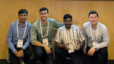
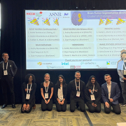
Meet the winners (left to right): Syed M. Anwar, Ph.D., M.S., principal investigator at Children’s National; Daniel Capellan Martin, M.Sc., Polytechnic University of Madrid; Abhijeet Parida, data scientist at Children’s National; and Austin Tapp, Ph.D., postdoctoral research fellow at Children’s National.

Using an award-winning artificial intelligence (AI) algorithm developed at Children’s National Hospital, researchers ranked first in the world in the Brain Tumor Segmentation-Africa (BraTS-Africa) challenge for their approach to identifying different parts of deadly gliomas. The details of their innovative method were recently published on arXiv, a curated research-sharing platform.

“Technology can bridge the gap in healthcare between high- and low-resource countries,” said Marius George Linguraru, D.Phil., M.A., M.Sc., the Connor Family Professor in Research and Innovation and principal investigator in the Sheikh Zayed Institute for Pediatric Surgical Innovation (SZI). “By tailoring methods we created at our hospital to fit the needs of specific regions, such as sub-Saharan Africa, our research helps improve medical imaging and diagnosis in challenging environments.”

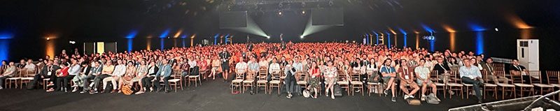
Dr. Linguraru was the program chair of the International Conference on Medical Image Computing and Computer Assisted Intervention (MICCAI) 2024 in Marrakesh, Morocco, the leading global meeting on AI in medical imaging.

Children’s National leads the way

Gliomas are a type of brain tumor with a high death rate and are especially difficult to diagnose in low- and middle-income countries. Given the increased need in Africa, researchers worldwide came together in Morocco to compete over the best way to accurately detect and measure tumors using MRI data and AI.

By applying advanced machine-learning techniques, the researchers adapted tools initially designed for well-resourced settings to work in countries with far fewer.

The study focused on transfer learning, a process in which an AI model is trained in advance on a large number of brain tumor images and then adjusted to work with smaller sets of new data. In this case, the models were adapted to work with local sub-Saharan African data using a strategy to combine different models’ strengths.

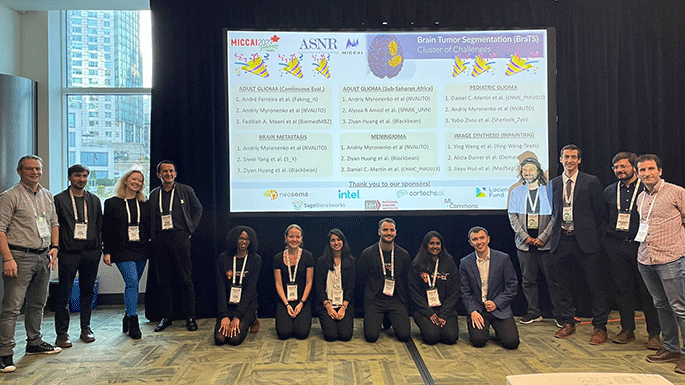
When tested, the approach achieved impressive accuracy scores. The Children’s National team, which included a colleague from the Polytechnic University of Madrid, ranked first in the BraTS-Africa 2024 challenge for identifying different parts of gliomas.

“To make the method widely available, the winning algorithm is shared online for others to use and improve upon,” Dr. Linguraru said. “My favorite part of these competitions is how they highlight the way innovation and collaboration can reduce global healthcare inequalities.”

The big picture

Children’s National researchers consistently lead global events using AI and advanced imaging to tackle complex healthcare challenges. In 2023, the team won a global contest to measure pediatric brain tumors at the MICCAI 2023 Conference. This year’s success in the BraTS-Africa challenge builds on this knowledge base and expands its use to adult gliomas.

At the Radiological Society of North America 2024 annual meeting, which drew 50,000 attendees, Zhifan Jiang, Ph.D., a staff scientist in the Precision Medical Imaging Lab at SZI, also won the Cum Laude Award for his scientific poster on applying AI to radiological images to predict severe outcomes for children with brain tumors caused by neurofibromatosis type 1.

“These achievements show how our science is leading the world in using AI for good,” Dr. Linguraru said. “Every day, we’re building on our knowledge of advanced imaging, brain tumors and AI to improve the diagnosis, measurement and treatment of deadly tumors — on a global scale.”

Attendees of the Brain Tumor Segmentation-Africa (BraTS-Africa) challenge

CellBuilder: A ready-made solution for cell & gene therapy manufacturing

A clean room at CNRI.

With CellBuilder and our global partnerships, Children’s National hopes to expand access to groundbreaking cell and gene therapy treatments as they take off in the next five years.

With cell and gene therapies poised to reshape cancer and rare disease treatments, researchers at Children’s National Hospital are pioneering ready-to-use solutions that will bring these cutting-edge therapies directly to hospitals and other treatment centers, shrinking the distance between doctors and patients.

“The next five years are going to be a period of tremendous growth for cell and gene therapy,” said Patrick Hanley, Ph.D., chief and director of the Cellular Therapy Program at Children’s National. “Currently, there’s no shortage of interest from the medical community, but there’s a shortage of people who can manufacture and administer this care. We’re looking for ways to get these treatments to the patients by providing other institutions the tools they need to launch these programs cost-effectively, safely and efficiently.”

Called CellBuilder, the starter kits for cell and gene therapy programs could transform the landscape for pediatric patients.

The big picture

Dr. Hanley and many members of the Children’s National team have been working in cell and gene therapy for more than a decade, gaining extensive experience in the technical and regulatory hurdles inherent in creating treatments that target diseases at their source.

In cell therapy, a specific cell type is modified and transferred to a patient with a payload that can target a disease or disorder. For example, T cells may be modified and delivered to patients to teach their immune systems to fight cancer.  In gene therapy, a patient’s genetic code is modified to treat or prevent diseases, such as sickle cell disease, cancers and other genetic disorders. This can be done by introducing a healthy copy of a gene, repairing a faulty gene or altering a gene’s function.

Children’s National has become a leader in manufacturing virus-specific T cells, one method of delivering cell therapies, and the Cellular Therapy Program has conducted consortium-led, multi-center trials. Many other healthcare sites across the country want to start programs offering this care at their facilities.

The holdup in the field

Starting a cell and gene therapy program from scratch can take years of effort, training and money. That’s why Jay Tanna, M.S., R.A.C., quality assurance manager of the Cellular Therapy Program at Children’s National, said the team is creating CellBuilder starter kits, which include the manufacturing protocol and the resources necessary to launch a cell therapy program almost instantly.

“With a suitable knowledge base, institutions can start their own cellular therapy program at the point of care, using our manufacturing protocols, vetted reagents and other key elements of the process,” Tanna said. “Of course, interested institutions would have to meet regulatory requirements and establish a clean room to manufacture these therapies. If they want to use CellBuilder to run a clinical trial, they can do that. If they want to take it to become a licensed product, they can do that, too.”

Children’s National has worked with more than five institutions to build their virus-specific T-cell program and is now using the kits to accelerate and commercialize the process to increase patient access. The lab has also entered into a memorandum of understanding with the Tokyo-based Hitachi Global Life Solutions, Inc., an innovative modular clean room manufacturer, with the goal of offering a bundled solution.

Why we’re excited

Dr. Hanley and his colleagues say that the partnerships Children’s National is forging as they consult with other experts in this field will expand access to cell and gene therapy across the country—and hopefully around the world.

“It used to be that, to get a CAR T cell, you would collect the cells at Children’s National, ship them to a company like Novartis, have the therapy manufactured there and then shipped back,” said Michael Keller, M.D., who co-led a first-of-its kind immunotherapy trial as the Translational Research Laboratory director at the Children’s National Cell Enhancement and Technologies for Immunotherapy Program. “It was expensive, time-consuming and limited patient access. Now, there’s growing interest in manufacturing at each site, just like you would with a bone marrow transplant.”

With CellBuilder and our global partnerships, Children’s National hopes to expand access to groundbreaking cell and gene therapy treatments as they take off in the next five years, extending lifetimes and improving the quality of life for children suffering from rare disorders.

“We’re trying to capture the momentum underway in the field by providing this kit so that institutions don’t have to know how to do everything,” Dr. Hanley said. “We provide all the knowledge, a reagent list and everything else they need—and they provide the care.”

Novel camera + machine learning = hope for more precise neurosurgery

collage of hyperspectral imaging (sHSI) camera and brain surgery

Researchers at Children’s National Hospital developed a compact imaging camera capable of seeing beyond the human visual spectrum to help segment healthy brain tissue from tumors during surgery. The groundbreaking technology will allow neurosurgeons to make more precise, real-time decisions in the operating room, rather than sending samples to pathology labs for biopsies.

In a manuscript published in Bioengineering, the team of engineers and neurosurgeons details how its snapshot hyperspectral imaging (sHSI) camera can be used to capture and process images of brain tissue, using the wide spectrum of light between visible and infrared wavelengths. That additional information — beyond the human eye — has the potential to allow for more accurate and complete tumor removal.

“In the hands of a neurosurgeon, this camera, when combined with machine learning, could dramatically improve outcomes for some of our most vulnerable brain tumor patients,” said Richard Jaepyeong Cha, Ph.D., an optical engineer and principal investigator at the Sheikh Zayed Institute of Pediatric Surgical Innovation. “We are able to attach the camera to a surgical microscope and process a significant amount of information from the patient while in the operating room. Not only could this lead to more complete tumor resection, it will also allow the surgeon to save as much healthy brain tissue as possible and reduce lifelong neurological complications.”

Why we’re excited

Brain tumors are the most common solid tumors in children, accounting for the highest number of pediatric cancer deaths globally each year. To develop a treatment plan, neurosurgeons need to understand the tumor’s features, including its type, grade of malignancy, location and its categorization as a primary or metastatic cancer. This information leads to decisions about how to remove or biopsy a tumor.

Under the current protocols, surgeons evaluate tumor margins in the operating room by examining the appearance of the brain tissue and sending out small samples to the pathology department for biopsies. This can lead to longer surgeries and difficult real-time surgical decisions. For instance, some low-grade tumors are visually indistinguishable from healthy brain tissue.

In four investigational cases approved by the hospital’s institutional research board, the sHSI camera was used in the operating room to help segment healthy pediatric brain tissue from tumors. Unlike the conventional red-green-blue (RGB) imaging cameras, which use only those three colors, HSI captures spectral data at each pixel of the image — a task too complex for the human eye — and sends it instantly for processing by an algorithm designed to assist in tumor segmentation.

What’s ahead

Despite the small dataset, the researchers were able to successfully segment healthy brain tissue from lesions with a high specificity during pediatric brain tumor resection procedures. Significant work remains to refine the technology and the machine learning behind it. Researchers also plan to integrate the sHSI camera into a laparoscope to visualize tumors that are not on the brain’s surface and collect data from more angles.

“As we develop these groundbreaking tools, we plan to continue to expand the dataset and refine the algorithm to make pediatric neurosurgery continually more precise,” said Naomi Kifle, M.S., research and development engineer at Children’s National and first author on the paper. “As our dataset grows, we hope to create a model that can distinguish healthy brain tissue, tumor and skull. This groundbreaking surgical tool shows significant promise.”

AI team wins international competition to measure pediatric brain tumors

Winners of the International Conference on Medical Image Computing and Computer Assisted Intervention
Children’s National Hospital scientists won first place in a global competition to use artificial intelligence (AI) to analyze pediatric brain tumor volumes, demonstrating the team’s ground-breaking advances in imaging and machine learning.

During the International Conference on Medical Image Computing and Computer Assisted Intervention (MICCAI), the Children’s National team demonstrated the most accurate algorithm to study the volume of brain tumors – the most common solid tumors affecting children and adolescents and a leading cause of disease-related death at this young age. The technology could someday help oncologists understand the extent of a patient’s disease, quantify the efficacy of treatments and predict patient outcomes.

“The Brain Tumor Segmentation Challenge inspires leaders in medical imaging and deep learning to try to solve some of the most vexing problems facing radiologists, oncologists, computer engineers and data scientists,” said Marius George Linguraru, D.Phil., M.A., M.Sc., the Connor Family Professor in Research and Innovation and principal investigator in the Sheikh Zayed Institute for Pediatric Surgical Innovation. “I am honored that our team won, and I’m even more thrilled for our clinicians and their patients, who need us to keep moving forward to find new ways to treat pediatric brain tumors.”

Why we’re excited

With roughly 4,000 children diagnosed yearly, pediatric brain tumors are consistently the most common type of pediatric solid tumor, second only to leukemia in pediatric malignancies. At the urging of Linguraru and one of his peers at the Children’s Hospital of Philadelphia, pediatric data was included in the international competition for the first time, helping to ensure that children are represented in medical and technological advances.

The contest required participants to use data from multiple institutions and consortia to test competing methods fairly. The Children’s National team created a method to tap into the power of two types of imaging and machine learning: 3D convolutional neural network and 3D Vision Transformer-based deep learning models. They identified regions of the brain affected by tumors, made shrewd data-processing decisions driven by the team’s experience in AI for pediatric healthcare and achieved state-of-the-art results.

The competition drew 18 teams who are leaders from across the AI and machine learning community. The runner-up teams were from NVIDIA and the University of Electronic Science and Technology of China.

The big picture

“Children’s National has an all-star lineup, and I am thrilled to see our scientists recognized on an international stage,” said interim Executive Vice President and Chief Academic Officer Catherine Bollard, M.D., M.B.Ch.B., director of the Center for Cancer for Immunology Research. “As we work to attack brain tumors from multiple angles, we continue to show our exceptional ability to create new and better tools for diagnosing, imaging and treating these devastating tumors.”

Harnessing children’s immune systems to fight their own brain tumors

Dalia Haydar, Pharm.D., Ph.D., principal investigator for the Program for Cell Enhancement and Technologies for Immunotherapies, recently joined Children’s National Hospital to help develop breakthrough treatments that hopefully will be a key in the fight against pediatric brain tumors. She brings her deep experience at St. Jude Children’s Research Hospital to the Center for Cancer and Immunology Research (CCIR) to help support the NexTGen team’s 10-year, $25-million Cancer Grand Challenges award.

Dalia – literally – has drive: She commutes 180 miles round trip from her home in Hershey, Pa., to her lab. She says she is grateful to be at one of the few research institutions in the world that is researching how to harness the power of CAR T-cell therapies to attack solid tumors in kids. While these therapies have been approved to treat leukemia and other blood cancers, solid tumors have proven far more stubborn. Haydar has tremendous hope that she and the team led by CCIR Director Catherine Bollard, M.D., M.B.Ch.B., will change that.

Q: Could you explain the importance of this research?

A: Unfortunately, once a patient is diagnosed with a brain tumor, especially a kid, there’s very little we can do. Using chemotherapy or radiation therapy has big disadvantages because of developmental delays and other side effects. We are hoping this kind of immunotherapy – where we take the patient’s own immune cells and engineer them in the lab to attack their cancer – will eradicate their very harsh and aggressive tumors, without causing significant adverse effects.

Q: How are researchers at Children’s National going to attack solid tumors with a treatment originally designed for blood cancers?

A: We have a lot of resources and expertise at Children’s National that we are trying to put together to develop a therapy that would cure brain tumors. Unfortunately, solid tumors are hard to treat and there are several challenges for any kind of immunotherapy. But right now, being at a place where all the necessary resources, support and expertise are available, we are hoping to address each of these challenges, and we are determined to do something in a meaningful timeframe to push that survival curve toward the advantage of those kids.

Q: How soon can this work be done?

A: Within two or three years, we are hopeful we’ll be able to identify the best working regimen of this CAR T-cell immunotherapy and investigate if it will work in a patient. I foresee, in the next 5 to 10 years, that we’re hopefully going to have such therapy for kids with brain tumors.

Q: What has surprised most you in your work?

A: There are so many challenges in developing immunotherapies for kids with brain tumors. First, if it works for adults, it doesn’t necessarily work for kids. Some of the tumors in kids are more aggressive. We need to understand the tumor itself, besides understanding the immunotherapy we’re developing.

The other challenge is CAR-T immunotherapy is not like a pill or taking radiotherapy that is standardized for several patients. It’s a very expensive therapy. It’s taking the patient’s own immune cell, like a bone marrow transplant. We put it in the lab, re-engineer the cells without transforming them into a cancer cell, enable those immune cells to attack the cancer, and then put them back into the patient. There are a lot of steps you need to take to make sure you don’t artificially harm those cells or introduce contamination.

One of the most intriguing challenges for me is how we make immunotherapy work for kids who have different kinds of brain tumors – a medulloblastoma versus a glioma versus an embryonal tumor. This is one of the challenges that keeps me on my toes, and I’m hoping to answer.

Q: What is the power of being in a multi-center environment like the Children’s National Research Institute?

A: We have to do enough science on the bench to support any proposal for the therapy to move to the clinic. The last thing we want to do is to investigate a drug or therapy in patients without really knowing how it works and the potential adverse effects. Being able to work with researchers at different stages of the bench-to-bedside spectrum, as well as being able to have access to patient samples and innovative preclinical models, helps push the science forward in a shorter time frame.

A lifetime of achievements: Roger Packer, M.D.

Roger Packer with patient

Over the years, Dr. Packer and his team in Washington, D.C., have made meaningful contributions to children all around the world diagnosed with childhood brain tumors, including medulloblastoma and gliomas.

Earlier in December, Roger Packer, M.D., senior vice president of the Center for Neurosciences and Behavioral Medicine at Children’s National Hospital, received the 2020 Lifetime Achievement Award from the International Symposium on Pediatric Neuro-Oncology at the meeting organized in Karuizawa, Japan. The prestigious recognition is a testament to the years of commitment and dedication Dr. Packer has devoted to the care of children with brain tumors and as such, have placed him as a top leader.

This award is a recognition of how the field has grown since the first International Symposium on Pediatric Neuro-Oncology Dr. Packer organized in Seattle in 1989. “It grew from a small gathering of investigators to now a multidisciplinary group of over 2,000 investigators,” Dr. Packer says.

Over the years, Dr. Packer and his team in Washington, D.C., have made meaningful contributions to children all around the world diagnosed with childhood brain tumors, including medulloblastoma and gliomas. These findings have contributed to an increase of the survival rate from 50% to over 80% for children with medulloblastoma. In addition, his contributions have led to newer molecular targeted therapies and improved the quality of life of children who are long-term survivors.

“The field, especially in the last decade, rapidly transitioned to a more biologically informed field,” Dr. Packer explains. “We are now utilizing new, exciting discoveries in biology and genetics to inform new approaches to treatment. This kind of transition gives us great hope for the future.”

In his early career, Dr. Packer worked with two neuro-oncology patients who died and would impact his decision to further study this field. At that time, there was minimal understanding of the nature of neuro-oncology diseases or how to best treat them. As a neurologist, he was frustrated by the lack of understanding and as a pediatrician, he was frustrated at the lack of ability to do success management.

“I saw this as a gap in my personal knowledge and found that the field was struggling to come up with new answers and new approaches,” he says. “But at the same time other, advances were being made in child cancer care, such as with leukemia. However, there was no wide focus on pediatric brain tumors.”

Combining his knowledge of neurology with his curiosity and relying on other leaders that surrounded him in the same field, Dr. Packer worked on driving this new work forward. Today, he is still heavily involved in the development of treatment protocols that are increasingly transitional for a variety of brain tumors, including low-grade and high-grade gliomas.

“With the help of our great colleagues at Children’s National, we continue to try to develop new means to treat these tumors, including immunological approaches and the incorporation in the use of novel means, such as low-intensity and high-intensity focused ultrasound,” he says. “We also have an excellent multidisciplinary team at Children’s National that has grown over the last decade some of whom are acknowledged national leaders in the fields of brain tumors, clinical research and clinical care. We also have a robust program focusing on the neurocognitive outcome of children and ways to intervene to ameliorate intellectual compromise and improve quality of life.”