New advances in AI help doctors understand pediatric brain tumors
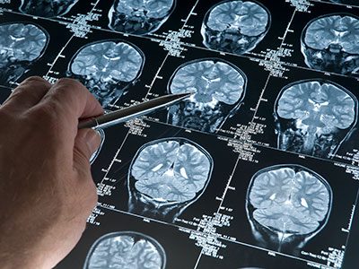
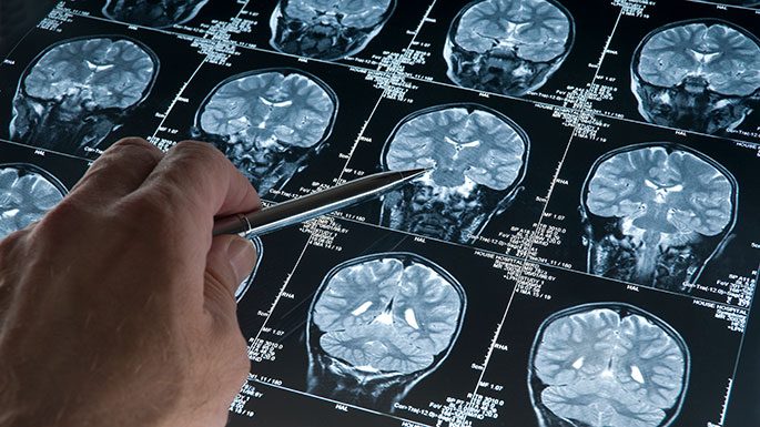
One of the first and most important steps in fighting brain tumors is to understand exactly where they are and how big they are – using magnetic resonance imaging (MRI) scans.
Brain tumors are the most common cause of cancer-related death in children. These tumors can be very difficult to treat because they often grow in very delicate areas of the brain. One of the first and most important steps in fighting these tumors is to understand exactly where they are and how big they are – using magnetic resonance imaging (MRI) scans.
Traditionally, doctors have had to look at these scans and manually draw outlines around the tumors. This takes a lot of time and can vary from one doctor to another. But what if artificial intelligence (AI) could help make this easier and more efficient?
A recent international research effort called BraTS-PEDs 2023 (short for Brain Tumor Segmentation in Pediatrics) explored exactly that. This challenge co-led by Children’s National Hospital and Children’s Hospital of Philadelphia invited research teams from around the world – including a team led by Marius George Linguraru, DPhil, MA, MSc – to develop the best AI tools for automatically finding and measuring brain tumors in children’s MRIs.
A global AI competition for pediatric care
The BraTS-PEDs 2023 challenge was the first of its kind focused specifically on children. Teams were given MRI scans from 167 pediatric patients with brain tumors collected by leading consortia – the Children’s Brain Tumor Network, DMG/DIPG Registry – and Collaborative Network for Neuro-oncology Clinical Trials and reviewed by volunteer neuroradiologists from the American Society of Neuroradiology. They used these scans to train their AI programs and then tested them on new cases to see how well they performed. Our team’s algorithm was the best at measuring these tumors.
Most teams used a special type of AI network called a “U-Net,” which is great at looking at images and figuring out shapes and boundaries. The challenge showed that AI can do a very good job at finding the tumor and measuring its volume. However, it was more difficult for AI to accurately mark the small, active parts inside the tumor, known as “enhancing tumor” regions.
These results are a huge step forward. Using AI can help doctors make faster, more precise decisions, and reduce differences between hospitals or between individual doctors.
Why this matters
When doctors can accurately map out a brain tumor, they can plan surgery better, target radiation therapy more precisely, and track how well treatments are working over time. This can lead to fewer side effects and better outcomes for children.
In some cases, timely and accurate measurements can even be the difference between life and death. Having advanced AI tools could mean that children get the right treatments faster and with more confidence.
Looking ahead
The research community is now working on including even more data from different hospitals around the world and making the data and algorithm public, like Children’s National has done here. They also plan to study more types of brain tumors and scans taken after surgery and treatments. In the future, AI could become a regular part of how doctors look at brain tumors — like an extra set of smart eyes that never get tired.
At Children’s National, leaders like Dr. Linguraru are helping turn this vision into reality, giving children with brain tumors a better chance at a healthy future. You can read the full study – BraTS-PEDs: Results of the Multi-Consortium International Pediatric Brain Tumor Segmentation Challenge 2023 – in the MELBA Journal.
Other authors from Children’s National include Roger J. Packer, MD, Brian Rood, MD, Miriam Bornhorst, MD, Xinyang Liu, PhD, Zhifan Jiang, PhD, and Syed Muhammad Anwar, PhD, MS.