

 https://innovationdistrict.childrensnational.org/wp-content/uploads/2025/05/Electrophysiology-team-feature.jpg
300
400
Innovation District
https://innovationdistrict.childrensnational.org/wp-content/uploads/2023/12/ID-mobile-header.png
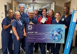
Innovation District2025-05-27 16:51:402026-01-12 16:18:37Leadless pacemakers, subcutaneous defibrillators successfully implanted in pediatric patients
https://innovationdistrict.childrensnational.org/wp-content/uploads/2025/05/Electrophysiology-team-feature.jpg
300
400
Innovation District
https://innovationdistrict.childrensnational.org/wp-content/uploads/2023/12/ID-mobile-header.png
Innovation District2025-05-27 16:51:402026-01-12 16:18:37Leadless pacemakers, subcutaneous defibrillators successfully implanted in pediatric patientsWhy it matters
Those hours can be critical for surgeries on a timeline, such as partial heart transplants. Recently, Dr. O’Hara and Yue-Hin Loke, MD, director of the 3D Cardiac Visualization Laboratory, worked together to provide computed tomography (CT) based 3D visualization for the first mitral valve partial heart transplant at Children’s National. The case required exceptionally precise measurements as the surgery included the first replacement of an artificial mechanical valve with living mitral valve from a donor heart. The valve from the donor heart and precise areas of extra heart tissue were needed to rebuild structures of the heart that were no longer present after the patient’s existing artificial valve was placed. The surgeon wanted a geometrical twin of the patient’s heart to better understand how the donor valve would be transplanted.
The ability to produce highly detailed models in the virtual space allows surgeons to see realistic, 3D detailed structures inside the heart which typically don’t show well using more routine cardiac imaging modalities such as transthoracic or transesophageal echocardiogram. The models also allow surgeons to customize views of complex structures and offer tailored visual orientations of areas in and around the heart.
Dr. Loke says the technology is often used by the surgeons to assist with planning for many different procedures when the heart anatomy is complex. One cardiac surgeon, for example, used 3D visualization for planning ventricular assist device cannula placement to account for, and avoid, other heart structures during implantation.

")
Children’s National leads the way
The application and speed of developing clinically relevant 3D models continues to grow. In early years, the team averaged 20 to 25 models each year. Today, they are on track to model closer to 50 models.
The key to their success, is the expertise of the embedded 3D lab and the dedicated engineer behind it, who reviews the computer-generated models and enhances specific areas of interest or focus. “He [Dr. O’Hara] is very engaged in the clinical space,” Dr. Loke says. “He talks to the surgeons directly, sees how things are done and can help think through what perspectives the visualization needs to include.”
That human aspect won’t be going away any time soon. Most existing machine learning models, like most other technology in the cardiac space, is based on the adult population. While some of the image rendering can be performed using machine learning, both Dr. Loke and Dr. O’Hara note that making the final product useful to pediatric cardiac surgical planning is “much more art than science.”
https://innovationdistrict.childrensnational.org/wp-content/uploads/2025/05/Electrophysiology-team-feature.jpg
300
400
Innovation District
https://innovationdistrict.childrensnational.org/wp-content/uploads/2023/12/ID-mobile-header.png
Innovation District2025-05-27 16:51:402026-01-12 16:18:37Leadless pacemakers, subcutaneous defibrillators successfully implanted in pediatric patients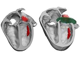 https://innovationdistrict.childrensnational.org/wp-content/uploads/2025/05/Heart-valve-modeling-feature.jpg
300
400
Innovation District
https://innovationdistrict.childrensnational.org/wp-content/uploads/2023/12/ID-mobile-header.png
Innovation District2025-05-12 11:45:572026-01-12 16:18:45Living tissue heart valve replaces mechanical mitral valve through partial heart transplant
https://innovationdistrict.childrensnational.org/wp-content/uploads/2025/05/Heart-valve-modeling-feature.jpg
300
400
Innovation District
https://innovationdistrict.childrensnational.org/wp-content/uploads/2023/12/ID-mobile-header.png
Innovation District2025-05-12 11:45:572026-01-12 16:18:45Living tissue heart valve replaces mechanical mitral valve through partial heart transplant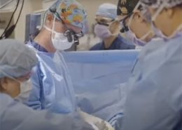 https://innovationdistrict.childrensnational.org/wp-content/uploads/2024/08/cardio-surgery-feature.jpg
300
400
Innovation District
https://innovationdistrict.childrensnational.org/wp-content/uploads/2023/12/ID-mobile-header.png
Innovation District2024-08-29 14:18:562026-01-12 16:18:39Long-term outcomes are key measure of CHD care quality and safety
https://innovationdistrict.childrensnational.org/wp-content/uploads/2024/08/cardio-surgery-feature.jpg
300
400
Innovation District
https://innovationdistrict.childrensnational.org/wp-content/uploads/2023/12/ID-mobile-header.png
Innovation District2024-08-29 14:18:562026-01-12 16:18:39Long-term outcomes are key measure of CHD care quality and safety https://innovationdistrict.childrensnational.org/wp-content/uploads/2023/03/heart-video-feature-1.gif
300
400
Innovation District
https://innovationdistrict.childrensnational.org/wp-content/uploads/2023/12/ID-mobile-header.png
Innovation District2023-03-13 12:47:452026-01-12 16:18:424D flow explained: Advanced imaging measures critical blood flow characteristics of single ventricle hearts
https://innovationdistrict.childrensnational.org/wp-content/uploads/2023/03/heart-video-feature-1.gif
300
400
Innovation District
https://innovationdistrict.childrensnational.org/wp-content/uploads/2023/12/ID-mobile-header.png
Innovation District2023-03-13 12:47:452026-01-12 16:18:424D flow explained: Advanced imaging measures critical blood flow characteristics of single ventricle hearts