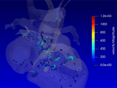
4D flow explained: Advanced imaging measures critical blood flow characteristics of single ventricle hearts
Yue-Hin “Tom” Loke, M.D., pediatric cardiologist and director of the 3D Cardiac Visualization Laboratory at Children’s National Hospital, uses magnetic resonance imaging and software rendering to create novel 4D flow images of children with single ventricle congenital heart disease.
“My research measures the degree of vortex formation (and) the degree of energy loss in the atrium as potential measurements of heart health and uses these measurements as a potential gauge of the heart health of children born with single ventricle conditions including hypoplastic left heart syndrome,” he says. “This information can be used to guide the management of the care for children with congenital heart disease. This technology provides valuable insight into how well the heart is working, especially before the Fontan procedure.”
Learn more about the approach and how it impacts clinical care decisions in the Children’s National Heart Institute.