Transforming pediatric hip surgery with robotics and 3D imaging
“This project is exciting as it targets more precise surgery with improved safety in terms of decreased radiation,” says Matthew Oetgen, MD, division chief of Orthopaedic Surgery and Sports Medicine at Children’s National.
What if surgeons could fix a child’s hip with pinpoint accuracy – using fewer X-rays and cutting-edge robotics? That’s the promise of a new National Institutes of Health funded project combining 3D imaging and robotic technology to improve the treatment of slipped capital femoral epiphysis (SCFE), a condition that affects the hip joint in growing children.
What’s been the hold-up in the field?
Right now, orthopedic surgeons place screws by hand using 2D X-ray images to guide them. To see the screw from different angles, they have to move the X-ray machine around. “The screw must be placed near the center of the femoral head, but not penetrate it,” says Kevin Cleary, PhD, associate director of engineering at the Sheikh Zayed Institute for Pediatric Surgical Innovation (SZI). This process takes time and can increase the amount of radiation the patient receives.
Even though better tools like 3D imaging and surgical robots exist, they aren’t used together in current surgical practice. “Individual procedures have nuances that require their own validated workflows,” says Tyler Salvador, a research engineer at Children’s National Hospital. In other words, each type of surgery is different, and doctors need proven steps before using new technology in the operating room.
How does this work move the field forward?
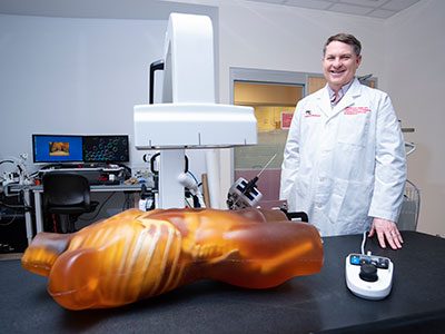
This project brings together low-dose 3D X-rays from nView with a small surgical robot called Micromate™ to help place screws more precisely during SCFE surgeries. “Our research group has been developing robotics, imaging, and related technologies to improve surgical procedures,” says Dr. Cleary. While these tools exist separately, putting them together in one system focused on bone surgery is new. Tyler Salvador adds, “This will provide a complete solution for precision SCFE implant placement and verification.”
“This project is exciting as it targets more precise surgery with improved safety in terms of decreased radiation,” says Matthew Oetgen, MD, division chief of Orthopaedic Surgery and Sports Medicine at Children’s National. “This is a paradigm-shifting effort that will improve outcomes while improving safety which is the holy grail of translational clinical research. It embodies the goal of the SZI — combining technical expertise with surgical leadership to improve outcomes in pediatric surgery.”
Children’s National leads the way
Children’s National is unique because the Sheikh Zayed Institute combines research and clinical care in one place. “Having the labs right inside the hospital helps us work closely with doctors,” says Salvador.
Together, this work is paving the way for safer, faster and more precise surgeries for children with hip problems. By combining advanced imaging and robotics, the team at Children’s National is helping shape the future of pediatric orthopedic care. In addition to this robotic hip pinning project, the Children’s National team is also behind two additional groundbreaking projects including robotic gallbladder removal and a kidney surgery initiative.
This project has been funded in whole with federal funds from the National Institutes of Health under Contract No. R01EB035559.