Family love and the right care for neurofibromatosis type 1 give Maddox a fresh start
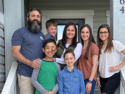
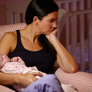
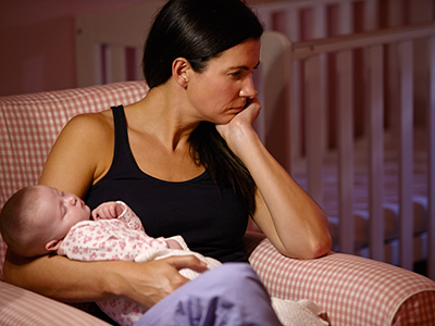
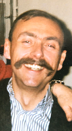
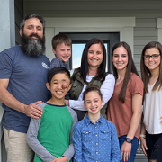
Maddox and his family in early 2020.
13-year-old Maddox Gibson is learning to cook. He says he wants to be a chef and wants to make meals for people who need it most — the homeless and the hungry.
It makes sense that he’s eager to help people who need it. As a young child growing up in a group home in his native country of China, he knows firsthand how important that support can be. In 2017 at age 10, he found his own endless supply of love and support when he met and was adopted by the Gibson family.
Zhen Chao, now called Maddox, was born in China with a genetic condition called neurofibromatosis type 1 that can cause painful or disfiguring tumors called plexiform neurofibromas. Zhen Chao had two on his head when he arrived — on his scalp and on his left optic nerve — which had been largely untreated for most of his life in China. On top of that, his right leg had been fractured and not fixed properly years before, causing him pain and weakness that left him wheelchair bound.
Adoptive mom Lindsey, a registered nurse, knew he would need special care to meet all the unique challenges he faced, and she’d done her homework — he needed the expertise of Miriam Bornhorst, M.D., and the Gilbert Family Neurofibromatosis Institute at Children’s National Hospital to help him thrive in his new life in the U.S. Since shortly after he came to the U.S., Lindsey has been driving Maddox the 6-plus hours from their home in North Carolina to Washington, D.C., regularly, to get care for all of his health challenges.
Maddox’s optic neurofibroma was too large when he arrived at Children’s National for a simple surgical removal. Due to her role as the lead investigator on a cutting edge clinical trial for the orphan drug selumetinib — a so-called MEK inhibitor that has shown early promise at reducing the cell growth of tumors like plexiform neurofibromas, Dr. Bornhorst enrolled Maddox in a compassionate use program for the drug, an opportunity that is not widely available. The drug was initially developed for something completely different — treatment of melanoma and non-small cell lung cancer in adults–but has been adapted through its FDA orphan drug designation for pediatric clinical trials in NF1. In the time since Maddox started taking it, it was approved for use in NF1 patients by the FDA.
The trial drug did its job — in late 2019, Maddox’s tumor had shrunk enough that chief neurosurgeon Robert Keating, M.D., and plastic surgeon Michael Boyajian, M.D., were able to successfully remove it. Follow-up procedures led by that team have also worked to repair the tissue that was impacted by the optic neurofibroma.
In addition to treatment of his neurofibromas, Maddox and his mom are able to see every service they need during one stay in D.C. The Neurofibromatosis Institute works closely across specialties, so his corrective surgery for his leg from Children’s chief of orthopaedics, Matthew Oetgen, M.D., MBA, in September 2019. He was assessed and prescribed physical therapy early in the process and even before surgery, so now he’s stronger than ever and walking. Learning difficulties, including autism and ADHD are common in NF1 patients, and so the NF Institute’s neuropsychology team has evaluated him and worked with the family to find resources and strategies near home that will support him. It should be noted, those learning difficulties only became apparent after Maddox taught himself English from scratch in only two years’ time with the help of his school’s ESOL program.
This kind of full spectrum care, from clinical assessment to surgical treatment and psychological supports, is crucial to the lives of patients with neurofibromatosis type 1 and is only available at a pediatric specialty care institution like Children’s National. The hospital has gathered some of the preeminent researchers, surgeons, and physicians within the NF Institute to make sure that the care families will travel hundreds of miles to receive is the best possible, using the latest evidence-based treatments for every challenge they face.
Though his care and follow-ups will continue at Children’s National Hospital and his condition may pose new challenges in the future, for now, Maddox is able to focus on exploring new things and doing what he loves — playing outdoors with his family, learning to cook and building with Legos.