Tag Archive for: urology

H-IPSE internalized by just a limited range of cells

Hepatocytes

A team led by Children’s National Hospital found that H-IPSE is internalized by just a limited range of cells, including hepatocytes.

Schistosoma mansoni is a parasite that hides out in snails, breaks free into waterways, and then infects humans, spending much of its life inside blood vessels, laying eggs and jeopardizing public health when those eggs are excreted in urine or feces. As parasitic diseases go, the ailment it causes, Schistosomiasis, is second only to malaria in global impact, according to the Centers for Disease Control and Prevention.

In order to elude the human host’s defenses, S. mansoni uses self-defense tactics that researchers are trying to better understand in order to outmaneuver the parasite. A research team led by Children’s National Hospital is trying to tease out the multiple steps that enable this parasite to reproduce and generate millions of eggs without killing its host.

The parasite’s eggs secrete a number of proteins, with IPSE as one of the most abundant, the team recently presented during the American Society of Tropical Medicine and Hygiene 2019 annual meeting. That protein binds immunoglobulin, which induces basophils and mast cells to release IL-4. After sequestering chemokines, H-IPSE infiltrates the cell nucleus (thus H-IPSE is called an infiltrin), modulating gene expression.

“H-IPSE tips the immune system balance, making it more likely to trigger a Th2 anti-inflammatory response,” says Michael Hsieh, M.D., Ph.D., director of transitional urology at Children’s National and the research project’s senior author. “It downregulates pro-inflammatory pathways, but we wanted to know more about which specific human cells it targets.”

Using Trypan Blue, a stain that selectively colors certain cells bright blue, they solved the mystery, finding that H-IPSE is internalized by just a limited range of cells. What’s more, some cell types, like urothelial cells and hepatocytes (the liver’s chief functioning cells, which activate innate immunity), are more susceptible than neurons, endothelial cells or immature dendritic cells.

In addition to Dr. Hsieh, presentation co-authors include Olivia Lamanna, Evaristus Mbanefo and Kenji Ishida, all of Children’s National; Franco Falcone, of University of Nottingham; and Theodore Jardetzky and Luke Pennington, of Stanford University.

Children’s National ranked No. 6 overall and No. 1 for newborn care by U.S. News

Children’s National in Washington, D.C., is the nation’s No. 6 children’s hospital and, for the third year in a row, its neonatology program is No.1 among all children’s hospitals providing newborn intensive care, according to the U.S. News Best Children’s Hospitals annual rankings for 2019-20.

This is also the third year in a row that Children’s National has been in the top 10 of these national rankings. It is the ninth straight year it has ranked in all 10 specialty services, with five specialty service areas ranked among the top 10.

“I’m proud that our rankings continue to cement our standing as among the best children’s hospitals in the nation,” says Kurt Newman, M.D., President and CEO for Children’s National. “In addition to these service lines, today’s recognition honors countless specialists and support staff who provide unparalleled, multidisciplinary patient care. Quality care is a function of every team member performing their role well, so I credit every member of the Children’s National team for this continued high performance.”

The annual rankings recognize the nation’s top 50 pediatric facilities based on a scoring system developed by U.S. News. The top 10 scorers are awarded a distinction called the Honor Roll.

“The top 10 pediatric centers on this year’s Best Children’s Hospitals Honor Roll deliver outstanding care across a range of specialties and deserve to be nationally recognized,” says Ben Harder, chief of health analysis at U.S. News. “According to our analysis, these Honor Roll hospitals provide state-of-the-art medical expertise to children with rare or complex conditions. Their rankings reflect U.S. News’ assessment of their commitment to providing high-quality, compassionate care to young patients and their families day in and day out.”

The bulk of the score for each specialty is based on quality and outcomes data. The process also includes a survey of relevant specialists across the country, who are asked to list hospitals they believe provide the best care for patients with challenging conditions.

Below are links to the five specialty services that U.S. News ranked in the top 10 nationally:

The other five specialties ranked among the top 50 were cardiology and heart surgery, diabetes and endocrinology, gastroenterology and gastro-intestinal surgery, orthopedics, and urology.

Experimental fertility preservation provides hope for young men

germ cells in testicular tissues

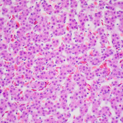
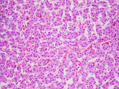
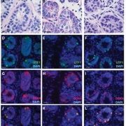
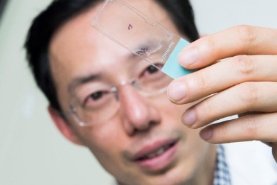
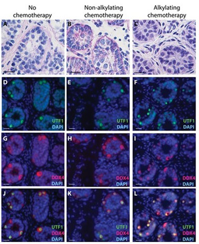
Confirming the presence of germ cells in testicular tissues obtained from patients. Undifferentiated embryonic cell transcription factor 1 (UTF1) is an established marker of undifferentiated spermatogonia as well as the pan-germ cell marker DEAD-box helicase 4 (DDX4). UTF1 (green) and/or DDX4 (red) immunostaining was confirmed in 132 out of 137 patient tissues available for research, including patients who had received previous non-alkylating (B, E, H, K) or alkylating (C, F, I, L) chemotherapy treatment. © The Author(s) 2019. Published by Oxford University Press on behalf of the European Society of Human Reproduction and Embryology.

Testicular tissue samples obtained from 189 males who were facing procedures that could imperil fertility were cryopreserved at one university, proving the feasibility of centralized processing and freezing of testicular tissue obtained from academic medical centers, including Children’s National, scattered around the world.

“It’s not surprising that the University of Pittsburgh would record the highest number of samples over the eight-year period (51 patients), given its role as the central processing facility for our recruiting network of academic medical centers,” says Michael Hsieh, M.D., Ph.D., director of transitional urology at Children’s National. “Children’s National recruited the third-highest number of patients, which really speaks to the level of collaboration I have with Jeff Dome’s team and their commitment to thinking about the whole patient and longer-term issues like fertility.”

An estimated 2,000 U.S. boys and young men each year receive treatments or have cancers or blood disorders that place them at risk for infertility. While older youths who have undergone puberty can bank their sperm prior to undergoing sterilizing doses of chemotherapy or radiation, there have been scant fertility preservation options for younger boys. However, some older adolescents and young men are too sick or stressed to bank sperm. For patients with no sperm to bank or who are too sick or stressed to bank sperm, the experimental procedure of freezing testicular tissue in anticipation that future cell- or tissue-based therapies can generate sperm is the only option.

Recent research in experimental models indicates that such testicular tissue biopsies contain stem cells, blank slate cells, hinting at the potential of generating sperm from biopsied tissue.

“This study demonstrates that undifferentiated stem and progenitor spermatogonia may be recovered from the testicular tissues of patients who are in the early stages of their treatment and have not yet received an ablative dose of therapy. The function of these spermatogonia was not tested,” writes lead author Hanna Valli-Pulaski, Ph.D., research assistant professor at the University of Pittsburgh, and colleagues in a study published online May 21, 2019, in Human Reproduction.

Right now, hematologists and oncologists discuss future treatment options with patients and families, as well as possible long-term side effects, including infertility. At Children’s National, they also mention the ongoing fertility preservation study and encourage families to speak with Dr. Hsieh. He meets with families, explains the study goals – which include determining better ways to freeze and thaw tissue and separating malignant cells from normal cells – what’s known about experimental fertility preservation and what remains unknown. Roughly half of patients decide to enroll.

“This study is unique in that there is definitely a potential direct patient benefit,” Dr. Hsieh adds. “One of the reasons the study is compelling is that it presents a message of hope to the families. It’s a message of survivorship: We’re optimistic we can help your child get through this and think about long-term issues, like having their own families.”

In this phase of the study, testicular tissue was collected from centers in the U.S. and Israel from January 2011 to November 2018 and cryopreserved. Patients designated 25% of the tissue sample to be used for the research study; 75 percent remains stored in liquid nitrogen at temperatures close to absolute zero for the patient’s future use. The fertility preservation patients ranged from 5 months old to 34 years old, with an average age of 7.9 years.

Thirty-nine percent of patients had started medical treatment prior requesting fertility preservation. Sixteen percent received non-alkylating chemotherapy while 23% received alkylating chemotherapy, which directly damages the DNA of cancer cells.

The research team found that the number of undifferentiated spermatogonia per seminiferous tubule increase steadily with age until about age 11, then rise sharply.

“We recommend that all patients be counseled and referred for fertility preservation before beginning medical treatments known to cause infertility. Because the decision to participate may be delayed, it is encouraging that we were able to recover undifferentiated spermatogonia from the testes of patients already in the early stages of chemotherapy treatments,” Dr. Hsieh says.

In addition to Dr. Hsieh, study co-authors include lead author, H. Valli-Pulaski, K.A. Peters, K. Gassei, S.R. Steimer, M. Sukhwani, B.P. Hermann, L. Dwomor, S. David, A.P. Fayomi, S.K. Munyoki, T. Chu, R. Chaudhry, G.M. Cannon, P.J. Fox, T.M. Jaffe, J.S. Sanfilippo, M.N. Menke and senior author, K.E. Orwig, all of University of Pittsburgh; E. Lunenfeld, M. Abofoul-Azab and M. Huleihel, Ben-Gurion University of the Negev; L.S. Sender, J. Messina and L.M. Klimpel, CHOC Children’s Hospital;  Y. Gosiengfiao, and E.E. Rowell, Ann & Robert H. Lurie Children’s Hospital of Chicago; C.F. Granberg, Mayo Clinic; P.P. Reddy, Cincinnati Children’s Hospital Medical Center; and J.I. Sandlow, Medical College of Wisconsin.

Financial support for the research covered in this post was provided by Eunice Kennedy Shriver National Institute for Child Health and Human Development under awards HD061289 and HD092084; Scaife Foundation; Richard King Mellon Foundation; University of Pittsburgh Medical Center; United States-Israel Binational Science Foundation and Kahn Foundation.

Two-stage orchiopexy: making sure families follow through

boy and his mom at the doctors

Sometime in the third trimester of pregnancy, a male fetus’ testes migrate from where they formed, in the abdomen, to where they’ll reside for the rest of his life, in the scrotum. In some baby boys, the testes take some additional time to make this journey, descending sometime before 6 months of age. But 5% of term male births will have an undescended testis (up to 30% in preterm boys), necessitating surgery to get them in the right place.

For testes that are in the abdomen, the surgical approach with the highest likelihood of moving the testis into the scrotal position without loss of the testis is a two stage, laparoscopic approach, explains Tanya Davis, M.D., a pediatric urologist at Children’s National Health System. First, surgeons divide the blood supply for the undescended testicle, clipping some vessels that are too short to extend to the scrotum and sparing others. After waiting four to six months to give these spared vessels time to grow and develop further, they perform a second procedure that repositions the testicle in the scrotum.

“Doing an orchiopexy in two parts is much less traumatic for the testicle,” Dr. Davis explains. “It significantly improves the chances that the testicle will end up with enough oxygenated blood to survive the procedure.”

“This surgery is pivotal for fertility – unless the testes are correctly positioned, they won’t develop normally to produce viable sperm. Proper placement is important for testis cancer screening in the future. Testes that are undescended have a higher risk of testis cancer and are unable to be easily screened for developing cancers since they can’t be examined,” Dr. Davis says.

But she and her colleagues – H. Gil Rushton, M.D., division chief of urology at Children’s National, and resident Campbell Grant, M.D., who will be a pediatric urology fellow at Cincinnati Children’s starting this summer – suspected that not everyone who received the first part of this procedure was completing the process.

“It felt like we were doing more of the first stage than the second stage,” Dr. Davis says. “We wanted to see whether what we suspected anecdotally was actually true.”

To investigate that question, the three researchers gathered medical records from all patients at Children’s who had the two-stage laparoscopic orchiopexy procedure over the past decade – 105 in all. They then looked to see who didn’t undergo the second stage, the length of the time interval between the two stages for those who’d had both parts, and whether there were any risk factors to taking longer than recommended to have the second stage or missing it altogether. They also planned to “recapture” any patients who never had the second stage to schedule it – a pivotal step toward not only preserving fertility and improving the ability to detect testis cancer in the future, but for making sure they didn’t receive the invasive first part of the procedure for nothing.

Their results, presented at George Washington University’s GW Research Days and Children’s National annual Research & Education Week, showed that the vast majority of patients seem to receiving both parts of two-stage laparoscopic orchiopexy at Children’s National: Only four of the 105 patients didn’t receive the second stage. “Three of those were lost to follow-up completely”, says Dr. Davis, “but they were able to recapture one patient, whose parents had been concerned about exposure to anesthesia twice – valuable insight for counseling other patients in the future on options for this procedure.”

In addition, while they found that most patients received the second stage during the recommended four-to-six month window after the first stage, a fraction went beyond that timeframe. According to Dr. Davis, the older age of the patient was the most significant risk factor for waiting too long.

“When a child is older, you might have to coordinate surgery around his school schedule or activities that affect the rest of the family, a concern that’s probably not as pressing for those who get this surgery done when their children are infants,” she adds.

The study has prompted Dr. Davis and her colleagues to institute a protocol to routinely contact patients three months after their first surgery to remind them to get their second procedure scheduled. In the meantime, she says, it’s a relief that fewer patients are missing the second stage than they’d suspected.

“We were happy to be wrong,” Dr. Davis says. “It shows that we’re doing a good job in terms of counseling patients to understand what’s wrong and what they need to do to correctly complete their course of treatment.”

Decoding cellular signals linked to hypospadias

DNA Molecule

“By advancing our understanding of the genetic causes and the anatomic differences among patients, the real goal of this research is to generate knowledge that will allow us to take better care of children with hypospadias,” Daniel Casella, M.D. says.

Daniel Casella, M.D., a urologist at Children’s National, was honored with an AUA Mid-Atlantic Section William D. Steers, M.D. Award, which provides two years of dedicated research funding that he will use to better understand the genetic causes for hypospadias.

With over 7,000 new cases a year in the U.S., hypospadias is a common birth defect that occurs when the urethra, the tube that transports urine out of the body, does not form completely in males.

Dr. Casella has identified a unique subset of cells in the developing urethra that have stopped dividing but remain metabolically active and are thought to represent a novel signaling center. He likens them to doing the work of a construction foreman. “If you’re constructing a building, you need to make sure that everyone follows the blueprints.  We believe that these developmentally senescent cells are sending important signals that define how the urethra is formed,” he says.

His project also will help to standardize the characterization of hypospadias. Hypospadias is classically associated with a downward bend to the penis, a urethra that does not extend to the head of the penis and incomplete formation of the foreskin. Still, there is significant variability among patients’ anatomy and to date, no standardized method for documenting hypospadias anatomy.

“Some surgeons take measurements in the operating room, but without a standardized classification system, there is no definitive way to compare measurements among providers or standardize diagnoses from measurements that every surgeon makes,” he adds. “What one surgeon may call ‘distal’ may be called ‘midshaft’ by another.” (With distal hypospadias, the urethra opening is near the penis head; with midshaft hypospadias, the urethra opening occurs along the penis shaft.)

“By advancing our understanding of the genetic causes and the anatomic differences among patients, the real goal of this research is to generate knowledge that will allow us to take better care of children with hypospadias,” he says.

Parents worry about lingering social stigma, since some boys with hypospadias are unable to urinate while standing, and in older children the condition can be associated with difficulties having sex. Surgical correction of hypospadias traditionally is performed when children are between 6 months to 1 year old.

When reviewing treatment options with family, “discussing the surgery and postoperative care is straight forward. The hard part of our discussion is not having good answers to questions about long-term outcomes,” he says.

Dr. Casella’s study hopes to build the framework to enable that basic research to be done.

“Say we wanted to do a study to see how patients are doing 15-20 years after their surgery.  If we go to their charts now, often we can’t accurately describe their anatomy prior to surgery.  By establishing uniform measurement baselines, we can accurately track long-term outcomes since we’ll know what condition that child started with and where they ended up,” he says.

Dr. Casella’s research project will be conducted at Children’s National under the mentorship of Eric Vilain, M.D., Ph.D., an international expert in sex and genitalia development; Dolores J. Lamb, Ph.D., HCLD, an established leader in urology based at Weill Cornell Medicine; and Marius George Linguraru, DPhil, MA, MSc, an expert in image processing and artificial intelligence.

Bladder cancer’s unique bacterial “fingerprint”

Michael H. Hsieh, M.D., Ph.D.

Michael H. Hsieh, M.D., Ph.D.

Decades ago, researchers thought that the native bacteria scattered throughout the human body—such as in the gut, the oral cavity and the skin—served little useful purpose. This microbiota, whose numbers at least match those of the cells in the body they live on and in, were considered mostly harmless hitchhikers.

More recently, research has revealed that these natural flora play key roles in maintaining and promoting health. In addition, studies have shown that understanding what a “typical” microbiome looks like and how it might change over time can provide an early warning system for some health conditions, including cancer.

Now, a small, multi-institutional study conducted in experimental models suggests that as bladder cancer progresses, it appears to be associated with a unique bacterial fingerprint within the bladder—a place thought to be bacteria-free except in the case of infection until just a few years ago. The finding opens the possibility of a new way to spot the disease earlier.

Bladder cancer is the fourth-most common malignancy among U.S. men but, despite its prevalence, mortality rates have remained stubbornly high. Patients often are diagnosed late, after bladder cancer has advanced. And, it remains difficult to discern which patients with non-invasive bladder cancer will go on to develop muscle-invasive disease.

Already, researchers know that patients with grade 4 oral squamous cell carcinoma, women with increasingly severe grades of cervical cancer and patients with cirrhosis who develop liver cancer have altered oral, vaginal and gut microbiomes, respectively.

New technological advances have led to identification of a diverse community of bacteria within the bladder, the urinary microbiome. Leveraging these tools, a research team that includes Children’s National Health System investigators studied whether an experimental model’s urinary bacterial community changed as bladder cancer progressed, evolving from a microbiome into a urinary “oncobiome.”

To test the hypothesis, the research team led by Michael H. Hsieh, M.D., Ph.D., a Children’s urologist, exposed an experimental model of bladder cancer to a bladder-specific cancer-causing agent, n-butyl-n-(4-hydroxybutyl) nitrosamine (BBN). Bladder cancers induced by BBN closely resemble human cancers in tissue structure at the microscopic level and by gene expression analyses. Ten of the preclinical models received a .05 percent concentration of BBN in their drinking water over five months and were housed together. Ten other experimental models received regular tap water and shared a separate, adjacent cage.

Researchers collected urine samples ranging from 10 to 100 microliters at the beginning of the longitudinal study, one week after it began, then once monthly. They isolated microbial DNA from the urine and quantified it to determine how much DNA was microbial. All of the bladders from experimental models exposed to BBN and two bladders from the control group were analyzed by a pathologist trained in bladder biology.

According to the study published online July 5, 2018, by the biology preprint server Biorxiv, they found a range of pathologies:

  • Five of the experimental models that received BBN did not develop cancer but had histology consistent with inflammation. Three had precancer on histology: urothelial dysplasia, hyperplasia or carcinoma in situ. Two developed cancer: invasive urothelial carcinomas, one of which had features of a squamous cell carcinoma.
  • The experimental model that developed invasive carcinoma had markedly different urinary bacteria at baseline, with Rubellimicrobium, a gram negative organism found in soil that has not been associated with disease previously, Escherichia and Kaistobacter, also found in soil, as the most prominent bacteria. By contrast, in the other experimental models the most common urinary bacteria were Escherichia, Prevotella, Veillonella, Streptococcus, Staphylococcus and Neisseria.
  • By month four, the majority of experimental models exposed to BBN had significantly higher proportion of Gardnerella and Bifidobacterium compared with their control group counterparts.

“Closely analyzing the urinary bacterial community among experimental models exposed to BBN, we saw distinct differences in microbial profiles by month four that were not present in earlier months,” Dr. Hsieh says. “While Gardnerella is associated with the development of cancer, Bifidobacterium has been shown to exert antitumor immunity, and its increasing abundance points to the need for additional research to understand its precise role in oncogenesis.”

Dr. Hsieh adds that although the study is small, its findings are of significance to children who are prone to developing urinary tract infections (UTIs), including children with spina bifida, due to the association between UTIs and bladder cancer. “This work is important because it not only suggests that the urinary microbiome could be used to diagnose bladder cancer, but that it could also perhaps predict cancer outcomes. If the urinary microbiome contributes to bladder carcinogenesis, it may be possible to favorably change the microbiome through antibiotics and/or probiotics in order to treat bladder cancer.”

In addition to Dr. Hsieh, co-authors include Catherine S. Forster, M.D., M.S., and Crystal Stroud, of Children’s National; James J. Cody, Nirad Banskota, Yi-Ju Hsieh and Olivia Lamanna, of the Biomedical Research Institute; Dannah Farah and Ljubica Caldovic, of The George Washington University; and Olfat Hammam, of Theodor Bilharz Research Institute.

Research reported in this news release was supported by the National Institutes of Health under award number R01 DK113504 and the Margaret A. Stirewalt Endowment.

Children’s urology fellow wins best basic science award

Rebecca Zee

Rebecca Zee, a Children’s urology fellow, was awarded the best basic science prize at the Societies for Pediatric Urology annual meeting for her abstract describing a novel treatment to prevent ischemia reperfusion injury following testicular torsion.

Occurring in 1 in 4,000 males, testicular torsion occurs when the testis twists along the spermatic cord, limiting blood supply to the testicle. Despite prompt surgical intervention and restoration of blood flow, up to 40 percent of patients experience testicular atrophy due to a secondary inflammatory response, or ischemia reperfusion injury. Cytisine, a nicotine analog that the Food and Drug Administration approved for smoking cessation, recently has been found to activate a novel anti-inflammatory cascade, limiting the post-reperfusion inflammatory response.

“Administration of cytisine was recently found to limit inflammation and preserve renal function following warm renal ischemia,” Zee says. “We hypothesized that cytisine would similarly prevent ischemia reperfusion injury and limit testicular atrophy following testicular torsion.”

Using an established experimental model, Zee and colleagues induced unilateral testicular torsion by anesthetizing the adult male experimental models and rotating their right testicles by 720 degrees for two hours. In the treatment arm, the preclinical models were given cytisine as a 1.5 mg/kg injection one hour before or one hour after creating the testicular torsion. Eighteen hours after blood flow was restored to the right testis, total leukocyte infiltration and inflammatory gene expression were evaluated. Thirty days later, the researchers measured testicular weight and evaluated pro-fibrotic genes.

“We found that the administration of cytisine significantly decreases long-term testicular atrophy and fibrosis following testicular torsion,” says Daniel Casella, M.D., a urologist at Children’s National Health System and the study’s senior author. “What is particularly exciting is that we found similar long-term outcomes in the group that was given cytisine one hour after the creation of testicular torsion. This scenario is much more clinically applicable, given that we would not be able to treat patients until they present with testicular pain,” Dr. Casella adds.

Additional research is needed to determine the optimal cytisine dosing and administration regimen, however the researchers are hopeful that they can transition their findings to a pilot clinical trial in the near future.

In addition to Zee and Dr. Casella, the multi-institutional team included Children’s co-authors Nazanin Omidi, Christopher Bayne, Michael Hsieh, M.D., and Evaristus Mbanefo, in addition to Elina Mukherjee and Sunder Sims-Lucas, Ph.D., from the University of Pittsburgh.

Financial support for this work was provided by the Joseph E. Robert Jr. Center for Surgical Care.

Finding new ways to fight hemorrhagic cystitis for cancer patients

Michael Hsieh

Children diagnosed with cancer face fear and uncertainty, a series of medical appointments, and multiple diagnostic tests and treatments.

Children diagnosed with cancer face fear and uncertainty, a series of medical appointments, and multiple diagnostic tests and treatments. On top of these challenges, says Children’s National Health System urologist Michael Hsieh, M.D., Ph.D., many patients contend with additional issues: Treatment side effects, discomforts, and dangers that nearly eclipse that of the cancer itself. One of the most common side effects is hemorrhagic cystitis (HC), a problem marked by extreme inflammation in the bladder that can lead to tremendous pain and bleeding.

HC often results from administering two common chemotherapy drugs, cyclophosphamide and ifosfamide, used to treat a wide variety of pediatric cancers, including leukemias and cancers of the eye and nerves. In the United States alone, nearly 400,000 patients of all ages receive these drugs annually. Of these, up to 40 percent develop some form of HC, from symptomatic disease characterized by pain and bloody urine to cellular changes to the bladder detected by microscopic analysis.

“Having to deal with therapy complications makes the cancer ordeal so much worse for our patients,” says Dr. Hsieh, Director of the Clinic for Adolescent and Adult Pediatric Onset Urology at Children’s National. “Being able to eliminate this extremely detrimental side effect once and for all could have an enormous impact on patients at our hospital and around the world.”

Preventing complications with mesna

The severity of side effects from cyclophosphamide and ifosfamide can vary from mild and fleeting to bladder bleeding so extensive that patients require multiple transfusions and surgery to remove blood clots that can obstruct urinary release, says Dr. Hsieh, who frequently treats patients with this condition. But HC isn’t inevitable, he adds. A drug called mesna has the potential to prevent this complication when prescribed before a patient receives chemotherapy.

The problem is for a fraction of patients, mesna simply doesn’t work. For others, mesna can cause its own serious side effects, such as life-threatening malfunctions of the heart’s electrical system or allergic reactions.

“These kids are often already very sick from their cancers and treatments, and then you compound it with these complications,” says Dr. Hsieh. “There’s a desperate need for alternatives to mesna.”

Looking at alternative treatments

In a new review of the scientific literature, published August 24 by Urology, senior author Dr. Hsieh and a colleague detail all the substitutes for this drug that researchers have examined over several years.

One of these is hyperhydration, or delivering extra fluid intravenously to help flush the bladder and keep dangerous chemotherapy drug metabolites from accumulating and causing damage. Hyperhydration, however, isn’t an option for some patients with kidney, lung, or liver problems, who can’t tolerate excess fluid.

Researchers also have invested heavily in antioxidants as alternative treatments. Because much of the damage caused by these chemotherapy agents is thought to result from a cascade of oxidizing free radicals that cyclophosphamide and ifosfamide launch in the bladder, antioxidants might prevent injury by halting the free radical attack. Antioxidants that researchers have explored for this purpose include cytokines, or immune-signaling molecules, known as interleukin-1 and tumor necrosis factor, and a compound called reduced glutathione. Other studies have tested plant-based antioxidants, including a component of red wine known as resveratrol; a compound called diallyl disulfide isolated from garlic oil; and extracts from Uncaria tomentosa, a woody vine commonly known as “cat’s claw” that grows in the jungles of Central and South America.

Researchers also have tested options that focus on reducing the intense inflammation that cyclophosphamide and ifosfamide cause in the bladder, including the corticoid steroid drug dexamethasone as well as another cytokine known as interleukin-4.

However, Dr. Hsieh says, studies have shown that each of these treatments is inferior to mesna. To truly combat HC, researchers not only need to find new drugs and methods that outperform mesna but also new ways to reverse HC after other measures fail—problems he’s working to solve in his own lab.

11 Children’s National surgeons and physicians to participate at WOFAPS 2016

a90dbd49-0ca5-42b7-b6e5-a4f1be664809-large

Pediatric surgeons, physicians, and scientists from around the world are meeting in the nation’s capital Oct. 8 to 11 , for the 5th World Congress of the World Federation of Associations of Pediatric Surgeons (WOFAPS) hosted by The Sheikh Zayed Institute for Pediatric Surgical Innovation at Children’s National Health System. It’s the first time that the meeting will be in North America. This year’s theme is “re-imagining children’s surgery through global innovation and integration.”

The 5th WOFAPS congress includes many scientific and research plenary sessions by pediatric surgical experts from around the world. Eleven Children’s National and Sheikh Zayed Institute surgeons and physicians are participating in panels covering different topics and areas of expertise including:

  • Minimally Invasive Surgery: Current State of Endoscopic & Minimally Invasive Bariatric Surgery
  • The Current Standards of Management & Controversies in Pediatric Tumors: Neuroblastoma & Wilms Tumor
  • Per Oral Endoscopic Myotomy (POEM) Techniques for Pediatric Achalasia: Approach, Techniques & Setting up Program
  • Hot Topics in Pediatric Urology: Controversies & Advances