Tag Archive for: tumor cells

A transient low-dose MEKi treatment in a pre-clinical model prevents NF1-OPG formation

Using targeted signaling pathway therapy to prevent pediatric glioma formation

Researchers at Children’s National Hospital identified a vulnerability in a developmental signaling pathway that can be hijacked to drive pediatric low-grade glioma (pLGG) formation, according to a pre-clinical study published in Developmental Cell. The study demonstrated that targeted treatment prevents tumor formation, long before irreversible damage to the optic nerve can cause permanent loss of vision. This finding will inform chemo-prevention therapeutic trials in the future.

Brain tumors are the most common solid tumors in children, the most prevalent of which are pLGGs. Approximately 10% to 15% of pLGGs arise in patients with the familial cancer predisposition syndrome known as neurofibromatosis type 1 (NF1). This is a genetic condition that increases risks of developing tumors along the nerves and in the brain.

Nearly 20% of children with NF1 develop pLGGs along the optic pathway, also known as NF1-associated optic pathway glioma (NF1-OPG). Despite many advances in cancer therapy, there are no definitive therapies available that prevent or alleviate the neurological deficits (i.e. vision loss) and that could improve the quality of life.

“The evidence presented can inform chemoprevention therapeutic trials for children with NF1-OPG,” said Yuan Zhu, Ph.D., scientific director and Gilbert Family Endowed professor at the Gilbert Family Neurofibromatosis Institute and associate director of the Center for Cancer and Immunology Research, both part of Children’s National. “This therapeutic strategy may also be applicable to children with the developmental disorders that are at high risk of developing pediatric tumors, such as other RASopathies.”

The mechanism of vulnerability to pLGGs during development is not fully understood. It has been implied that the cell population of origin for this debilitating tumor is transiently proliferative during development. The NF1 gene produces a protein that helps regulate normal cell proliferation, survival and differentiation by inhibiting MEK/ERK signaling. When there is loss of function in NF1, it abnormally activates the MEK/ERK signaling pathway and leads to tumor formation.

Certain cells that exist transiently during the normal development of the brain and optic nerve are vulnerable to tumor formation because they depend on the MEK/ERK signaling. In this study, researchers in Zhu’s lab identified cells that were MEK/ERK pathway dependent and grew during a transient developmental window as the lineage-of-origin for NF1-OPG in the optic nerve. The researchers used a genetically engineered pre-clinical model to design a transient, low-dose chemo-preventative strategy, which prevented these tumors entirely.

“When we provided a dose-dependent inhibition of MEK/ERK signaling, it rescued the emergence and increase of brain lipid binding protein-expressing (BLBP+) migrating GPs glial progenitors, preventing NF1-OPG formation,” wrote Jecrois et al. “Equally importantly, the degree of ERK inhibition required for preventing NF1-OPG formation also greatly improved the health and survival of the NF1-deficient model.”

Ongoing clinical trials using MEK inhibitors (MEKi) are being performed for children as young as 1 month old. Thus, it becomes increasingly feasible to design a chemo-preventative trial using a MEKi to treat children with NF1. These treatment paradigms may have the potential to not only prevent OPG formation, but also other NF1-associated and RASopathies-associated developmental defects and tumors.

A transient low-dose MEKi treatment in a pre-clinical model prevents NF1-OPG formation

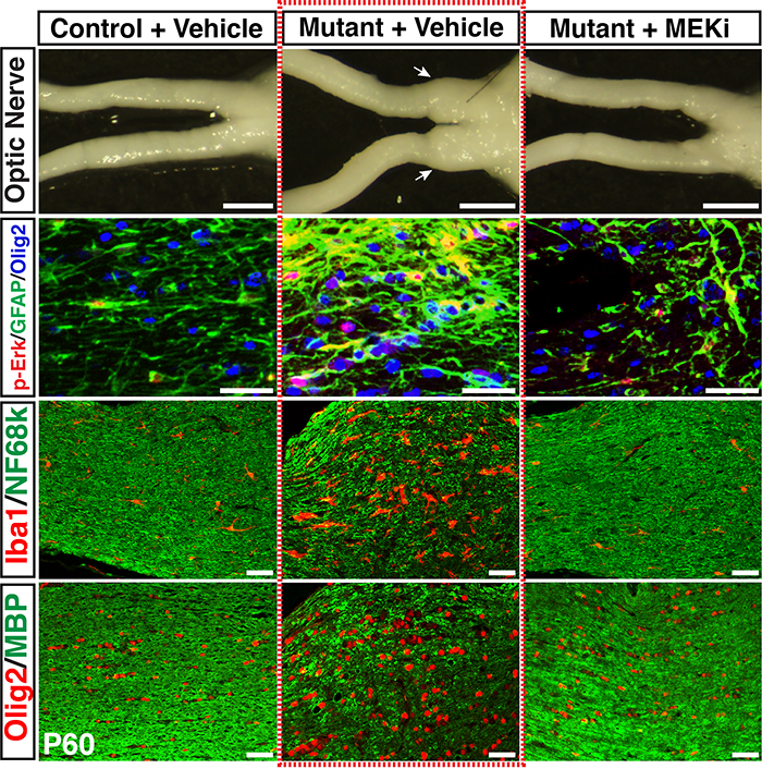
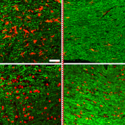
A transient low-dose MEKi treatment in a pre-clinical model prevents NF1-OPG formation. The middle panels highlighted by a red dashed box show an OPG in the optic nerve (arrows, top), exhibiting abnormal triply-labeled tumor cells, inflammation and nerve damage (the bottom three panels), which are absent in the normal (left panels) or MEKi-treated Nf1-deficient optic nerves (right panels). [Credit: Jecrois et al., Developmental Cell, (2021)]

Catherine Bollard

Engineering TGFB receptor to enhance NK cells and fight neuroblastoma

Catherine Bollard

“In this study, we have genetically engineered cord blood derived NK cells so that they are not only resistant to the devastating effects of TGFb, but they are not able to become activated in the presence of TGFb,” said, Catherine Bollard, M.B.Ch.B., M.D.

Catherine Bollard, M.B.Ch.B., M.D., and her research team published results showing potential efficacy of a novel cell therapy for treatment of pediatric patients with relapsed/refractory neuroblastoma.

The research paper, entitled, “Engineering the TGFβ receptor to Enhance the Therapeutic Potential of Natural Killer Cell as an Immunotherapy for Neuroblastoma,” was published on April 29, 2019 by Clinical Cancer Research and is being recognized for the potential efficacy of the “off the shelf” treatment for patients with relapsed/refractory neuroblastoma.

The researcher’s approach allows them to manipulate Natural Killer (NK) cells, expand and reinfuse them within a patient so they can fight cancer and disease.

“In this study, we have genetically engineered cord blood derived NK cells so that they are not only resistant to the devastating effects of TGFb, but they are not able to become activated in the presence of TGFb,” said, Dr. Bollard, who is the senior corresponding author of the study and director of the Center for Cancer and Immunology Research at the Children’s Research Institute. “In other words, turning the negative effects of TGFb into positive effects enhances the persistence and anti-tumor activity of these tumor-killing NK cells in vivo.”

NK cells are highly cytolytic, and their potent antitumor effects can be rapidly triggered by a lack of human leukocyte antigen (HLA) expression on interacting target cells, as in the case for a majority of solid tumors, including neuroblastoma. With neuroblastoma being a leading cause of pediatric cancer-related deaths, it presents as an ideal candidate for NK cell therapy.

“This manuscript encompasses a significant portion of work, in which we generated genetically-modified NK cells as an enhanced form of immunotherapy for neuroblastoma,” said Rachel Burga, Ph.D., lead author and graduate of the Institute for Biomedical Sciences at George Washington and Children’s National Health System.  “We’re very excited to share our pre-clinical findings which demonstrate the efficacy of approaches to “hijack” the TGFb receptor and target TGFb in the tumor microenvironment.”

She added that the approach will allow for the NK cells to simultaneously resist the immune suppression in the microenvironment and initiate activation to increase their ability to target tumor cells.

Pre-clinical testing and research for this trial began in 2016 and ended in 2019. “The idea came from a Department of Defense award given to Dr. Bollard and Dr. Cruz and they took the idea and reduced it to practice and showed feasibility for pre-clinical trial,” said Rohan Fernandes, Ph.D., assistant professor in the Department of Medicine at George Washington University and senior author on the manuscript.

Fernandes added that the timeframe to start the clinical trial is within the next two to four years at Children’s National.

Additional authors include Rachel A. Burga, Ph.D., Eric Yvon, Rohan Fernandes, Conrad Russell Cruz, and Catherine M. Bollard, M.B.Ch.B., M.D.

Brian Rood

Improving the understanding of medulloblastoma

Brian Rood

Brian Rood, M.D., employed quantitative proteomics to tumor samples that led to novel therapeutic targets for Medulloblastoma and other tumors.

In a recently published study, Brian Rood, M.D., a neuro-oncologist at Children’s National Health System, employed quantitative proteomics to tumor samples, a technique that could lead to novel therapeutic targets for medulloblastoma and other tumors in the future.

Currently, many experts use genomic characterization to understand the genetic makeup of cancer cells, which has deepened the field’s collective knowledge of tumor biology. However, it has remained challenging to infer specific information about how the tumors will respond and consequently develop more effective therapies. Medulloblastoma is the most common pediatric, malignant brain tumor. Through Dr. Rood’s research using proteomic analysis, he was able to identify and measure the protein makeup of medulloblastoma, which led to a potential pathway for clinical intervention to treat this life-threatening cancer. The findings were published online June 7, 2018, in Acta Neuropathologica Communications.

“The goal of this research was to find out how these tumor cells function at the protein level, which may ultimately help the field identify drug therapies to stop them,” says Dr. Rood. “The genes of a cancer cell are like a blueprint for a building, but the blueprints aren’t always followed in a cancer cell: Not every active gene will produce its corresponding protein. Proteins do the work of the cell, and understanding them will provide a better overall understanding of a cancer cell’s biology.”

Dr. Rood compared proteomic and genomic data to confirm that genetics do not accurately predict the quantity of proteins. By directly quantitating the proteins and comparing them between different subgroups of the disease, they were able to identify protein-based pathways driving tumor biology. With this information, Dr. Rood was able to demonstrate that medulloblastoma depends on a crucial pathway, the eukaryotic initiation factor 4F protein synthesis pathway, resulting in the identification of a potential target for new treatments in medulloblastoma.

Ultimately, Dr. Rood found that proteomic analysis complements genomic characterization and the two can be used together to create a more complete understanding of tumor biology. Going forward, he hopes proteomic analysis will become common practice for studying all tumors, allowing tumors to be categorized and grouped together by protein makeup to help the field identify more effective therapies for all tumors.