Tag Archive for: stroke

The endovascular embolic hemispherectomy team.

New hemimegalencephaly procedure is all about teamwork

Children’s National experts pioneered a novel approach of inducing strokes to stop seizures and improve neurodevelopmental outcomes in newborns under three months old with hemimegalencephaly (HME). The procedure, called an endovascular embolic hemispherectomy, can be safely used to provide definitive treatment of HME-related epilepsy in neonates and young infants. Monica Pearl, M.D., neurointerventional radiologist, and Panagiotis Kratimenos, M.D., Ph.D., neonatologist, discuss why having a multidisciplinary team skilled at this procedure is the reason we’re the only center in the world capable of providing this treatment.

baby with brain monitor

The history behind the novel hemimegalencephaly procedure

Traditionally, when a baby is diagnosed with hemimegalencephaly (HME), doctors turn to a hemispherectomy at 3 months of age, which involves surgically removing half of a baby’s brain. At Children’s National Hospital, our doctors pioneered the endovascular embolic hemispherectomy, an approach using induced controlled strokes to eliminate the affected part of the brain, halting seizures. Monica Pearl, M.D., neurointerventional radiologist, and Tammy Tsuchida, M.D., Ph.D., neonatal neurologist, talk about this life-changing procedure.

Angelique and family pose in front of their house

Inducing strokes to better treat babies with hemimegalencephaly

When a family from Texas received a shocking diagnosis for their newborn daughter, they knew there was one place they needed to go – Children’s National Hospital in Washington, D.C. At birth, Angelique was diagnosed with a rare and devastating condition known as hemimegalencephaly (HME) which causes uncontrollable and frequent seizures. Monica Pearl, M.D., neurointerventional radiologist, and the team at Children’s National have pioneered an approach to treat HME, where they induce controlled strokes to eliminate the affected part of the brain, halting seizures in their tracks. They’re the only team in the world doing this work. Angelique’s parents knew the clock was ticking — every day they waited meant irreversible damage to their daughter’s developing brain.

doctor looking at brain MRIs

NINDS awards $10 million for pediatric concussion research

doctor looking at brain MRIs

Researchers will use advanced brain imaging and blood tests to explore biological markers—changes in blood pressure, heart rate and pupil reactivity—that could predict which children will develop persistent symptoms after concussion.

The National Institute of Neurological Disorders and Stroke has awarded a $10-million grant to the Four Corners Youth Consortium, a group of academic medical centers studying concussions in school-aged children. Led in part by the Safe Concussion Outcome Recovery and Education (SCORE) program at Children’s National Hospital, the project is named Concussion Assessment, Research and Education for Kids, or CARE4Kids.

Researchers will use advanced brain imaging and blood tests to explore biological markers—changes in blood pressure, heart rate and pupil reactivity—that could predict which children will develop persistent symptoms after concussion. The five-year CARE4Kids study will enroll more than 1,300 children ages 11-18 nationwide.

The five-year study will be led by Gerard Gioia, Ph.D., division chief of Neuropsychology at Children’s National Hospital, Frederick Rivara, M.D., M.P.H., at Seattle Children’s Center for Child Health, Behavior and Development and University of Washington’s Medicine’s Department of Pediatrics, and Dr. Chris Giza at University of California, Los Angeles (UCLA).

“We will be gathering innovative data to help answer the critical question asked by every patient: ‘When can I expect to recover from this concussion?’” said Dr. Gioia. “We have a great team and are excited to have been selected to study this important issue.”

Christopher G. Vaughan, Psy.D., neuropsychologist, and Raquel Langdon, M.D., neurologist, both at Children’s National, will join Dr. Gioia as principal investigators of the study at this site.

Every year, more than 3 million Americans are diagnosed with concussions. Symptoms continue to plague 30 percent of patients three months after injury—adolescents face an even higher risk of delayed recovery. Chronic migraine headaches, learning and memory problems, exercise intolerance, sleep disturbances, anxiety and depressed mood are common.

“Providing individualized symptom-specific treatments for youth with a concussion has been a longstanding aim of the SCORE program,”Dr. Vaughan said. “This project will lead to a better understanding of the specific markers for which children may have a longer recovery. With this knowledge, we can start individualized treatments earlier in the process and ultimately help to reduce the number of children who experienced prolonged effects after concussion.”

The grant was announced on September 9, 2021.

In Washington, D.C., an estimated 240 children ages 11 to 18, will participate in the study.

The study will unfold in two phases. The first part will evaluate children with concussion to identify a set of biomarkers predictive of persistent post-concussion symptoms. To validate the findings, the next stage will confirm that these biomarkers accurately predict prolonged symptoms in a second group of children who have been diagnosed with concussion. The goal is to develop a practical algorithm for use in general clinical practice for doctors and other health professionals caring for pediatric patients.

Institutions currently recruiting patients for the study include Children’s National Hospital, UCLA Mattel Children’s Hospital, Seattle Children’s, the University of Washington, University of Rochester, University of Texas Southwestern Medical Center and Wake Forest School of Medicine. Indiana University, the National Institute of Nursing Research, University of Arkansas, University of Southern California and the data coordinating center at the University of Utah are also involved in the project.

Earlier research conducted by the Four Corners Youth Consortium that led to this project was funded by private donations from Stan and Patti Silver, the UCLA Steve Tisch BrainSPORT Program and the UCLA Easton Clinic for Brain Health; Children’s National Research Institute; as well as from the Satterberg Foundation to Seattle Children’s Research Institute; and an investment from the Sports Institute at UW Medicine.

Blood Clot or thrombus

Endovascular therapy for acute stroke in children

Blood Clot or thrombus

Endovascular therapies for acute childhood stroke remain controversial and little evidence exists to determine the minimum age and size cut-off for thrombectomy in children. In a recent study published in the Journal of NeuroInterventional Surgery, Monica S. Pearl, M.D., director of Neurointerventional Radiology Program at Children’s National Hospital, and other experts found an increasing number of reports suggesting the feasibility of thrombectomy in at least some children by experienced operators.

When compared with adults, technical modifications may be necessary in children owing to differences in vessel sizes, tolerance of blood loss, safety of contrast and radiation exposure, and differing stroke etiologies. Dr. Pearl and experts reviewed critical considerations for neurologists and neurointerventionalists when treating pediatric stroke with endovascular therapies.

Additional study authors from Children’s National include: Dana Harrar, M.D., Ph.D., and Carlos Castillo Pinto, M.D., F.A.A.P.

Read the full study in the Journal of NeuroInterventional Surgery.

Artificial Intelligence concept image

Thrombectomy can be efficient and safe in childhood stroke, new study finds

Artificial Intelligence concept image

A recent study adds to the growing evidence that mechanical thrombectomy can be effective and safe not only in adults, but also in childhood stroke.

Previous randomized trials proved the effectiveness of thrombectomy for large intracranial vessel occlusions in adults only. However, a recent retrospective study led by Monica S. Pearl, M.D., Neurointerventional Radiology Program director at Children’s National Hospital, finds that thrombectomy can be safely performed in carefully selected cases of childhood stroke. The study further shows that treated children have good neurological outcomes.

In the findings, Dr. Pearl and other leading experts discussed specific circumstances and important considerations to take into account when managing a child with acute ischemic stroke due to a large vessel occlusion.

“We are raising the bar for the expected level of care for children with acute ischemic stroke,” said Dr. Pearl. “Care should be multidisciplinary and involve stroke neurology, neuroradiology, neurointerventional radiology, neurosurgery, cardiology, hematology and ICU teams.”

Prior to the study, clear guidelines for patient selection, thrombectomy technique and periprocedural care did not exist for the pediatric population despite the proven success of mechanical thrombectomy in adults.

Through a case-based approach encompassing a broad range of ages and clinical presentations, Dr. Pearl and other leading experts presented select cases of acute ischemic stroke in children and discussed the nuances, risks, benefits and management plan for each child.

Many of the clinical scenarios highlighted unanswered questions in the management and treatment of children with acute ischemic stroke due to large vessel occlusion. The study adds to the growing evidence that mechanical thrombectomy can be effective and safe not only in adults, but also in childhood stroke.

“It’s exciting to be shaping management for children with acute ischemic stroke,” said Dr. Pearl. “We are serving as the model for individualized, patient-centered care with multidisciplinary specialists and institutional collaboration caring for children with acute ischemic stroke.”

However, Dr. Pearl and experts encourage caution because etiology in childhood stroke differs substantially from that in acute ischemic stroke in adults, with potentially major impact on procedure success and safety.

The mission of the Neurointerventional Radiology Program, a new effort at Children’s National, is to provide exceptional family-centered care and cutting-edge diagnostic and endovascular treatment options for children with neurovascular disorders. Dr. Pearl serves as the program’s full time, dedicated neurointerventional radiologist, a specialized expertise found only in a handful of other pediatric hospitals in the country.

You can find the full study published in JAHA. Learn more about the Children’s National Research Institute Center for Neuroscience Research.

brain network illustration

$2.5M to protect the brain from metabolic insult

brain network illustration

The brain comprises only 2% of the body’s volume, but it uses more than 20% of its energy, which makes this organ particularly vulnerable to changes in metabolism.

More than 30 million Americans have diabetes, with the vast majority having Type 2 disease. Characterized by insulin resistance and persistently high blood sugar levels, poorly controlled Type 2 diabetes has a host of well-recognized complications: compared with the general population, a greatly increased risk of kidney disease, vision loss, heart attacks and strokes and lower limb amputations.

But more recently, says Nathan A. Smith, MS, Ph.D., a principal investigator in Children’s National Research Institute’s Center for Neuroscience Research, another consequence has become increasingly apparent. With increasing insulin resistance comes cognitive damage, a factor that contributes significantly to dementia diagnoses as patients age.

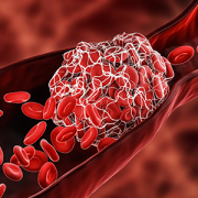
The brain comprises only 2% of the body’s volume, but it uses more than 20% of its energy, Smith explains – which makes this organ particularly vulnerable to changes in metabolism. Type 2 diabetes and even prediabetic changes in glucose metabolism inflict damage upon this organ in mechanisms with dangerous synergy, he adds. Insulin resistance itself stresses brain cells, slowly depriving them of fuel. As blood sugar rises, it also increases inflammation and blocks nitric oxide, which together narrow the brain’s blood vessels while also increasing blood viscosity.

When the brain’s neurons slowly starve, they become increasingly inefficient at doing their job, eventually succumbing to this deprivation. These hits don’t just affect individual cells, Smith adds. They also affect connectivity that spans across the brain, neural networks that are a major focus of his research.

While it’s well established that Type 2 diabetes significantly boosts the risk of cognitive decline, Smith says, it’s been unclear whether this process might be halted or even reversed. It’s this question that forms the basis of a collaborative Frontiers grant, $2.5 million from the National Science Foundation split between his laboratory; the lead institution, Stony Brook University; and Massachusetts General Hospital/Harvard Medical School.

Smith and colleagues at the three institutions are testing whether changing the brain’s fuel source from glucose to ketones – byproducts from fat metabolism – could potentially save neurons and neural networks over time. Ketones already have shown promise for decades in treating some types of epilepsy, a disease that sometimes stems from an imbalance in neuronal excitation and inhibition. When some patients start on a ketogenic diet – an extreme version of a popular fat-based diet – many can significantly decrease or even stop their seizures, bringing their misfiring brain cells back to health.

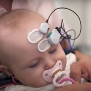
Principal Investigator Smith and his laboratory at the Children’s National Research Institute are using experimental models to test whether ketones could protect the brain against the ravages of insulin resistance. They’re looking specifically at interneurons, the inhibitory cells of the brain and the most energy demanding. The team is using a technique known as patch clamping to determine how either insulin resistance or insulin resistance in the presence of ketones affect these cells’ ability to fire.

They’re also looking at how calcium ions migrate in and out of the cells’ membranes, a necessary prerequisite for neurons’ electrical activity. Finally, they’re evaluating whether these potential changes to the cells’ electrophysiological properties in turn change how different parts of the brain communicate with each other, potentially restructuring the networks that are vital to every action this organ performs.

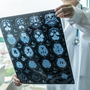
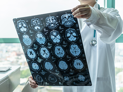
Colleagues at Athinoula A. Martinos Center for Biomedical Imaging at Massachusetts General Hospital and Harvard Medical School, led by Principal Investigator Eva-Maria Ratai, Ph.D.,  will perform parallel work in human subjects. They will use imaging to determine how these two fuel types, glucose or ketones, affect how the brain uses energy and produces the communication molecules known as neurotransmitters. They’re also investigating how these factors might affect the stability of neural networks using techniques that investigate the performance of these networks both while study subjects are at rest and performing a task.

Finally, colleagues at the Laufer Center for Physical and Quantitative Biology at Stony Brook University, led by Principal Investigator Lilianne R. Mujica-Parodi, Ph.D., will use results generated at the other two institutions to construct computational models that can accurately predict how the brain will behave under metabolic stress: how it copes when deprived of fuel and whether it might be able to retain healthy function when its cells receive ketones instead of glucose.

Collectively, Smith says, these results could help retain brain function even under glucose restraints. (For this, the research team owes a special thanks to Mujica-Parodi, who assembled the group to answer this important question, thus underscoring the importance of team science, he adds.)

“By supplying an alternate fuel source, we may eventually be able to preserve the brain even in the face of insulin resistance,” Smith says.

Suresh Magge

Sudden blindness leads to unusual diagnosis

Suresh Magge

Suresh N. Magge, M.D., and his colleagues at Children’s National recently published the details of an unusual case of advanced moyamoya disease in the journal Stroke.

When Children’s National Health System Neurosurgeon Suresh N. Magge, M.D., met his new patient, the 16-year-old had suddenly lost her vision in both eyes.

To discover the reason for this abrupt loss of vision, her doctors ran a battery of tests. An ophthalmologist found no problems with her eyes.  Her optic nerves, which run signals generated from the eyes to the brain, also appeared to work normally. However, a computerized tomography scan and magnetic resonance imaging showed the unmistakable signs of a stroke in her occipital lobe, the portion of the brain responsible for interpreting signals relayed from the optic nerves.

“Her brain basically wasn’t seeing what her eyes saw,” Dr. Magge explains.

Delving deeper, her Children’s National care team found the reason why their young patient had suffered a stroke by using a cerebral angiogram, an imaging test that shows the blood vessels in and around the brain. The teen had moyamoya disease, a rare condition that causes blood vessels in the brain to narrow, often leading otherwise healthy adults and children to have strokes.

According to the National Institutes of Health, moyamoya is Japanese for “puff of smoke,” so named because of the telltale signs this condition presents on an angiogram. When arteries in the brain narrow, brain tissue becomes “thirsty” for more blood, Dr. Magge explains, leading its cells to produce chemicals that prompt new blood vessels to grow. These new collateral blood vessels often grow in a thin tangle that looks like smoke on an angiogram. Generally, however, they do not supply sufficient oxygenated blood to meet the brain’s needs, leaving it starved for oxygen. Eventually, the blood supply can get so low that patients suffer transient ischemic attacks, “mini-strokes” that temporarily deprive the brain tissue of oxygen, or full-blown strokes typically characterized by weakness, speech problems, facial paralysis or other problems.

Dr. Magge’s patient had little warning before her stroke occurred. The first major symptom that led her to seek medical attention was abrupt blindness, which Dr. Magge says is a highly unusual occurrence for a moyamoya diagnosis. That’s why he and colleagues decided to publish the details of her case as a teaching report April 14, 2017 in the journal Stroke.

The Children’s National co-authors wrote that once their patient was diagnosed with a stroke due to advanced moyamoya disease, with blood vessels severely narrowed throughout her brain, the first order of business was stabilizing her symptoms and making sure she did not have further strokes. Her blood pressure was stabilized, and she was started on aspirin therapy to decrease her risk of further strokes. She took time to recover as much as possible from her original stroke.

A few weeks later, Dr. Magge and his neurosurgery colleagues performed a type of surgery to revascularize – or restore blood flow – to areas of the brain that were still healthy but at risk of having subsequent strokes. The surgical procedure, known as pial synangiosis, reconfigures the brain’s blood vessels to make sure that these vulnerable areas of the brain have a sufficient blood supply.

Years later, Dr. Magge says, his patient is doing well, except for the original blindness, a permanent consequence of the stroke to her occipital lobe before her diagnosis. She has not had new strokes since the revascularization surgery. She will need aspirin therapy and periodic neurological checkups for the rest of her life, Dr. Magge explains, to make sure that the blood supply to her brain remains stable.

Children’s experts use a team approach to treat patients with complex care needs: Neurologists, neurosurgeons, intensivists, hematologists, anesthesiologists, neuroradiologists and nurses leverage their combined expertise with moyamoya disease to treat the complexities of this condition.

“A team approach is essential to deliver the best outcomes to children with life-changing diseases,” Dr. Magge says. “We try to help kids get back to living full and healthy lives.”

Blood Transfusion

Hydroxycarbamide effective in sickle cell stroke prevention

Blood Transfusion

Hydroxycarbamide treatment is on par with blood transfusions for preventing stroke in patients with sickle cell anemia.

PDF Version

What’s known

Strokes are common and devastating complications for patients with sickle cell anemia, often leading to severe and lifelong motor and neurocognitive problems for people with this congenital blood disorder. Results of a clinical trial published in 1998 showed that having regular blood transfusions could reduce the risk of having a first stroke by 90 percent in children with sickle cell anemia. Since then, doctors have employed this prophylactic treatment widely. However, blood transfusions can be painful, inconvenient and carry substantial risks themselves — including the potential of blood-borne infections, iron overload and immune-related reactions to blood products. Finding a way to reduce stroke risk without over-relying on blood transfusions could substantially benefit patients with sickle cell anemia.

What’s new

A team of researchers, including Naomi L.C. Luban, M.D., a Children’s National Health System hematologist and laboratory medicine specialist, tested transfusions against a drug treatment called hydroxycarbamide in a clinical trial to see if the pharmaceutical intervention could reduce strokes at least as well as transfusions. The clinical trial, known as “TCD With Transfusions Changing to Hydroxyurea (TWiTCH),” assigned 60 patients with sickle cell anemia who had abnormally high transcranial Doppler (TCD) flow velocities—a measure of blood flow in the brain that suggests elevated risk of stroke—to receive hydroxycarbamide instead of transfusions. The research team compared the outcomes for these patients with 61 other patients who received standard prophylactic transfusions. Over the 24-month study period, neither group experienced any strokes, although three transient ischemic attacks (a temporary blockage of blood flow in the brain) occurred in each group. These comparable findings suggest that hydroxycarbamide treatment, also known as hydroxyurea, is on par with transfusions for preventing strokes in patients with sickle cell anemia.

Questions for future research

Q: Does hydroxycarbamide offer a long-term way for patients with sickle cell anemia to avoid transfusions?
Q: Could hydroxycarbamide help patients with sickle cell anemia who already have suffered a stroke or who have had severe problems with blood vessels in their brains that impair blood flow?
Q: Which other treatments can help patients avoid the myriad complications that accompany sickle cell anemia?

Source: Hydroxycarbamide versus chronic transfusion for maintenance of transcranial doppler flow velocities in children with sickle cell anemia—TCD With Transfusions Changing to Hydroxyurea (TWiTCH): A multicentre, open-label, phase 3, non-inferiority trial.” Ware, R.E. B. R. Davis, W. H. Schultz, R.C. Brown, B. Aygun, S. Sarnaik, I. Odame, B. Fuh, A. George, W. Owen, L. Luchtman-Jones, Z.R. Rogers, L. Hilliard, C. Gauger, C. Piccone, M.T. Lee, J.L. Kwiatkowski, S. Jackson, S.T. Miller, C. Roberts, M.M. Heeney, T.A. Kalfa, S. Nelson, H. Imran, K. Nottage, O. Alvarez, M. Rhodes, A.A. Thompson, J.A. Rothman, K.J. Helton, D. Roberts, J. Coleman, M.J. Bonner, A. Kutlar, N. Patel, J. Wood, L. Piller, P. Wei, J. Luden, N.A. Mortier, S.E. Stuber, N. L. C. Luban, A.R. Cohen, S. Pressel and R.J. Adams. Published by The Lancet on Feb. 13, 2016.