Tag Archive for: stem cell

microglia cells damage the myelin sheath of neuron axons

Katrina Adams, Ph.D., awarded fellowship to help restore functions in MS patients

microglia cells damage the myelin sheath of neuron axons

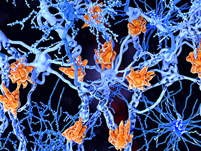
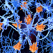
Multiple sclerosis is a demyelinating disease in which the insulating covers of nerve cells are damaged. Microglia cells (orange) attack the oligodendrocytes that form the insulating myelin sheath around neuron axons, leading to the destruction of the myelin sheath and to the loss of nerve function.

For her contributions to Multiple Sclerosis (MS) research, Katrina Adams, Ph.D., postdoctoral researcher at Children’s National Hospital, received the career transition fellowship from The National Multiple Sclerosis Society. The $600,000 fellowship will support a two-year period of advanced postdoctoral training in MS research and the first three years of research support in a new faculty appointment.

MS symptoms, including vision loss, pain, fatigue and reduced motor coordination, result from the demyelination of neuronal axons that transport critical information across the brain and spinal cord. Demyelination is the loss of myelin protein, which is normally produced by oligodendrocyte cells.

In the healthy brain, oligodendrocytes repair demyelinated areas by replacing damaged or lost myelin in a process called remyelination. Recent evidence has shown that oligodendrocytes display differences in their molecular and functional properties. One source of new oligodendrocytes in the adult brain is neural stem cells, which have been shown to generate oligodendrocytes that contribute to remyelination.

“The goal of this project is to determine whether neural stem cell-derived oligodendrocytes are distinct from other oligodendrocytes, both in the healthy brain and in MS,” said Adams. “I aim to understand the molecular mechanisms that regulate generation of oligodendrocytes from neural stem cells, with the goal of identifying signals that could be targeted in MS patients to promote remyelination.”

Remyelination is very limited in MS patients and current therapies for MS have very little impact on promoting remyelination.

This study will take advantage of the state-of-the-art facilities for single-cell analysis, transcriptomics, microscopy, and animal research in Children’s Research Institute at Children’s National. Adams also added that her postdoctoral mentor, Vittorio Gallo, Ph.D., interim chief academic officer and interim director of the Children’s National Research Institute, and principal investigator for the DC-IDDRC, has renowned expertise in glial biology, animal models of MS and white matter injury.

“This research will be the first to directly compare neural stem cell-derived oligodendrocytes with other resident oligodendrocytes in MS brain samples,” said Adams. “The results of this study will provide critical insight into the role that neural stem cells play in repair of MS demyelinated lesions.”

Adams received her doctorate in molecular biology from the University of California, Los Angeles where she used pluripotent stem cells to study motor neuron development. She currently investigates signaling pathways that regulate neural stem and progenitor cell maintenance and differentiation in the developing postnatal and adult brain, with a focus on the Endothelin-1 pathway. She is interested in understanding how stem and progenitor cells respond to disease or injury, such as in Multiple Sclerosis, with the hope of identifying new therapeutic targets.

illustration of the brain

New research provides glimpse into landscape of the developing brain

illustration of the brain

Stem and progenitor cells exhibit diversity in early brain development that likely contributes to later neural complexity in the adult cerebral cortex, this according to a new study in Science Advances. This research expands on existing ideas about brain development, and could significantly impact the clinical care of neurodevelopmental diseases in the future.

Stem and progenitor cells exhibit diversity in early brain development that likely contributes to later neural complexity in the adult cerebral cortex, this according to a study published Nov. 6, 2020, in Science Advances. Researchers from the Center for Neuroscience Research (CNR) at Children’s National Hospital say this research expands on existing ideas about brain development, and could significantly impact the clinical care of neurodevelopmental diseases in the future. The study was done in collaboration with a research team at Yale University led by Nenad Sestan, M.D, Ph.D.

“Our study provides a new glimpse into the landscape of the developing brain. What we are seeing are new complex families of cells very early in development,” says Tarik Haydar, Ph.D., director of CNR at Children’s National, who led this study. “Understanding the role of these cells in forming the cerebral cortex is now possible in a way that wasn’t possible before.”

The cerebral cortex emerges early in development and is the seat of higher-order cognition, social behavior and motor control. While the rich neural diversity of the cerebral cortex and the brain in general is well-documented, how this variation arises is relatively poorly understood.

“We’ve shown in our previous work that neurons generated from different classes of cortical stem and progenitor cells have different functional properties,” says William Tyler, Ph.D., CNR research faculty member and co-first author of the study. “Part of the reason for doing this study was to go back and try to classify all the different progenitors that exist so that eventually we can figure out how each contributes to the diversity of neurons in the adult brain.”

Using a preclinical model, the researchers were able to identify numerous groups of cortical stem and precursor cells with distinct gene expression profiles. The team also found that these cells showed early signs of lineage diversification likely driven by transcriptional priming, a process by which a mother cell produces RNA for the sole purpose of passing it on to its daughter cells for later protein production.

Tarik Haydar

“Our study provides a new glimpse into the landscape of the developing brain. What we are seeing are new complex families of cells very early in development,” says Tarik Haydar, Ph.D., director of CNR at Children’s National, who led this study. “Understanding the role of these cells in forming the cerebral cortex is now possible in a way that wasn’t possible before.”

Using novel trajectory reconstruction methods, the team observed distinct developmental streams linking precursor cell types to particular excitatory neurons. After comparing the dataset of the preclinical model to a human cell database, notable similarities were found, such as the surprising cross-species presence of basal radial glial cells (bRGCs), an important type of progenitor cell previously thought to be found mainly in the primate brain.

“At a very high level, the study is important because we are directly testing a fundamental theory of brain development,” says Zhen Li, Ph.D., CNR research postdoctoral fellow and co-first author of the study. The results add support to the protomap theory, which posits that early stem and progenitor diversity paves the way for later neuronal diversity and cortical complexity. Furthermore, the results also hold exciting translational potential.

“There is evidence showing that neurodevelopmental diseases affect different populations of the neural stem cells differently,” says Dr. Li. “If we can have a better understanding of the complexity of these neural stem cells there is huge implication of disease prevention and treatment in the future.”

“If we can understand how this early landscape is affected in disorders, we can predict the resulting changes to the cortical architecture and then very narrowly define ways that groups of cells behave in these disorders,” adds Dr. Haydar. “If we can understand how the cortex normally achieves its complex architecture, then we have key entry points into improving the clinical course of a given disorder and improving quality of life.”

Future topics the researchers hope to study include the effects of developmental changes on brain function, the origin and operational importance of bRGCs, and the activity, connections and cognitive features enabled by different families of neurons.

t-cells

Tailored T-cell therapies neutralize viruses that threaten kids with PID

t-cells

Tailored T-cells specially designed to combat a half dozen viruses are safe and may be effective in preventing and treating multiple viral infections, according to research led by Children’s National Hospital faculty.

Catherine Bollard, M.B.Ch.B., M.D., director of the Center for Cancer and Immunology Research at Children’s National and the study’s senior author, presented the teams’ findings Nov. 8, 2019, during a second-annual symposium jointly held by Children’s National and the National Institute of Allergy and Infectious Diseases (NIAID), part of the National Institutes of Health (NIH). Children’s National and NIAID formed a research partnership in 2017 to develop and conduct collaborative clinical research studies focused on young children with allergic, immunologic, infectious and inflammatory diseases. Each year, they co-host a symposium to exchange their latest research findings.

According to the NIH, more than 200 forms of primary immune deficiency diseases impact about 500,000 people in the U.S. These rare, genetic diseases so impair the person’s immune system that they experience repeated and sometimes rare infections that can be life threatening. After a hematopoietic stem cell transplantation, brand new stem cells can rebuild the person’s missing or impaired immune system. However, during the window in which the immune system rebuilds, patients can be vulnerable to a host of viral infections.

Because viral infections can be controlled by T-cells, the body’s infection-fighting white blood cells, the Children’s National first-in-humans Phase 1 dose escalation trial aimed to determine the safety of T-cells with antiviral activity against a half dozen opportunistic viruses: adenovirus, BK virus, cytomegalovirus (CMV), Epstein-Barr virus (EBV), Human Herpesvirus 6 and human parainfluenza-3 (HPIV3).

Eight patients received the hexa-valent, virus-specific T-cells after their stem cell transplants:

  • Three patients were treated for active CMV, and the T-cells resolved their viremia.
  • Two patients treated for active BK virus had complete symptom resolution, while one had hemorrhagic cystitis resolved but had fluctuating viral loads in their blood and urine.
  • Of two patients treated prophylactically, one developed EBV viremia that was treated with rituximab.

Two additional patients received the T-cell treatments under expanded access for emergency treatment, one for disseminated adenoviremia and the other for HPIV3 pneumonia. While these critically ill patients had partial clinical improvement, they were being treated with steroids which may have dampened their antiviral responses.

“These preliminary results show that hexaviral-specific, virus-specific T-cells are safe and may be effective in preventing and treating multiple viral infections,” says Michael Keller, M.D., a pediatric immunologist at Children’s National and the lead study author. “Of note, enzyme-linked immune absorbent spot assays showed evidence of antiviral T-cell activity by three months post infusion in three of four patients who could be evaluated and expansion was detectable in two patients.”

In addition to Drs. Bollard and Keller, additional study authors include Katherine Harris M.D.; Patrick J. Hanley Ph.D., assistant research professor in the Center for Cancer and Immunology; Allistair Abraham, M.D., a blood and marrow transplantation specialist; Blachy J. Dávila Saldaña, M.D., Division of Blood and Marrow Transplantation; Nan Zhang Ph.D.; Gelina Sani BS; Haili Lang MS; Richard Childs M.D.; and Richard Jones M.D.

###

Children’s National-NIAID 2019 symposium presentations

“Welcome and introduction”
H. Clifford Lane, M.D., director of NIAID’s Division of Clinical Research

“Lessons and benefits from collaboration between the NIH and a free-standing children’s hospital”
Marshall L. Summar, M.D., director, Rare Disease Institute, Children’s National

“The hereditary disorders of PropionylCoA and Cobalamin Metabolism – past, present and future”
Charles P. Venditti, M.D., Ph.D., National Human Genome Research Institute Collaboration

“The road(s) to genetic precision therapeutics in pediatric neuromuscular disease: opportunities and challenges”
Carsten G. Bönnemann, M.D., National Institute of Neurological Disorders and Stroke

“Genomic diagnostics in immunologic diseases”
Helen Su, M.D., Ph.D., National Institute of Allergy and Infectious Diseases

“Update on outcomes of gene therapy clinical trials for X-SCID and X-CGD and plans for future trials”
Harry Malech, M.D., National Institute of Allergy and Infectious Diseases

“Virus-specific T-cell therapies: broadening applicability for PID patients”
Catherine Bollard, M.D., Children’s National 

“Using genetic testing to guide therapeutic decisions in Primary Immune Deficiency Disease”
Vanessa Bundy, M.D., Ph.D., Children’s National 

Panel discussion moderated by Lisa M. Guay-Woodford, M.D.
Drs. Su, Malech, Bollard and Bundy
Morgan Similuk, S.C.M., NIAID
Maren Chamorro, Parent Advocate

“Underlying mechanisms of pediatric food allergy: focus on B cells
Adora Lin, M.D., Ph.D., Children’s National 

“Pediatric Lyme outcomes study – interim update”
Roberta L. DeBiasi, M.D., MS, Children’s National 

“Molecular drivers and opportunities in neuroimmune conditions of pediatric onset”
Elizabeth Wells, M.D., Children’s National 


Epstein Barr virus

Fighting lymphoma with targeted T-cells

Epstein-Barr virus

The Epstein-Barr virus (EBV) is best known as the cause of mononucleosis, the ubiquitous “kissing disease” that most people contract at some point in their life. But in rare instances, this virus plays a more sinister role as the impetus of lymphomas, cancers that affect the white blood cells known as lymphocytes.

The Epstein-Barr virus (EBV) is best known as the cause of mononucleosis, the ubiquitous “kissing disease” that most people contract at some point in their life. But in rare instances, this virus plays a more sinister role as the impetus of lymphomas, cancers that affect the white blood cells known as lymphocytes. EBV-associated lymphomas account for about 40% of Hodgkin lymphomas, 20% of diffuse large B-cell lymphomas, and more than 90% of natural killer/T-cell lymphomas. This latter type of lymphoma typically has a very poor prognosis even with the “standard of care” lymphoma treatments such as chemotherapy and/or radiation.

When these interventions fail, the only curative approach is an allogeneic  hematopoietic stem cell transplant from a healthy donor, a treatment that’s tough on patients’ bodies and carries significant risks, says Lauren P. McLaughlin, M.D., a pediatrician specializing in hematology and oncology at Children’s National in Washington, D.C. Patients who receive these allogenic transplants are immune-compromised until the donor cells engraft; the grafts can attack patients’ healthy cells in a phenomenon called graft versus host disease; and if patients relapse or don’t respond to this treatment, few options remain.

To help improve outcomes, Dr. McLaughlin and colleagues tested an addition to the allogeneic hematopoietic stem cell transplant procedure for patients with EBV-associated lymphomas: infusion of a type of immune cell called T cells specifically trained to fight cells infected with EBV.

Dr. McLaughlin, along with Senior Author Catherine M. Bollard, M.D., M.B.Ch.B., director of the Center for Cancer and Immunology Research and the Program for Cell Enhancement and Technologies for Immunotherapy at Children’s National, and colleagues tested this therapy in 26 patients treated at Children’s National or Baylor College of Medicine. They published these results online on Sept. 27, 2018, in the journal Blood. The study was a Phase I clinical trial, meaning that the therapy was tested primarily for safety, with efficacy as a secondary aim.

Seven patients who received the therapy had active disease that had not responded to conventional therapies. The other 19 were patients deemed to be at high risk for relapse.

Before each patient received their stem cell transplant, their donors gave an additional blood sample to generate the cancer-fighting T cells. Over the next 8 to 10 weeks, the researchers painstakingly manufactured the immune cells known as T-cells that specifically targeted EBV, growing these cells into numbers large enough for clinical use. Then, as early as 30 days after transplant, the researchers infused these T-cells into patients administering at least two doses, spaced two weeks apart.

Over the next several weeks, the researchers at CNMC and Baylor College of Medicine monitored patients with comprehensive exams to see how they fared after these transplants. The results showed that adverse effects from the treatment were exceedingly rare. There were no immediate infusion-related toxicities to the T-cell therapy and only one incident of dose-limiting toxicity.

This therapy may be efficacious, depending on the individual patients’ circumstances, Dr McLaughlin adds. For those in complete remission but at high risk of relapsing, the two-year survival rate was 78%, suggesting that the administration of this novel T-cell therapy may give the immune system a boost to prevent the lymphoma from returning after transplant. For patients with active T-cell lymphomas, two-year survival rates were 60%. However, even these lower rates are better than the historical norm of 30-50%, suggesting that the targeted T-cell therapies could help fight disease in patients with this poor prognosis lymphoma.

Dr. McLaughlin, the study’s lead author and a Lymphoma Research Foundation grantee, notes that researchers have more work to do before this treatment becomes mainstream. For example, this treatment will need to be tested in larger populations of patients with EBV-related lymphoma to determine who would derive the most benefit, the ideal dose and dose timing. It also may be possible to extend targeted T-cell treatments like this to other types of cancers. In the future, Dr. McLaughlin adds, it may be possible to develop T-cells that could be used “off the shelf”—in other words, they wouldn’t need to come from a matched donor and would be ready to use whenever a recipient needs them. Another future goal is using this therapy as one of the first lines of treatment rather than as a last resort.

“Our ultimate goal is to find a way to avoid chemotherapy and/or radiation therapy while still effectively treating a patient’s cancer,” she says. “Can you use the immune system to do that job? We’re working to answer that question.”

In addition to Drs. McLaughlin and Bollard, study co-authors include Rayne Rouce, Stephen Gottschalk, Vicky Torrano, George Carrum, Andrea M. Marcogliese, Bambi Grilley, Adrian P. Gee, Malcolm K. Brenner, Cliona M. Rooney and Helen E. Heslop, all of Baylor College of Medicine; Meng-Fen Wu from the Dan L. Duncan Comprehensive Cancer Center; and Fahmida Hoq and Patrick J. Hanley, Ph.D. from Children’s National in Washington, D.C.

germ cells in testicular tissues

Experimental fertility preservation provides hope for young men

germ cells in testicular tissues

Confirming the presence of germ cells in testicular tissues obtained from patients. Undifferentiated embryonic cell transcription factor 1 (UTF1) is an established marker of undifferentiated spermatogonia as well as the pan-germ cell marker DEAD-box helicase 4 (DDX4). UTF1 (green) and/or DDX4 (red) immunostaining was confirmed in 132 out of 137 patient tissues available for research, including patients who had received previous non-alkylating (B, E, H, K) or alkylating (C, F, I, L) chemotherapy treatment. © The Author(s) 2019. Published by Oxford University Press on behalf of the European Society of Human Reproduction and Embryology.

Testicular tissue samples obtained from 189 males who were facing procedures that could imperil fertility were cryopreserved at one university, proving the feasibility of centralized processing and freezing of testicular tissue obtained from academic medical centers, including Children’s National, scattered around the world.

“It’s not surprising that the University of Pittsburgh would record the highest number of samples over the eight-year period (51 patients), given its role as the central processing facility for our recruiting network of academic medical centers,” says Michael Hsieh, M.D., Ph.D., director of transitional urology at Children’s National. “Children’s National recruited the third-highest number of patients, which really speaks to the level of collaboration I have with Jeff Dome’s team and their commitment to thinking about the whole patient and longer-term issues like fertility.”

An estimated 2,000 U.S. boys and young men each year receive treatments or have cancers or blood disorders that place them at risk for infertility. While older youths who have undergone puberty can bank their sperm prior to undergoing sterilizing doses of chemotherapy or radiation, there have been scant fertility preservation options for younger boys. However, some older adolescents and young men are too sick or stressed to bank sperm. For patients with no sperm to bank or who are too sick or stressed to bank sperm, the experimental procedure of freezing testicular tissue in anticipation that future cell- or tissue-based therapies can generate sperm is the only option.

Recent research in experimental models indicates that such testicular tissue biopsies contain stem cells, blank slate cells, hinting at the potential of generating sperm from biopsied tissue.

“This study demonstrates that undifferentiated stem and progenitor spermatogonia may be recovered from the testicular tissues of patients who are in the early stages of their treatment and have not yet received an ablative dose of therapy. The function of these spermatogonia was not tested,” writes lead author Hanna Valli-Pulaski, Ph.D., research assistant professor at the University of Pittsburgh, and colleagues in a study published online May 21, 2019, in Human Reproduction.

Right now, hematologists and oncologists discuss future treatment options with patients and families, as well as possible long-term side effects, including infertility. At Children’s National, they also mention the ongoing fertility preservation study and encourage families to speak with Dr. Hsieh. He meets with families, explains the study goals – which include determining better ways to freeze and thaw tissue and separating malignant cells from normal cells – what’s known about experimental fertility preservation and what remains unknown. Roughly half of patients decide to enroll.

“This study is unique in that there is definitely a potential direct patient benefit,” Dr. Hsieh adds. “One of the reasons the study is compelling is that it presents a message of hope to the families. It’s a message of survivorship: We’re optimistic we can help your child get through this and think about long-term issues, like having their own families.”

In this phase of the study, testicular tissue was collected from centers in the U.S. and Israel from January 2011 to November 2018 and cryopreserved. Patients designated 25% of the tissue sample to be used for the research study; 75 percent remains stored in liquid nitrogen at temperatures close to absolute zero for the patient’s future use. The fertility preservation patients ranged from 5 months old to 34 years old, with an average age of 7.9 years.

Thirty-nine percent of patients had started medical treatment prior requesting fertility preservation. Sixteen percent received non-alkylating chemotherapy while 23% received alkylating chemotherapy, which directly damages the DNA of cancer cells.

The research team found that the number of undifferentiated spermatogonia per seminiferous tubule increase steadily with age until about age 11, then rise sharply.

“We recommend that all patients be counseled and referred for fertility preservation before beginning medical treatments known to cause infertility. Because the decision to participate may be delayed, it is encouraging that we were able to recover undifferentiated spermatogonia from the testes of patients already in the early stages of chemotherapy treatments,” Dr. Hsieh says.

In addition to Dr. Hsieh, study co-authors include lead author, H. Valli-Pulaski, K.A. Peters, K. Gassei, S.R. Steimer, M. Sukhwani, B.P. Hermann, L. Dwomor, S. David, A.P. Fayomi, S.K. Munyoki, T. Chu, R. Chaudhry, G.M. Cannon, P.J. Fox, T.M. Jaffe, J.S. Sanfilippo, M.N. Menke and senior author, K.E. Orwig, all of University of Pittsburgh; E. Lunenfeld, M. Abofoul-Azab and M. Huleihel, Ben-Gurion University of the Negev; L.S. Sender, J. Messina and L.M. Klimpel, CHOC Children’s Hospital;  Y. Gosiengfiao, and E.E. Rowell, Ann & Robert H. Lurie Children’s Hospital of Chicago; C.F. Granberg, Mayo Clinic; P.P. Reddy, Cincinnati Children’s Hospital Medical Center; and J.I. Sandlow, Medical College of Wisconsin.

Financial support for the research covered in this post was provided by Eunice Kennedy Shriver National Institute for Child Health and Human Development under awards HD061289 and HD092084; Scaife Foundation; Richard King Mellon Foundation; University of Pittsburgh Medical Center; United States-Israel Binational Science Foundation and Kahn Foundation.

Nobuyuki Ishibashi

Children’s receives NIH grant to study use of stem cells in healing CHD brain damage

Nobuyuki Ishibashi

“Bone marrow stem cells are used widely for stroke patients, for heart attack patients and for those with developmental diseases,” explains Nobuyuki Ishibashi, M.D. “But they’ve never been used to treat the brains of infants with congenital heart disease. That’s why we are trying to understand how well this system might work for our patient population.”

The National Institutes of Health (NIH) awarded researchers at Children’s National Health System $2.6 million to expand their studies into whether human stem cells could someday treat and even reverse neurological damage in infants born with congenital heart disease (CHD).

Researchers estimate that 1.3 million infants are born each year with CHD, making it the most common major birth defect. Over the past 30 years, advances in medical technology and surgical practices have dramatically decreased the percentage of infants who die from CHD – from a staggering rate of nearly 100 percent just a few decades ago to the current mortality rate of less than 10 percent.

The increased survival rate comes with new challenges: Children with complex CHD are increasingly diagnosed with significant neurodevelopmental delay or impairment. Clinical studies demonstrate that CHD can reduce oxygen delivery to the brain, a condition known as hypoxia, which can severely impair brain development in fetuses and newborns whose brains are developing rapidly.

Nobuyuki Ishibashi, M.D., the study’s lead investigator with the Center for Neuroscience Research and director of the Cardiac Surgery Research Laboratory at Children’s National, proposes transfusing human stem cells in experimental models through the cardio-pulmonary bypass machine used during cardiac surgery.

“These cells can then identify the injury sites,” says Dr. Ishibashi. “Once these cells arrive at the injury site, they communicate with endogenous tissues, taking on the abilities of the damaged neurons or glia cells they are replacing.”

“Bone marrow stem cells are used widely for stroke patients, for heart attack patients and for those with developmental diseases,” adds Dr. Ishibashi. “But they’ve never been used to treat the brains of infants with congenital heart disease. That’s why we are trying to understand how well this system might work for our patient population.”

Dr. Ishibashi says the research team will focus on three areas during their four-year study – whether the stem cells:

  • Reduce neurological inflammation,
  • Reverse or halt injury to the brain’s white matter and
  • Help promote neurogenesis in the subventricular zone, the largest niche in the brain for creating the neural stem/progenitor cells leading to cortical growth in the developing brain.

At the conclusion of the research study, Dr. Ishibashi says the hope is to develop robust data so that someday an effective treatment will be available and lasting neurological damage in infants with congenital heart disease will become a thing of the past.