Tag Archive for: schistosomiasis

Micrograph of human parasite Schistosoma mansonii

Diagnosing and monitoring of urogenital schistosomiasis

Micrograph of human parasite Schistosoma mansonii

Urogenital schistosomiasis (UGS) is caused by egg-laying S. haematobium worms dwelling within the veins draining the main pelvic organs, including the bladder, uterus, and cervix.

Although urogenital schistosomiasis remains a major global challenge, Michael Hsieh, M.D., Ph.D., director of Transitional Urology at Children’s National Hospital, and other experts including Eglal Shalaby Rana, M.D., from Children’s National, show in a new study published in Advances in Parasitology that newer refinements in associated technologies may lead to improvements in patient care.

In addition, application of investigational imaging methods, such as confocal laser endomicroscopy and two-photon microscopy in urogenital schistosomiasis, are likely to contribute to the understanding of this infection’s pathogenesis.

Read the full study in Advances in Parasitology.

Schistosoma

Parasitic eggs trigger upregulation in genes associated with inflammation

Schistosoma

Of the 200 million people around the globe infected with Schistosomiasis, about 100 million of them were sickened by the parasite Schistosoma haematobium.

Of the 200 million people around the globe infected with Schistosomiasis, about 100 million of them were sickened by the parasite Schistosoma haematobium. As the body reacts to millions of eggs laid by the blood flukes, people can develop fever, cough and abdominal pain, according to the Centers for Disease Control and Prevention. Schistosomiasis triggered by S. haematobium can also include hematuria, bladder calcification and bladder cancer.

Despite the prevalence of this disease, there are few experimental models specifically designed to study it, and some tried-and-true preclinical models don’t display the full array of symptoms seen in humans. It’s also unclear how S. haematobium eggs deposited in the host bladder modulate local tissue gene expression.

To better understand the interplay between the parasite and its human host, a team led by Children’s National Hospital injected 6,000 S. haematobium eggs into the bladder wall of seven-week-old experimental models.

After four days, they isolated RNA for analysis, comparing differences in gene expression in various treatment groups, including those that had received the egg injection and experimental models whose bladders were not exposed to surgical intervention.

Using the Database for Annotation, Visualization and Integrated Discovery (DAVID) – a tool that helps researchers understand the biological meaning of a long list of genes – the team identified commonalities with other pathways, including malaria, rheumatoid arthritis and the p53 signaling pathway, the team recently presented during the American Society of Tropical Medicine and Hygiene 2019 annual meeting. Some 325 genes were differentially expressed, including 34 genes in common with previous microarray data.

“Of particular importance, we found upregulation in genes associated with inflammation and fibrosis. We also now know that the body may send it strongest response on the first day it encounters a bolus of eggs,” says Michael Hsieh, M.D., Ph.D., director of transitional urology at Children’s National, and the research project’s senior author. “Next, we need to repeat these experiments and further narrow the list of candidate genes to key genes associated with immunomodulation and bladder cancer.”

In addition to Dr. Hsieh, presentation co-authors include Lead Author Kenji Ishida, Children’s National; Evaristus Mbanefo and Nirad Banskota, National Institutes of Health; James Cody, Vigene Biosciences; Loc Le, Texas Tech University; and Neil Young, University of Melbourne.

Financial support for research described in this post was provided by the National Institutes of Health under award No. R01-DK113504.

clatharin cage viewed by electron microscopy

IPSE infiltrates nuclei through clathrin-mediated endocytosis

clatharin cage viewed by electron microscopy

IPSE, one of the important proteins excreted by the parasite Schistosoma mansoni, infiltrates human cellular nuclei through clathrin-coated vesicles, like this one.

IPSE, one of the important proteins excreted by the parasite Schistosoma mansoni infiltrates human cellular nuclei through clathrin-mediated endocytosis (a process by which cells absorb metabolites, hormones and proteins), a research team led by Children’s National Hospital reported during the American Society of Tropical Medicine and Hygiene 2019 annual meeting.

Because the public health toll from the disease this parasite causes, Schistosomiasis, is second only to malaria in global impact, research teams have been studying its inner workings to help create the next generation of therapies.

In susceptible host cells – like urothelial cells, which line the urinary tract – IPSE modulates gene expression, increasing cell proliferation and angiogenesis (formation of new blood vessels). On a positive note, neurons appear better able to fend off its nucleus-infiltrating ways.

“We know that IPSE contributes to the severity of symptoms in Schistosomiasis, which leads some patients to develop bladder cancer, which develops from the urothelial lining of the bladder. Our team’s carefully designed experiments reveal IPSE’s function in the urothelium and point to the potential of IPSE playing a therapeutic role outside of the bladder,” says Michael Hsieh, M.D., Ph.D., director of transitional urology at Children’s National and the research project’s senior author.

In addition to Dr. Hsieh, research co-authors include Evaristus Mbanefo, Ph.D.; Kenji Ishida, Ph.D.; Austin Hester, M.D.; Catherine Forster, M.D.; Rebecca Zee, M.D., Ph.D.; and Christina Ho, M.D., all of Children’s National; Franco Falcone, Ph.D., University of Nottingham; and Theodore Jardetzky, Ph.D., and Luke Pennington, M.D., Ph.D., candidate, both of Stanford University.

Financial support for research described in this post was provided by the National Institutes of Health under award No. R01-DK113504.

Hepatocytes

H-IPSE internalized by just a limited range of cells

Hepatocytes

A team led by Children’s National Hospital found that H-IPSE is internalized by just a limited range of cells, including hepatocytes.

Schistosoma mansoni is a parasite that hides out in snails, breaks free into waterways, and then infects humans, spending much of its life inside blood vessels, laying eggs and jeopardizing public health when those eggs are excreted in urine or feces. As parasitic diseases go, the ailment it causes, Schistosomiasis, is second only to malaria in global impact, according to the Centers for Disease Control and Prevention.

In order to elude the human host’s defenses, S. mansoni uses self-defense tactics that researchers are trying to better understand in order to outmaneuver the parasite. A research team led by Children’s National Hospital is trying to tease out the multiple steps that enable this parasite to reproduce and generate millions of eggs without killing its host.

The parasite’s eggs secrete a number of proteins, with IPSE as one of the most abundant, the team recently presented during the American Society of Tropical Medicine and Hygiene 2019 annual meeting. That protein binds immunoglobulin, which induces basophils and mast cells to release IL-4. After sequestering chemokines, H-IPSE infiltrates the cell nucleus (thus H-IPSE is called an infiltrin), modulating gene expression.

“H-IPSE tips the immune system balance, making it more likely to trigger a Th2 anti-inflammatory response,” says Michael Hsieh, M.D., Ph.D., director of transitional urology at Children’s National and the research project’s senior author. “It downregulates pro-inflammatory pathways, but we wanted to know more about which specific human cells it targets.”

Using Trypan Blue, a stain that selectively colors certain cells bright blue, they solved the mystery, finding that H-IPSE is internalized by just a limited range of cells. What’s more, some cell types, like urothelial cells and hepatocytes (the liver’s chief functioning cells, which activate innate immunity), are more susceptible than neurons, endothelial cells or immature dendritic cells.

In addition to Dr. Hsieh, presentation co-authors include Olivia Lamanna, Evaristus Mbanefo and Kenji Ishida, all of Children’s National; Franco Falcone, of University of Nottingham; and Theodore Jardetzky and Luke Pennington, of Stanford University.

Love is in the air and, for parasites, inside our bodies

Michael H. Hsieh

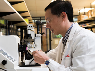
As featured in a PBS video, schistosome worms form lifelong bonds and females produce thousands of eggs daily only when they live inside human hosts, says Michael H. Hsieh, M.D., Ph.D.

“Love is in the air, the sea, the earth and all over and inside our bodies,” the PBS Valentine’s Day-themed video begins. As the public television station notes, what humans consider romance can look vastly different for creatures big and small, including serenading mice, spiders who wrap their gifts in silk and necking giraffes.

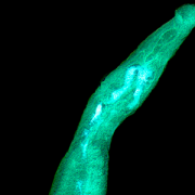
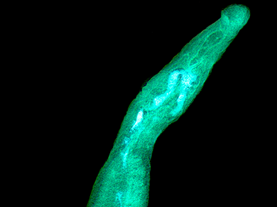
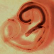
The “spooning” parasites segment of the video is where viewers see research conducted by Michael H. Hsieh, M.D., Ph.D., director of the Clinic for Adolescent and Adult PedIatric OnseT UroLogy at Children’s National Health System, and video filmed in his lab.

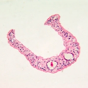
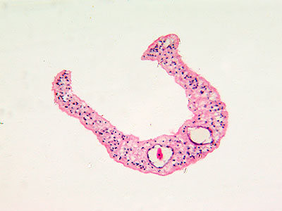
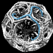
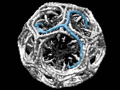
Schistosomiasis, a chronic infection with schistosome worms, is a distinctly one-sided love affair. As shown in Dr. Hsieh’s video clips, the male worm is shorter and fatter and equipped with a groove, a love canal where the longer, thinner female lodges, enabling the pair to mate for decades. This lifelong bond and the thousands of eggs it produces daily can only occur when the worms are inside the human host, Dr. Hsieh says.

While the video stresses Valentine’s Day romance, there are few rosy outcomes for humans who are the subject of the schistosome worms’ attention.

“Heavily and chronically infected individuals can have lots of problems,” Dr. Hsieh says. “This is a stunting and wasting health condition that prevents people from reaching their growth potential, impairs their academic performance and leaves them sapped of the energy needed to exercise or work. It truly perpetuates a cycle of poverty, particularly for affected children.”

Even the potential bright spot in this sobering story, the ability of the body’s immune system to fend off the parasitic worms, is only partly good news.

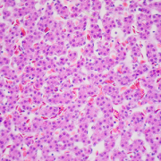
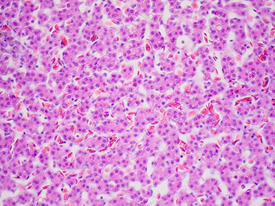
Schistosome worms have co-evolved with their human hosts, learning to take advantage of human vulnerabilities. Take the immune system. If it kicks too far into overdrive in trying to wall off the eggs from the rest of the body, it can interfere with organ function and trigger liver failure, kidney failure and early onset of bladder cancer, he says.

However, Dr. Hsieh and other schistosomiasis researchers are working on ways to positively harness the human immune response to schistosome worms, including developing diagnostics, drugs and vaccines. He says he and his colleagues would “love” to eliminate schistosomiasis as a global scourge.