Tag Archive for: Schistosoma mansoni

clatharin cage viewed by electron microscopy

IPSE infiltrates nuclei through clathrin-mediated endocytosis

clatharin cage viewed by electron microscopy

IPSE, one of the important proteins excreted by the parasite Schistosoma mansoni, infiltrates human cellular nuclei through clathrin-coated vesicles, like this one.

IPSE, one of the important proteins excreted by the parasite Schistosoma mansoni infiltrates human cellular nuclei through clathrin-mediated endocytosis (a process by which cells absorb metabolites, hormones and proteins), a research team led by Children’s National Hospital reported during the American Society of Tropical Medicine and Hygiene 2019 annual meeting.

Because the public health toll from the disease this parasite causes, Schistosomiasis, is second only to malaria in global impact, research teams have been studying its inner workings to help create the next generation of therapies.

In susceptible host cells – like urothelial cells, which line the urinary tract – IPSE modulates gene expression, increasing cell proliferation and angiogenesis (formation of new blood vessels). On a positive note, neurons appear better able to fend off its nucleus-infiltrating ways.

“We know that IPSE contributes to the severity of symptoms in Schistosomiasis, which leads some patients to develop bladder cancer, which develops from the urothelial lining of the bladder. Our team’s carefully designed experiments reveal IPSE’s function in the urothelium and point to the potential of IPSE playing a therapeutic role outside of the bladder,” says Michael Hsieh, M.D., Ph.D., director of transitional urology at Children’s National and the research project’s senior author.

In addition to Dr. Hsieh, research co-authors include Evaristus Mbanefo, Ph.D.; Kenji Ishida, Ph.D.; Austin Hester, M.D.; Catherine Forster, M.D.; Rebecca Zee, M.D., Ph.D.; and Christina Ho, M.D., all of Children’s National; Franco Falcone, Ph.D., University of Nottingham; and Theodore Jardetzky, Ph.D., and Luke Pennington, M.D., Ph.D., candidate, both of Stanford University.

Financial support for research described in this post was provided by the National Institutes of Health under award No. R01-DK113504.

Hepatocytes

H-IPSE internalized by just a limited range of cells

Hepatocytes

A team led by Children’s National Hospital found that H-IPSE is internalized by just a limited range of cells, including hepatocytes.

Schistosoma mansoni is a parasite that hides out in snails, breaks free into waterways, and then infects humans, spending much of its life inside blood vessels, laying eggs and jeopardizing public health when those eggs are excreted in urine or feces. As parasitic diseases go, the ailment it causes, Schistosomiasis, is second only to malaria in global impact, according to the Centers for Disease Control and Prevention.

In order to elude the human host’s defenses, S. mansoni uses self-defense tactics that researchers are trying to better understand in order to outmaneuver the parasite. A research team led by Children’s National Hospital is trying to tease out the multiple steps that enable this parasite to reproduce and generate millions of eggs without killing its host.

The parasite’s eggs secrete a number of proteins, with IPSE as one of the most abundant, the team recently presented during the American Society of Tropical Medicine and Hygiene 2019 annual meeting. That protein binds immunoglobulin, which induces basophils and mast cells to release IL-4. After sequestering chemokines, H-IPSE infiltrates the cell nucleus (thus H-IPSE is called an infiltrin), modulating gene expression.

“H-IPSE tips the immune system balance, making it more likely to trigger a Th2 anti-inflammatory response,” says Michael Hsieh, M.D., Ph.D., director of transitional urology at Children’s National and the research project’s senior author. “It downregulates pro-inflammatory pathways, but we wanted to know more about which specific human cells it targets.”

Using Trypan Blue, a stain that selectively colors certain cells bright blue, they solved the mystery, finding that H-IPSE is internalized by just a limited range of cells. What’s more, some cell types, like urothelial cells and hepatocytes (the liver’s chief functioning cells, which activate innate immunity), are more susceptible than neurons, endothelial cells or immature dendritic cells.

In addition to Dr. Hsieh, presentation co-authors include Olivia Lamanna, Evaristus Mbanefo and Kenji Ishida, all of Children’s National; Franco Falcone, of University of Nottingham; and Theodore Jardetzky and Luke Pennington, of Stanford University.

schistosome blood fluke

Therapy derived from parasitic worms downregulates proinflammatory pathways

schistosome blood fluke

A therapy derived from the eggs of the parasitic Schistosoma helps to protect against one of chemotherapy’s debilitating side effects by significantly downregulating major proinflammatory pathways, reducing inflammation.

A therapy derived from the eggs of parasitic worms helps to protect against one of chemotherapy’s debilitating side effects by significantly downregulating major proinflammatory pathways and reducing inflammation, indicates the first transcriptome-wide profiling of the bladder during ifosfamide-induced hemorrhagic cystitis.

The experimental model study findings were published online Feb. 7, 2019, in Scientific Reports.

With hemorrhagic cystitis, a condition that can be triggered by anti-cancer therapies like the chemotherapy drug ifosfamide and other oxazaphosphorines, the lining of the bladder becomes inflamed and begins to bleed. Existing treatments on the market carry their own side effects, and the leading therapy does not treat established hemorrhagic cystitis.

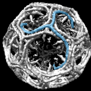
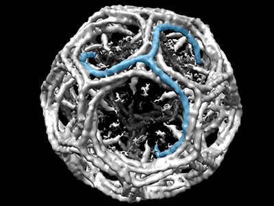
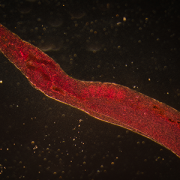
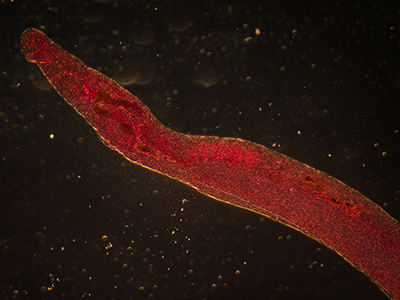
Around the world, people can become exposed to parasitic Schistosoma eggs through contaminated freshwater. Once inside the body, the parasitic worms mate and produce eggs; these eggs are the trigger for symptoms like inflammation. To keep their human hosts alive, the parasitic worms tamp down excess inflammation by secreting a binding protein with anti-inflammatory properties.

With that biological knowledge in mind, a research team led by Michael H. Hsieh, M.D., Ph.D., tested a single dose of IPSE, an Interleukin-4 inducing, Schistosoma parasite-derived anti-inflammatory molecule and found that it reduced inflammation, bleeding and urothelial sloughing that occurs with ifosfamide-related hemorrhagic cystitis.

In this follow-up project, experimental models were treated with ifosfamide to learn more about IPSE’s protective powers.

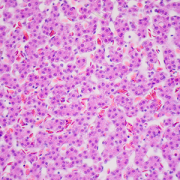
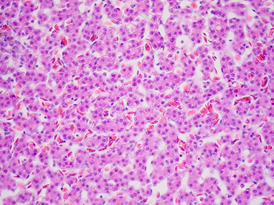
The preclinical models were given either saline or IPSE before the ifosfamide challenge. The bladders of the experimental models treated with ifosfamide had classic symptoms, including marked swelling (edema), dysregulated contraction, bleeding and urothelial sloughing. In contrast, experimental models “pre-treated” with IPSE were shielded from urothelial sloughing and inflammation, the study team found.

Transcriptional profiling of the experimental models’ bladders found the IL-1-B TNFa-IL-6 proinflammatory cascade via NFkB and STAT3 pathways serving as the key driver of inflammation. Pretreatment with IPSE slashed the overexpression of Il-1b, Tnfa and Il6 by 50 percent. IPSE drove significant downregulation of major proinflammatory pathways, including the IL-1-B TNFa-IL-6 pathways, interferon signaling and reduced (but did not eliminate) oxidative stress.

“Taken together, we have identified signatures of acute-phase inflammation and oxidative stress in ifosfamide-injured bladder, which are reversed by pretreatment with IPSE,” says Dr. Hsieh, a urologist at Children’s National Health System and the study’s senior author. “These preliminary findings reveal several pathways that could be therapeutically targeted to prevent ifosfamide-induced hemorrhagic cystitis in humans.”

When certain chemotherapy drugs are metabolized by the body, the toxin acrolein is produced and builds up in urine. 2-mercaptoethane sulfonate Na (MESNA) binds to acrolein to prevent urotoxicity. By contrast, IPSE targets inflammation at the source, reversing inflammatory changes that damage the bladder.

“Our work demonstrates that there may be therapeutic potential for naturally occurring anti-inflammatory molecules, including pathogen-derived factors, as alternative or complementary therapies for ifosfamide-induced hemorrhagic cystitis,” Dr. Hsieh adds.

In addition to Dr. Hsieh, study co-authors include Lead Author Evaristus C. Mbanefo and Rebecca Zee, Children’s National; Loc Le, Nirad Banskota and Kenji Ishida, Biomedical Research Institute; Luke F. Pennington and Theodore S. Jardetzky, Stanford University; Justin I. Odegaard, Guardant Health; Abdulaziz Alouffi, King Abdulaziz City for Science & Technology; and Franco H. Falcone, University of Nottingham.

Financial support for the research described in this report was provided by the Margaret A. Stirewalt Endowment, the National Institute of Diabetes and Digestive and Kidney Diseases under award R01DK113504, the National Institute of Allergy and Infectious Diseases under award R56AI119168 and a Urology Care Foundation Research Scholar Award.