Tag Archive for: pediatric orthopaedics

Dr. Laura Tosi talks to a patient

Refining criteria for childhood skeletal fragility and osteoporosis

Dr. Laura Tosi talks to a patient

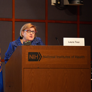
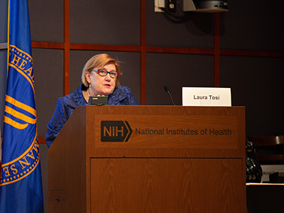
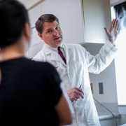
Orthopaedic surgeon Laura Tosi, M.D., presented information about bone fractures and skeletal fragility in children at this year’s POSNA Annual Meeting.

It’s true that broken bones are often a typical part of childhood, says international bone health expert Laura Tosi, M.D., an orthopaedic surgeon at Children’s National Hospital. But for some children, a single bone fracture under the right circumstances may be a signal that a child needs a closer look to rule out underlying skeletal fragility.

Dr. Tosi presented on this topic as part of the Pediatric Orthopaedic Society of North America’s (POSNA) 2020 Annual Meeting. The presentations were conducted virtually this year due to COVID-19.

“We know that between 27 and 40% of girls, and 42 to 51 percent of boys will have at least one fracture during childhood,” she says. “What we have also seen over time is that almost 40 percent of children who have one fracture will have more. How do we tell which children with a fracture may need our help to avoid future ones?”

During her session, Dr. Tosi discussed how adding more nuance to clinical evaluation criteria for childhood fractures can help identify which children should be evaluated for conditions affecting bone density.

To widen the scope and make sure an underlying bone density issue is detected and treated as early as possible, Dr. Tosi says there are some specific findings that should suggest the need for further exploration:

  • Does the child have an a priori risk for a fragility fracture due to a genetic bone disorder(such as osteogenesis imperfects (aka brittle bone disease) or immobility caused by a disorder such as spina bifida, cerebral palsy or muscular dystrophy?
  • Is there a mismatch between the fracture severity and level of trauma that led to the injury?
  • Does the child’s history include any of four factors known to be associated with increased fracture risk: early age at the time of the first fracture, intolerance to cow’s milk, low dietary calcium intake or high BMI values.
  • Does the child have a vertebral compression fracture?
  • Is there a family history of frequent fractures (which may indicate a previously unidentified genetic condition)

Dr. Tosi also laid out specific evaluation steps for a skeletal fragility condition when a child’s fracture meets criteria, including:

  • Family, nutrition and exercise histories
  • A detailed physical exam
  • Complete radiograph review, including previously existing films and bone densitometry
  • Rule out rickets and child abuse
  • A complete lab work up

“It can be extremely challenging to identify if a child’s first bone fracture is a result of typical childhood activity or something else,” says Dr. Tosi. “But the risks of waiting to evaluate a fracture that meets some of the criteria above may mean we are delaying a treatment that might improve bone density and prevent a future fracture altogether — which is always what we’d hope to do.”

In the past, bone health experts felt that the word “osteoporosis” should not be used in children and pushed for the term “low bone density for age.”  That perspective has begun to change thanks to important advances in our understanding of the genetic basis of bone fragility, the important role of chronic conditions and how the use of bone-active medications can significantly reduce fracture risk and improve function in certain conditions.

She then spoke about the benefits of early detection for conditions causing skeletal fragility by presenting compelling evidence of the resiliency of a child’s bones when they are managed appropriately.

She noted that she’s seen significant bone remodeling in patients with serious bone degeneration due to osteogenesis imperfecta and leukemia, for example, thanks to early detection and treatment.

“Our knowledge of bone density and bone health is improving, but is still imperfect,” she concluded. “But as we learn more, and are able to appropriately identify and treat kids with skeletal fragility or osteoporosis earlier, we can continue to refine how we evaluate and care for all of them.”

Laura Tosi

Technology bridges knowledge gaps in rare bone disease care

Laura Tosi

Dr. Tosi and colleagues presented on the NIH Rare Disease Day 2020 panel, Nontraditional Approaches to Improving Access for Rare Diseases.

As part of the global observance of Rare Disease Day in February, the NCATS and NIH Clinical Center hosted a special event to raise awareness about rare diseases, the people they affect and NIH research collaborations under way to address scientific challenges and advance new treatments.

This year, Children’s National Hospital orthopaedic surgeon Laura Tosi, M.D., took part in an afternoon panel, Nontraditional Approaches to Improving Access for Rare Diseases, where she outlined her work as the faculty chair of the Rare Bone Disease TeleECHO, a virtual meeting that allows care providers and experts to come together via the Zoom platform, discuss diagnosis of specific disorders and present cases for group input.

Dr. Tosi and the Rare Bone Disease Alliance have called Project ECHO Rare Bone Disease a game changer for care of these complex conditions. Rare bone disorders are only about 5% of all birth defects but include 461 skeletal disorders caused by 437 genes – making it difficult for any physician to see enough cases of any one disorder to correctly diagnose and treat it.

“Most doctors are like me, a pediatric orthopaedic surgeon. I need to know a lot of different rare diseases and it’s hard to keep everybody on the cutting edge,” Dr. Tosi says. “Even though we have found the genes for most of the disorders, the phenotypic overlaps, shortage of specialists and the multi-disciplinary needs of so many of the patients add to the challenges.”

So 7 months ago, Dr. Tosi joined together with colleagues at the Rare Bone Disease Alliance and the Osteogenesis Imperfecta Foundation to launch Project ECHO Rare Bone Disease. The now monthly telehealth meeting engages a distinguished faculty of experts from around the world and from across the spectrum of care for these rare bone disorders, including specialists in genetics, endocrinology, orthopaedics and others.

Project ECHO is a specific model for bridging distance and creating a network of professionals, with the goal of leveling the playing field for all by making vital information accessible to everyone, regardless of their location. In healthcare the model transcends traditional “telemedicine,” however. The program, launched from the University of New Mexico, self-describes itself as “telementoring, a guided practice where the participating clinician retains responsibility for the patient” but is able to discuss diagnosis and therapeutic recommendations with a set of esteemed faculty via a regular virtual meeting series.

In the case of the Rare Bone Disease TeleECHO, the ECHO’s faculty decided on two major foci for the curriculum. Half of the content is about how to make the right diagnosis and the other half shares the latest information about specific diseases. The sessions also offer free CME to attendees.

Dr. Tosi says that while finding cases to discuss can sometimes be challenging when it comes to rare bone diseases, she takes responsibility on herself to make sure the content is robust each month. So far the meetings have attracted between 40 and 90 participants per session – a great engagement rate for such a young teleECHO program.

“I believe ECHO advances knowledge of healthcare and democratizes it by offering universal accessibility across the globe,” Tosi notes.

The Rare Bone Disease Alliance, which consists of 12 organizations, experts and patient families working together, is now deciding what’s next for the Rare Bone Disease TeleECHO. They may develop disorder-specific ECHOs, are studying the frequency of the sessions and how best to improve participation for all sessions. The idea is to increase access to this expertise even further, as it could have critical impacts on patients worldwide living with these rare diseases.

In terms of key take-aways from the panel of experts at Rare Disease Day, the hope is that more disease groups might leverage this type of technology to connect people in nontraditional ways. Doing so has the potential to ensure that everyone with a rare disease receives the best support and care possible because their doctors have the knowledge they need when they need it.

Watch more sessions from the NIH’s Rare Disease Day 2020.

Dr. Lauri Tosi examines a patient

Building patient-centered outcomes research in osteogenesis imperfecta

Dr. Lauri Tosi examines a patient

Children’s orthopaedic surgeon Laura Tosi, M.D., is the co-lead on a program to improve patient-centered outcomes research and education in osteogenesis imperfecta that recently received a Eugene Washington Engagement Award of $250,000 from the Patient-Centered Outcomes and Research Institute (PCORI).

Children’s orthopaedic surgeon Laura Tosi, M.D., is the co-lead on a program to improve patient-centered outcomes research and education in osteogenesis imperfecta (OI) that recently received a Eugene Washington Engagement Award of $250,000 from the Patient-Centered Outcomes and Research Institute (PCORI). Dr. Tosi serves as project co-lead alongside colleagues Tracy Hart, project lead, from the Osteogenesis Imperfecta Foundation (OIF) and Bryce Reeve, Ph.D., co-project lead, director of the Center for Health Measurement at Duke University.

The project, which will be housed at the Osteogenesis Imperfecta Foundation, will run for two years and seeks to:

  • Create a community of stakeholders (patients/caregivers/clinicians/researchers) who are trained or training in patient-centered outcomes research, with specific attention to priority topics identified by the OI community.
  • Expand communications and education strategies related to patient-centered outcomes research to enhance the care of the OI community.
  • Establish and extend the capacity among patients, caregivers, clinicians and researchers in OI to participate in both patient-centered outcomes research and comparative effectiveness activities.
  • Develop an OI-specific toolkit focused on disseminating evidence-based clinical care recommendations to stakeholders and care providers, based on sustainable input from the OI community.
  • Extended the reach of these activities to support other rate bone disease communities.

Osteogenesis imperfecta is a group of genetic disorders causing connective tissue dysfunction and bone fragility. It is the most common of nearly 450 rare skeletal disorders and affects an estimated 25,000 to 50,000 people in the U.S. Collecting the patient’s perspective about natural history, clinical best practices, quality of life and research priorities is challenging because, like so many rare diseases, the affected population is relatively small and  geographically dispersed.

“We hope this project will give us the ability to develop a set of best practices for care and research based on research that incorporates the patient’s point-of-view,” says Dr. Tosi. “I’m excited to work with this team and begin to change how we think about and care for OI patients and their families.”

The traction sisters

Spinal-halo-gravity traction times three

The traction sisters

Three girls received spinal-halo-gravity traction at the same time at Children’s National prior to surgery for acute idiopathic scoliosis.

Washington, D.C.’s ABC affiliate, WJLA, recently featured a story about three girls who received treatment for acute idiopathic scoliosis through the Children’s National Spinal Fusion Surgical Home, a comprehensive and effective program that has demonstrated reductions in pain medication usage and medical stays following posterior spinal fusion surgery.

All three girls had extremely severe curvatures of the spine requiring a month long inpatient stay for spinal halo-gravity traction prior to surgical intervention. Spinal curves severe enough to require traction are rare, and often impede a child’s quality of life just as severely – eating, breathing and moving are difficult. Given the long hospital stay required and the challenges of asking a child to stay in a traction frame 23 hours out of every day, the orthopaedic surgery team tries to coordinate cases so that when possible, patients can support each other throughout the process. This was the first time, however, that the team had three traction patients on similar trajectories on the unit at the same time.

Spinal halo-gravity traction can reduce the degree of surgical intervention necessary by accomplishing some pre-operative gradual straightening of the spine prior to spinal fusion procedures. For severe spinal deformities this has been shown to improve the safety and effectiveness of the final surgical procedure.

Matthew Oetgen, M.D., discusses an image of a patient’s spine.

Eliminating unnecessary radiation exposure from spinal radiography

Matthew Oetgen, M.D., discusses an image of a patient’s spine.

Chief of Orthopaedics and Sports Medicine Matthew Oetgen, M.D., discusses an image of a patient’s spine.

If a child arrives at the pediatric orthopaedic specialist for an idiopathic scoliosis evaluation without an adequate radiographic image of his or her spine, it’s often necessary to order yet another imaging study for accurate assessment.

A study published in the Journal of the American Academy of Orthopaedic Surgeons found that in a 6 month period, almost half (43 percent) of patients referred for evaluation required a repeat radiograph due to missing or poor quality existing images.

“Repeating the radiograph means these kids received another exposure to radiation, too,” says Matthew Oetgen, M.D., the study’s lead author and chief of Orthopaedic Surgery and Sports Medicine at Children’s National Health System. “It’s frustrating because in many cases, a simple change in how the initial radiograph was taken could have prevented the need for more imaging studies.”

Dr. Oetgen and the study authors note that there is currently no standardized protocol for spinal radiography of suspected idiopathic scoliosis. However, a few basic criteria could greatly reduce the number of repeat images necessary. Radiographic images that allow for proper evaluation of idiopathic scoliosis and reduce radiation exposure include:

  • A full coronal view of the spine including skull base and pelvis
  • The iliac crest as an indicator of skeletal maturity
  • A full-length lateral view of the spine

Study authors also reinforced the need to do everything possible to reduce radiation exposure for children through proper use of protective shielding for reproductive organs and digital radiograph technology.

“Orthopaedic surgeons and pediatricians share the responsibility to ensure children are exposed to as little iatrogenic radiation as possible,” Dr. Oetgen concludes. “All physicians should be sure that the radiology facilities they refer patients to for spinal radiography employ every technology and safety measure available to limit radiation exposure. Additionally, we can and should work with radiologists to define evaluation criteria and improve what’s captured by radiography on the first try.”