Tag Archive for: Pediatric Academic Societies 2019 Annual Meeting

Early lipids in micropreemies’ diets can boost brain growth

preterm brain scans

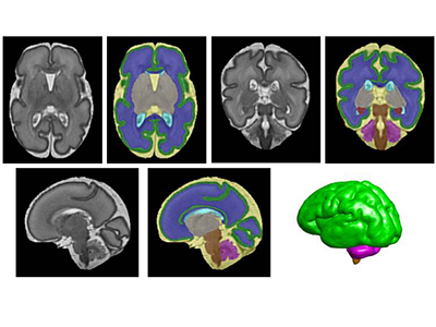
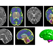
Segmentation of a preterm brain T2-weighted MRI image at 30 gestational weeks [green=cortical grey matter; blue=white matter; grey=deep grey matter; cyan=lateral ventricle; purple=cerebellum; orange=brainstem; red=hippocampus; yellow=cerebrospinal fluid].

Dietary lipids, already an important source of energy for tiny preemies, also provide a much-needed brain boost by significantly increasing global brain volume as well as increasing volume in regions involved in motor activities and memory, according to research presented during the Pediatric Academic Societies 2019 Annual Meeting.

“Compared with macronutrients like carbohydrates and proteins, lipid intake during the first month of life is associated with increased overall and regional brain volume for micro-preemies,” says Catherine Limperopoulos, Ph.D., director of MRI Research of the Developing Brain at Children’s National and senior author. “Using non-invasive magnetic resonance imaging, we see increased volume in the cerebellum by 2 weeks of age. And at four weeks of life, lipids increase total brain volume and boost regional brain volume in the cerebellum, amygdala-hippocampus and brainstem.”

The cerebellum is involved in virtually all physical movement and enables coordination and balance. The amygdala processes and stores short-term memories. The hippocampus manages emotion and mood. And the brainstem acts like a router, passing messages from the brain to the rest of the body, as well as enabling essential functions like breathing, a steady heart rate and swallowing.

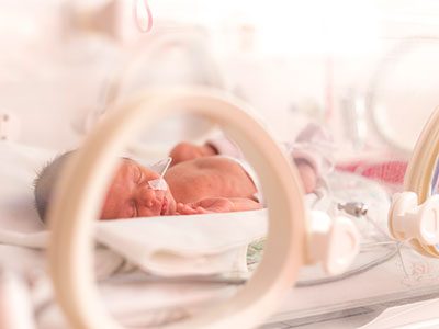
According to the Centers for Disease Control and Prevention, about 1 in 10 U.S. babies is born preterm, or before 37 weeks gestation. Regions of the brain that play vital roles in complex cognitive and motor activities experience exponential growth late in pregnancy, making the developing brains of preterm infants particularly vulnerable to injury and impaired growth.

Children’s research faculty examined the impact of lipid intake in the first month of life on brain volumes for very low birth weight infants, who weighed 1,500 grams or less at birth. These micro-preemies are especially vulnerable to growth failure and neurocognitive impairment after birth.

The team enrolled 68 micro-preemies who were 32 weeks gestational age and younger when they were admitted to Children’s neonatal intensive care unit during their first week of life. They measured cumulative macronutrients – carbohydrates, proteins, lipids and calories – consumed by these newborns at 2 and 4 weeks of life. Over years, Limperopoulos’ lab has amassed a large database of babies who were born full-term; this data provides unprecedented insights into normal brain development and will help to advance understanding of brain development in high-risk preterm infants.

“Even after controlling for average weight gain and other health conditions, lipid intake was positively associated with cerebellar and brainstem volumes in very low birthweight preterm infants,” adds Katherine M. Ottolini, the study’s lead author.

According to Limperopoulos, Children’s future research will examine the optimal timing and volume of lipids to boost neurodevelopment for micro-preemies.

Pediatric Academic Societies 2019 Annual Meeting presentation

  • “Early lipid intake improves brain growth in premature infants.”
    • Saturday, April 27, 2019, 1:15-2:30 p.m. (EST)

Katherine M. Ottolini, lead author; Nickie Andescavage, M.D., Attending, Neonatal-Perinatal Medicine and co-author; Kushal Kapse, research and development staff engineer and co-author; and Catherine Limperopoulos, Ph.D., director of MRI Research of the Developing Brain and senior author, all of Children’s National.

In HIE lower heart rate variability signals stressed newborns

newborn in incubator

In newborns with hypoxic-ischemic encephalopathy (HIE), lower heart rate variability correlates with autonomic manifestations of stress shortly after birth, underscoring the value of this biomarker, according to Children’s research presented during the Pediatric Academic Societies 2019 Annual Meeting.

Tethered to an array of machines that keep their bodies nourished, warm and alive, newborns with health issues can’t speak. But Children’s research teams are tapping into what the machinery itself says, looking for insights into which vulnerable infants are most in need of earlier intervention.

Heart rate variability – or the variation between heartbeats – is a sign of health. Our autonomic nervous system constantly sends signals to adjust our heart rate under normal conditions. We can measure heart rate variability non-invasively, providing a way to detect potential problems with the autonomic nervous system as a sensitive marker of health in critically ill newborns,” says An N. Massaro, M.D., co-Director of Research for the Division of Neonatology at Children’s National, and the study’s senior author. “We’re looking for validated markers of brain injury in babies with HIE, and our study helps to support heart rate variability as one such valuable physiological biomarker.”

In most newborns, the autonomic nervous system reliably and automatically receives information about the body and the outside world and, in response, controls essential functions like blood pressure, body temperature, how quickly the baby breathes and how rapidly the newborn’s heart beats. The sympathetic part stimulates body processes, while the parasympathetic part inhibits body processes. When the nervous system’s internal auto-pilot falters, babies can suffer.

The Children’s team enrolled infants with HIE in the prospective, observational study. (HIE is brain damage that occurs with full-term babies who experience insufficient blood and oxygen flow to the brain around the time they are born.) Fifteen percent had severe encephalopathy. Mean age of babies in the observational study was 38.9 weeks gestation. Their median Apgar score at five minutes was 3; the 0-9 Apgar range indicates how ready newborns are for the rigors of life outside the womb.

The team analyzed heart rate variability metrics for three time periods:

  • The first 24 to 27 hours of life
  • The first three hours after babies undergoing therapeutic cooling were rewarmed and
  • The first three hours after babies’ body temperature had returned to normal.

They correlated the relationship between heart rate variability for 68 infants during at least one of these time periods with the stress z-score from the NICU Network Neurobehavioral Scale. The scale is a standardized assessment of newborn’s neurobehavioral integrity. The stress summary score indicates a newborn’s overall stress response, and six test items specifically relate to autonomic function.

“Alpha exponent and root mean square in short timescales, root mean square in long timescales, as well as low and high frequency powers positively correlated with stress scores and, even after adjusting for covariates, remained independently associated at 24 hours,” says Allie Townsend, the study’s lead author.

Pediatric Academic Societies 2019 Annual Meeting presentation

  • “Heart rate variability (HRV) measures of autonomic nervous system (ANS) function relates to neonatal neurobehavioral manifestations of stress in newborn with hypoxic-ischemic encephalopathy (HIE).”
    • Monday, April 29, 2019, 5:45 p.m. (EST)

Allie Townsend, lead author; Rathinaswamy B. Govindan, Ph.D., staff scientist, Advanced Physiological Signals Processing Lab and co-author; Penny Glass, Ph.D., director, Child Development Program and co-author; Judy Brown, co-author; Tareq Al-Shargabi, M.S., co-author; Taeun Chang, M.D., director, Neonatal Neurology and Neonatal Neurocritical Care Program and co-author; Adré J. du Plessis, M.B.Ch.B., MPH, chief of the Division of Fetal and Transitional Medicine and co-author; An N. Massaro, M.D., co-Director of Research for the Division of Neonatology and senior author, all of Children’s National.

Placental function linked to brain injuries associated with autism

Claire Marie Vacher

“We saw long-term cerebellar white matter alterations in male experimental models, and behavioral testing revealed social impairments and increased repetitive behaviors, two hallmark features of ASD,” says Claire-Marie Vacher, Ph.D., lead study author.

Allopregnanolone (ALLO), a hormone made by the placenta late in pregnancy, is such a potent neurosteroid that disrupting its steady supply to the developing fetus can leave it vulnerable to brain injuries associated with autism spectrum disorder (ASD), according to Children’s research presented during the Pediatric Academic Societies 2019 Annual Meeting.

In order to more effectively treat vulnerable babies, the Children’s research team first had to tease out what goes wrong in the careful choreography that is pregnancy. According to the Centers for Disease Control and Prevention, about 1 in 10 babies is born preterm, before 37 weeks of gestation. Premature birth is a major risk factor for ASD.

The placenta is an essential and understudied organ that is shared by the developing fetus and the pregnant mother, delivering oxygen, glucose and nutrients and ferrying out waste products. The placenta also delivers ALLO, a progesterone derivative, needed to ready the developing fetal brain for life outside the womb.

ALLO ramps up late in gestation. When babies are born prematurely, their supply of ALLO stops abruptly. That occurs at the same time the cerebellum – a brain region essential for motor coordination, posture, balance and social cognition– typically undergoes a dramatic growth spurt.

“Our experimental model demonstrates that losing placental ALLO alters cerebellar development, including white matter development,” says Anna Penn, M.D., Ph.D., a neonatologist in the divisions of Neonatology and Fetal Medicine, and a developmental neuroscientist at Children’s National. “Cerebellar white matter development occurs primarily after babies are born, so connecting a change in placental function during pregnancy with lingering impacts on later brain development is a particularly striking result.”

The research team created a novel experimental model in which the gene encoding the enzyme responsible for producing ALLO is deleted in the placenta. They compared these preclinical models with a control group and performed whole brain imaging and RNAseq gene expression analyses for both groups.

“We saw long-term cerebellar white matter alterations in male experimental models, and behavioral testing revealed social impairments and increased repetitive behaviors, two hallmark features of ASD,” says Claire-Marie Vacher, Ph.D., lead study author. “These male-specific outcomes parallel the increased risk of brain injury and ASD we see in human babies born prematurely.”

ALLO binds to specific GABA receptors, which control most inhibitory signaling in the nervous system.

“Our findings provide a new way to frame poor placental function: Subtle but significant changes in utero may set in motion neurodevelopmental disorders that children experience later in life,” adds Dr. Penn, the study’s senior author. “Future directions for our research could include identifying new targets in the placenta or brain that could be amenable to hormone supplementation, opening the potential for earlier treatment for high-risk fetuses.”

Pediatric Academic Societies 2019 Annual Meeting presentation

  • “Placental allopregnanolone loss alters postnatal cerebellar development and function.”
    • Sunday, April 28, 2019, 5:15 p.m. to 5:30 p.m. (EST)

Claire-Marie Vacher, Ph.D., lead author; Jackie Salzbank, co-author; Helene Lacaille, co-author; Dana Bakalar, co-author; Jiaqi O’Reilly, co-author; and Anna Penn, M.D., Ph.D., a neonatologist in the divisions of Neonatology and Fetal Medicine, developmental neuroscientist and senior study author.

Firearm injuries disproportionately affect African American kids in DC Wards 7 and 8

Katie Donnelly

“Because the majority of patients in our analyses were injured through accidental shootings, this particular risk factor can help to inform policy makers about possible interventions to prevent future firearm injury, disability and death,” says Katie Donnelly, M.D.

Firearm injuries disproportionately impact African American young men living in Washington’s Wards 7 and 8 compared with other city wards, with nearly one-quarter of injuries suffered in the injured child’s home or at a friend’s home, according to a hot spot analysis presented during the Pediatric Academic Societies 2019 Annual Meeting.

“We analyzed the addresses where youths were injured by firearms over a nearly 12-year period and found that about 60 percent of these shootings occurred in Ward 7 or Ward 8, lower socioeconomic neighborhoods when compared with Washington’s six other Wards,” says Monika K. Goyal, M.D., MSCE, assistant chief of Children’s Division of Emergency Medicine and Trauma Services and the study’s senior author. “This granular detail will help to target resources and interventions to more effectively reduce firearm-related injury and death.”

In the retrospective, cross-sectional study, the Children’s research team looked at all children aged 18 and younger who were treated at Children’s National for firearm-related injuries from Jan. 1, 2006, to May 31, 2017. During that time, 122 children injured by firearms in Washington were treated at Children’s National, the only Level 1 pediatric trauma center in the nation’s Capitol:

  • Nearly 64 percent of these firearm-related injuries were accidental
  • The patients’ mean age was 12.9 years old
  • More than 94 percent of patients were African American and
  • Nearly 74 percent were male.

Of all injuries suffered by children, injuries due to firearms carry the highest mortality rates, the study authors write. About 3 percent of patients in Children’s study died from their firearm-related injuries. Among surviving youth:

  • Patients had a mean Injury Severity Score of 5.8. (The score for a “major trauma” is greater than 15.)
  • 54 percent required hospitalization, with a mean hospitalization of three days
  • Nearly 28 percent required surgery, with 14.8 percent transferred directly from the emergency department to the operating room and
  • Nearly 16 percent were admitted to the intensive care unit.

“Regrettably, firearm injuries remain a major public health hazard for our nation’s children and young adults,” adds Katie Donnelly, M.D., emergency medicine specialist and the study’s lead author. “Because the majority of patients in our analyses were injured through accidental shootings, this particular risk factor can help to inform policy makers about possible interventions to prevent future firearm injury, disability and death.”

Pediatric Academic Societies 2019 Annual Meeting poster presentatio

  • “Pediatric firearm-related injuries and outcomes in the District of Columbia.”
    • Monday, April 29, 2019, 5:45 p.m. to 7:30 p.m. (EST)

Katie Donnelly, M.D., emergency medicine specialist and lead author; Shilpa J. Patel, M.D., MPH, emergency medicine specialist and co-author; Gia M. Badolato, co-author; James Jackson, co-author; and Monika K. Goyal, M.D., MSCE, assistant chief of Children’s Division of Emergency Medicine and Trauma Services and senior author.

Other Children’s research related to firearms presented during PAS 2019 includes:

April 27, 8 a.m.: “Protect kids, not guns: What pediatric providers can do to improve firearm safety.” Gabriella Azzarone, Asad Bandealy, M.D.; Priti Bhansali, M.D.; Eric Fleegler; Monika K. Goyal, M.D., MSCE;  Alex Hogan; Sabah Iqbal; Kavita Parikh, M.D.; Shilpa J. Patel, M.D., MPH; Noe Romo; and Alyssa Silver.

April 29, 5:45 p.m.: “Emergency department visits for pediatric firearm-related injury: By intent of injury.” Shilpa J. Patel, M.D., MPH; Gia M. Badolato; Kavita Parikh, M.D.; Sabah Iqbal; and Monika K. Goyal, M.D., MSCE.

April 29, 5:45 p.m.: “Assessing the intentionality of pediatric firearm injuries using ICD codes.” Katie Donnelly, M.D.; Gia M. Badolato; James Chamberlain, M.D.; and Monika K. Goyal, M.D., MSCE.

April 30, 9:45 a.m.: “Defining a research agenda for the field of pediatric firearm injury prevention.” Libby Alpern; Patrick Carter; Rebecca Cunningham, Monika K. Goyal, M.D., MSCE; Fred Rivara; and Eric Sigel.

Breastfeeding boosts metabolites important for brain growth

Catherine Limperopoulos

“Proton magnetic resonance spectroscopy, a non-invasive imaging technique that describes the chemical composition of specific brain structures, enables us to measure metabolites that may play a critical role for growth and explain what makes breastfeeding beneficial for newborns’ developing brains,” says Catherine Limperopoulos, Ph.D.

Micro-preemies who primarily consume breast milk have significantly higher levels of metabolites important for brain growth and development, according to sophisticated imaging conducted by an interdisciplinary research team at Children’s National.

“Our previous research established that vulnerable preterm infants who are fed breast milk early in life have improved brain growth and neurodevelopmental outcomes. It was unclear what makes breastfeeding so beneficial for newborns’ developing brains,” says Catherine Limperopoulos, Ph.D., director of MRI Research of the Developing Brain at Children’s National. “Proton magnetic resonance spectroscopy, a non-invasive imaging technique that describes the chemical composition of specific brain structures, enables us to measure metabolites essential for growth and answer that lingering question.”

According to the Centers for Disease Control and Prevention, about 1 in 10 U.S. infants is born preterm. The Children’s research team presented their findings during the Pediatric Academic Societies 2019 Annual Meeting.

The research-clinicians enrolled babies who were very low birthweight (less than 1,500 grams) and 32 weeks gestational age or younger at birth when they were admitted to Children’s neonatal intensive care unit in the first week of life. The team gathered data from the right frontal white matter and the cerebellum – a brain region that enables people to maintain balance and proper muscle coordination and that supports high-order cognitive functions.

Each chemical has its own a unique spectral fingerprint. The team generated light signatures for key metabolites and calculated the quantity of each metabolite. Of note:

  • Cerebral white matter spectra showed significantly greater levels of inositol (a molecule similar to glucose) for babies fed breast milk, compared with babies fed formula.
  • Cerebellar spectra had significantly greater creatine levels for breastfed babies compared with infants fed formula.
  • And the percentage of days infants were fed breast milk was associated with significantly greater levels of both creatine and choline, a water soluble nutrient.

“Key metabolite levels ramp up during the times babies’ brains experience exponential growth,” says Katherine M. Ottolini, the study’s lead author. “Creatine facilitates recycling of ATP, the cell’s energy currency. Seeing greater quantities of this metabolite denotes more rapid changes and higher cellular maturation. Choline is a marker of cell membrane turnover; when new cells are generated, we see choline levels rise.”

Already, Children’s National leverages an array of imaging options that describe normal brain growth, which makes it easier to spot when fetal or neonatal brain development goes awry, enabling earlier intervention and more effective treatment. “Proton magnetic resonance spectroscopy may serve as an important additional tool to advance our understanding of how breastfeeding boosts neurodevelopment for preterm infants,” Limperopoulos adds.

Pediatric Academic Societies 2019 Annual Meeting presentation

  • “Improved cerebral and cerebellar metabolism in breast milk-fed VLBW infants.”
    • Monday, April 29, 2019, 3:30–3:45 p.m. (EST)

Katherine M. Ottolini, lead author; Nickie Andescavage, M.D., Attending, Neonatal-Perinatal Medicine and co-author; Kushal Kapse, research and development staff engineer and co-author; Sudeepta Basu, M.D., neonatologist and co-author; and Catherine Limperopoulos, Ph.D., director of MRI Research of the Developing Brain and senior author, all of Children’s National.

Looking for ‘help’ signals in the blood of newborns with HIE

An Massaro

“This data support our hypothesis that a panel of biomarkers – not a one-time test for a single biomarker – is needed to adequately determine the risk and timing of brain injury for babies with HIE,” says An N. Massaro, M.D.

Measuring a number of biomarkers over time that are produced as the body responds to inflammation and injury may help to pinpoint newborns who are more vulnerable to suffering lasting brain injury due to disrupted oxygen delivery and blood flow, according to research presented during the Pediatric Academic Societies 2019 Annual Meeting.

Hypoxic-ischemic encephalopathy (HIE) happens when blood and oxygen flow are disrupted around the time of birth and is a serious birth complication for full-term infants. To lessen the chance of these newborns suffering permanent brain injury, affected infants undergo therapeutic cooling, which temporarily lowers their body temperatures.

“Several candidate blood biomarkers have been investigated in HIE but we still don’t have one in clinical use.  We need to understand how these markers change over time before we can use them to direct care in patients,” says An N. Massaro, M.D., co-director of the Neonatal Neurocritical Care Program at Children’s National and the study’s senior author. “The newborns’ bodies sent out different ‘help’ signals that we detected in their bloodstream, and the markers had strikingly different time courses. A panel of plasma biomarkers has the potential to help us identify infants most in need of additional interventions, and to help us understand the most optimal timing for those interventions.”

Past research has keyed in on inflammatory cytokines and Tau protein as potential biomarkers of brain injury for infants with HIE who are undergoing therapeutic cooling. The research team led by Children’s faculty wanted to gauge which time periods to measure such biomarkers circulating in newborns’ bloodstreams. They enrolled 85 infants with moderate or severe HIE and tapped unused blood specimens that had been collected as cooling began, as well as 12, 24, 72 and 96 hours later. The infants’ mean gestational age was 38.7 weeks, their mean birth weight was about 7 pounds (3.2 kilograms), and 19% had severe brain disease (encephalopathy).

Cytokines – chemicals like Interleukin (IL) 6, 8 and 10 that regulate how the body responds to infection, inflammation and trauma – peaked in the first 24 hours of cooling for most of the newborns. However, the highest measure of Tau protein for the majority of newborns was during or after the baby’s temperature was restored to normal.

“After adjusting for clinical severity of encephalopathy and five-minute Apgar scores, IL-6, IL-8 and IL-10 predicted adverse outcomes, like severe brain injury or death, as therapeutic hypothermia began. By contrast, Tau protein measurements predicted adverse outcomes during and after the infants were rewarmed,” Dr. Massaro says.

IL-6 and IL-8 proteins are pro-inflammatory cytokines while IL-10 is considered anti-inflammatory.  These chemicals are released as a part of the immune response to brain injury. Tau proteins are abundant in nerve cells and stabilize microtubules.

“This data support our hypothesis that a panel of biomarkers – not a one-time test for a single biomarker – is needed to adequately determine the risk and timing of brain injury for babies with HIE,” she adds.

Pediatric Academic Societies 2019 Annual Meeting presentation

  • “Serial plasma biomarkers of brain injury in infants with hypoxic ischemic encephalopathy (HIE) treated with therapeutic hypothermia (TH).”
    • Saturday, April 27, 2019, 6 p.m. (EST)

Meaghan McGowan, lead author; Alexandra C. O’Kane, co-author; Gilbert Vezina, M.D.,  director, Neuroradiology Program and co-author; Tae Chang, M.D., director, Neonatal Neurology Program and co-author; and An N. Massaro, M.D., co-director of the Neonatal Neurocritical Care Program and senior author; all of Children’s National; and co-author Allen Everett, of Johns Hopkins School of Medicine.

Parents of older, healthier newborns with less social support less resilient

Ololade Okito

“We know that having a child hospitalized in the NICU can be a high-stress time for families,” says Ololade Okito, M.D., lead author of the cross-sectional study. “The good news is that as parental resiliency scores rise, we see a correlation with fewer symptoms of depression and anxiety.

Parents of older, healthier newborns who had less social support were less resilient during their child’s hospitalization in the neonatal intensive care unit (NICU), a finding that correlates with more symptoms of depression and anxiety, according to Children’s research presented during the Pediatric Academic Societies 2019 Annual Meeting.

Resiliency is the natural born, yet adaptable ability of people to bounce back in the face of significant adversity. Published research indicates that higher resilience is associated with reduced psychological distress, but the phenomenon had not been studied extensively in parents of children hospitalized in a NICU.

“We know that having a child hospitalized in the NICU can be a high-stress time for families,” says Ololade Okito, M.D., lead author of the cross-sectional study. “The good news is that as parental resiliency scores rise, we see a correlation with fewer symptoms of depression and anxiety. Parents who feel they have good family support also have higher resilience scores.”

The project is an offshoot of a larger study examining the impact of peer mentoring by other NICU parents who have experienced the same emotional rollercoaster ride as their tiny infants sometimes thrived and other times struggled.

The research team enrolled 35 parents whose newborns were 34 weeks gestation and younger and administered a battery of validated surveys, including:

  • The Connor-Davidson Resilience Scale
  • State-Trait Anxiety Inventory
  • Multidimensional Scale of Perceived Social Support
  • Edinburgh Postnatal Depression Scale and
  • Parent Stress Scale – NICU

Forty percent of these parents had high resilience scores; parents whose infants were a mean of 27.3 gestational weeks and who had more severe health challenges reported higher resilience. Another 40% of these parents had elevated depressive symptoms, while 31% screened positive for anxiety. Parental distress impairs the quality of parent-child interactions and long-term child development, the research team writes.

“Higher NICU-related stress correlates with greater symptoms of depression and anxiety in parents,” says Lamia Soghier, M.D., MEd, medical director of Children’s neonatal intensive care unit and the study’s senior author. “Specifically targeting interventions to these parents may help to improve their resilience, decrease the stress of parenting a child in the NICU and give these kids a healthier start to life.”

Pediatric Academic Societies 2019 Annual Meeting presentation

  • “Parental resilience and psychological distress in the neonatal intensive care unit (PARENT) study”
    • Tuesday, April 30, 2019, 7:30 a.m. (EST)

Ololade Okito, M.D., lead author; Yvonne Yui, M.D., co-author; Nicole Herrera, MPH, co-author; Randi Streisand, Ph.D., chief, Division of Psychology and Behavioral Health, and co-author; Carrie Tully, Ph.D., clinical psychologist and co-author; Karen Fratantoni, M.D., MPH, medical director, Complex Care Program, and co-author; and Lamia Soghier, M.D., MEd, medical unit director, neonatal intensive care unit, and senior author; all of Children’s National.