Tag Archive for: Oetgen

Megan Young

Orthopaedic Surgeon named 2019 LLRS Traveling Fellow

Megan Young

Megan Young, M.D.

Megan Young, M.D., has been named a 2019 Limb Lengthening and Reconstruction Society (LLRS) Traveling Fellow.

The fellowship provides leading education in limb lengthening and reconstruction, trains future leaders of LLRS and establishes mentor relationships between current society members and new surgeons.

“We are beyond proud that Dr. Young was selected for this highly competitive opportunity,” says Matthew Oetgen, M.D., chief of Orthopaedics and Sports Medicine. “This is only the latest recognition Dr. Young has received for her growing expertise in limb lengthening and complex lower extremity reconstructions.”

During her fellowship in August 2019, Dr. Young will travel to multiple limb lengthening and reconstruction surgical centers to share ideas and exchange best practices with orthopaedic surgeons at every career stage – from trainees to seasoned veterans. She will present her key takeaways from the experience at the society’s 2020 annual meeting.

Dr. Young has a special interest in caring for children with lower extremity deformity and limb length discrepancies and has developed a Limb Lengthening program at Children’s, which offers patients and their families comprehensive treatment options for these complex conditions using leading edge technology.

Matthew Oetgen examines a patient

Surgical home program for spinal fusion achieves long-term success

Matthew Oetgen examines a patient

“Our primary goal was to improve the value of care for children with scoliosis and their families,” says Dr. Oetgen. “Even better, we’ve shown that this model can be used consistently over time to maintain the benefits it delivers to this patient population.”

“Creating an effective process that benefits patients, is sustainable long term and doesn’t increase costs is one of the most challenging parts of any new procedure, both in health care and beyond,” says Matt Oetgen, M.D., chief of Orthopaedic Surgery and Sports Medicine at Children’s National.

Dr. Oetgen’s team accomplished this feat when building the Children’s National Spinal Fusion Surgical Home. The team used LEAN process mapping at the outset to engage a broad group of care providers who established a collaborative environment that empowered and engaged everyone to take ownership over a new care pathway for every patient who undergoes posterior spinal fusion surgery at the hospital.

This unique model designed using proven business process development tools has allowed patients require fewer pain medications after surgery and have shorter stays in the hospital. Even better, the team has maintained the integrity of the pathway consistently over a longer period of time than any other pediatric spinal fusion care model to date.

“Our primary goal was to improve the value of care for children with scoliosis and their families,” says Dr. Oetgen, who was the study’s lead author. “Even better, we’ve shown that this model can be used consistently over time to maintain the benefits it delivers to this patient population.”

The team conducted a retrospective analysis of prospective data from all patients (213) undergoing posterior spinal fusion at Children’s National Health System from 2014 to 2017, a period of time that captures nearly one year  before implementation of the new pathway and 2.5 years after implementation. The outcomes were reported in the Journal of Bone and Joint Surgery.

As pressure builds to increase the value of care, many hospital systems are trying standardized care pathways for many complex conditions, in an effort to decrease care variability, improve outcomes and decrease cost. Previous research has shown the effectiveness of a variety of standardized pathways with wide ranging goals for spinal fusion procedures, however, most published studies have focused only on the initial success of these pathways. This study is the first to look at the implementation over a period of 2.5 years to gauge whether the process and its effectiveness could be maintained long term.

The authors attribute physician buy-in across disciplines and strict adherence to pathway processes as key to the success of this model. In addition, the team created standardized educational procedures for onboarding new care providers and implemented standardized electronic order sets for both orthopaedic and anesthesia services to make the pathway easy to maintain with little deviation over time. Lean process mapping at the outset included a broad group of care providers who established a collaborative environment that empowered and engaged the entire team to take ownership over the new process.

“We used proven business models for culture change that were critical to the success of this program,” Dr. Oetgen says. “We’re proud of the model we have created and think it would work well in other pediatric hospitals with similar patient populations.”

Making the grade: Children’s National is nation’s Top 5 children’s hospital

Children’s National rose in rankings to become the nation’s Top 5 children’s hospital according to the 2018-19 Best Children’s Hospitals Honor Roll released June 26, 2018, by U.S. News & World Report. Additionally, for the second straight year, Children’s Neonatology division led by Billie Lou Short, M.D., ranked No. 1 among 50 neonatal intensive care units ranked across the nation.

Children’s National also ranked in the Top 10 in six additional services:

For the eighth year running, Children’s National ranked in all 10 specialty services, which underscores its unwavering commitment to excellence, continuous quality improvement and unmatched pediatric expertise throughout the organization.

“It’s a distinct honor for Children’s physicians, nurses and employees to be recognized as the nation’s Top 5 pediatric hospital. Children’s National provides the nation’s best care for kids and our dedicated physicians, neonatologists, surgeons, neuroscientists and other specialists, nurses and other clinical support teams are the reason why,” says Kurt Newman, M.D., Children’s President and CEO. “All of the Children’s staff is committed to ensuring that our kids and families enjoy the very best health outcomes today and for the rest of their lives.”

The excellence of Children’s care is made possible by our research insights and clinical innovations. In addition to being named to the U.S. News Honor Roll, a distinction awarded to just 10 children’s centers around the nation, Children’s National is a two-time Magnet® designated hospital for excellence in nursing and is a Leapfrog Group Top Hospital. Children’s ranks seventh among pediatric hospitals in funding from the National Institutes of Health, with a combined $40 million in direct and indirect funding, and transfers the latest research insights from the bench to patients’ bedsides.

“The 10 pediatric centers on this year’s Best Children’s Hospitals Honor Roll deliver exceptional care across a range of specialties and deserve to be highlighted,” says Ben Harder, chief of health analysis at U.S. News. “Day after day, these hospitals provide state-of-the-art medical expertise to children with complex conditions. Their U.S. News’ rankings reflect their commitment to providing high-quality care.”

The 12th annual rankings recognize the top 50 pediatric facilities across the U.S. in 10 pediatric specialties: cancer, cardiology and heart surgery, diabetes and endocrinology, gastroenterology and gastrointestinal surgery, neonatology, nephrology, neurology and neurosurgery, orthopedics, pulmonology and urology. Hospitals received points for being ranked in a specialty, and higher-ranking hospitals receive more points. The Best Children’s Hospitals Honor Roll recognizes the 10 hospitals that received the most points overall.

This year’s rankings will be published in the U.S. News & World Report’s “Best Hospitals 2019” guidebook, available for purchase in late September.

Benjamin Martin and Anjna Melwani

Children’s National orthopaedic surgery experts prepare for the 2018 POSNA annual meeting

The Pediatric Orthopaedic Society of North America (POSNA) will hold its 2018 annual meeting May 9-12, 2018 in Austin, TX. POSNA is dedicated to improving the care of children with musculoskeletal disorders through education, research and advocacy. Along with 1,400 othopeadic surgeons, physicians and other health care professionals, experts from Children’s National will attend and participate in the following activities:

  • Matthew Oetgen, M.D., M.B.A., Division Chief of Orthopaedic Surgery and Sports Medicine, along with hospitalists Rita Fleming, M.D., and Anjna Melwani, M.D., will give a presentation on quality, safety and value titled, “Hospitalist co-management of pediatric orthopedic patients improves outcomes and quality processes.”
  • Danielle Putur, M.D., Miguel Pelton, M.D., Niharika Patel, M.P.H., and Emily Niu, M.D., will present a poster titled, “ACL growth with age in the skeletally immature: an MRI study.”
  • Benjamin Martin, M.D., will present a poster titled, “The effectiveness of intrathecal morphine compared to oral methadone for postoperative pain control after posterior spinal fusion for adolescent idiopathic scoliosis.”
Benjamin Martin, M.D., and Anjna Melwani, M.D., are among the experts from Children’s National who will be presenting at the POSNA annual meeting.

Benjamin Martin, M.D., and Anjna Melwani, M.D., are among the experts from Children’s National who will be presenting at the POSNA annual meeting.

As a newly elected POSNA board member, Dr. Oetgen will also preside over the clinical award session, as well as chair the Spine Subspecialty Day, which is designed to update surgeons on current, cutting-edge topics and provide tips and tricks on a range of issues related to adolescent idiopathic scoliosis and moderate a discussion at this year’s meeting.

Additionally, Benjamin Martin, M.D., recently won the 2017 POSNA Clinical Trials Planning Grant – “The Treatment of Pediatric Diaphyseal Femur Fractures: A Clinical Trials Planning Grant.”

Visit the POSNA website to find out more information on this year’s conference.

Matthew Oetgen

3D printed implant used to repair knee cartilage

Matthew Oetgen

“Our preliminary study shows this novel 3D printed material is able to allow ingrowth from the bone, so the body started to grow into the material to help fix it in place,” says Matthew Oetgen, M.D., M.B.A. “These are the first step requirements for an implant like this to be acceptable for treating lesions.”

Every year, an estimated 1 million children tear the articulate cartilage that lines their knees. Unfortunately, these types of injuries are extremely hard to repair because of the cartilage’s poor healing qualities and unique physiochemical properties.

Now, a new study by Children’s National Health System researchers has found that a three dimensional (3D) printed synthetic implant can be successfully used as a scaffold to encourage the healing and repair of articulate cartilage lesions.

Three bones meet in the knee joint: the femur, the tibia and the patella. The surface of these bones is covered with articulate cartilage, which can be damaged by injury or by normal wear and tear. Because articulate cartilage has poor healing qualities, these injuries will rarely heal or regenerate on their own, especially in younger and more active patients.

“These are active 12 to 19 year olds, so it can really affect relatively normal kids,” says Matthew Oetgen, M.D., M.B.A., Division Chief of Orthopaedic Surgery and Sports Medicine at Children’s National. “While there are many ways to repair these lesions — from implanting autogenous cells to using grafts to fill the defect — none of these options are perfect, and they all have some down sides.”

To facilitate repair of these injuries, a team of researchers led by Dr. Oetgen received a grant from the Pediatric Orthopaedic Society of North America (POSNA) to design a 3D printed implant that promotes bone and cartilage growth.

To make the implant, the team used nanoporous thermoplastic polyurethane (TPU), a biodegradable material that is highly elastic and yet strong, very much like the native cartilage in the osteochondral region. TPU is also porous, which allows blood and nutrient flow through the implant.

“The implant is designed to allow native cells to repair the lesions with normal articular cartilage and not scar tissues like some repairs,” says Dr. Oetgen.

The implant itself has a stratified structure: an upper region that contains micro channels to allow for increased perfusion; a middle zone with a nanoporous structure that mimics porous cartilage and encourages stem cell recruitment, growth and differentiation; and a lower region, or articular surface, that allows for smooth transition from the articulating surface to the implant surface and minimizes adverse interactions between the articulate cartilage and the meniscus.

When tested in vitro, the implant was able to support the growth of stem cells and vascular cells, and structurally mature vascularized bone was formed around the implant after 10 days. In animal models with full thickness osteochondral lesions the implant did just as well: The scaffold was able to promote bone, soft tissue and vascular growth without eliciting an immune response.

“Our preliminary study shows this novel 3D printed material is able to allow ingrowth from the bone, so the body started to grow into the material to help fix it in place,” says Dr. Oetgen. “These are the first step requirements for an implant like this to be acceptable for treating lesions.”

Because of the ease with which 3D printing can be scaled up, Dr. Oetgen is hopeful that the implant will one day become a viable option for repairing articulate cartilage injuries. He plans on trying the implants in a larger animal model and on larger lesions, and is also looking at custom printing for the implants to match natural lesion shapes and sizes.

Femoral fracture

POSNA grant addresses variations in femoral fracture treatment

Femoral fracture

While there are plenty of options for treating pediatric femoral diaphyseal fractures, doctors don’t have a lot of specific guidance on the optimal regimen for each patient age, fracture location and fracture pattern.

Pediatric femoral diaphyseal fractures are some of the most common types of long bone fractures. There are many effective ways to treat these injuries, but unfortunately this assortment of options also leads to variations in cost and clinical outcome for patients and makes it difficult to develop clinical trials exploring the treatment of pediatric femur fractures.

To address this issue, a Children’s National research team led by Matthew Oetgen, M.D., M.B.A., Division Chief of Orthopaedic Surgery and Sports Medicine, received a $30,000 grant from the Pediatric Orthopaedic Society of North America (POSNA) to design a multi-centered, randomized, controlled clinical trial for the treatment of pediatric diaphyseal femur fractures. The team’s ultimate goal is to submit the resulting trial design to an extramural agency for study funding.

While there are plenty of options for treating pediatric femoral diaphyseal fractures, doctors don’t have a lot of specific guidance on the optimal regimen for each patient age, fracture location and fracture pattern. As a result, many treatment decisions are based on surgeon preference, regional variation in care and previous training or experience.

Another issue that arises in the treatment of diaphyseal femur fractures is the impact on the patient’s family. In general, femur fractures are caused by significant trauma that affects both the patient and the family members. On top of this, families are faced with issues such as extended hospitalization, the need for wheelchairs and walkers, pain control, missed school and secondary surgeries for removal of implants. Often, families are left to their own devices to resolve these issues, many of which are more impactful than the injury itself.

Dr. Oetgen believes that a well-planned and well-structured randomized clinical trial guided by patient and family concerns as well as expert surgical opinion has the potential to improve both treatment and care of femoral diaphyseal fracture patients.

“It is no longer good enough to design studies that only look at healing time for femur fractures,” explains Dr. Oetgen. “These injuries have such significant secondary impacts for the families of these patients, we need to determine which treatment is optimal for both fracture healing and is easiest for the families to tolerate. This grant will allow us to consider all of these outcomes in designing a study to find the best treatment for these injuries.”

To aid in the design of their clinical trial, Dr. Oetgen and his team will:

  1. Conduct an extensive literature review on the impact and treatment of pediatric femur fractures.
  2. Survey a diverse group of pediatric orthopaedic surgeons to establish areas of agreement, opposition and equipoise on the surgical treatment of pediatric femur fractures, and use that information to form a consensus opinion on the optimal design of the clinical trial.
  3. Solicit input from non-physician stakeholders (families, parents, payers, state Medicaid representatives, patient advocacy groups, professional organizations) on the important aspects of care in pediatric femur fracture treatment.

The team expects to have the study design competed by February 2019.

Clubfoot

Assessing clubfoot recurrence rates and causes

Clubfoot

A Children’s National research team performed a structured literature review to determine the reported rates of clubfoot correction and recurrence after the Ponseti technique and to identify factors that contribute to these issues.

Clubfoot, or talipes equinovarus, is a congenital foot deformity that affects the bones, muscles, tendons and blood vessels in the feet. It occurs in approximately 1 to 3 of every 1,000 births and is traditionally treated with the nonsurgical Ponseti technique, which uses manipulation and casting to correct the condition. Unfortunately, recurrence of clubfoot after treatment is somewhat common.

A Children’s National research team led by Matthew Oetgen, M.D., Division Chief of Orthopaedic Surgery and Sports Medicine, recently performed a structured literature review to determine the reported rates of clubfoot correction and recurrence after the Ponseti technique and to identify factors that contribute to these issues.

Ponseti treatment is generally administered during the first few weeks of life in order to take advantage of the elasticity of tissues forming the ligaments, joint capsules and tendons. These structures are stretched with weekly, gentle manipulations, and a plaster cast is applied after each session to retain the degree of correction obtained and to soften the ligaments. Over the course of six to eight weeks, the displaced bones are brought into the correct alignment. In order to maintain this alignment, braces are then worn for 23 hours a day for up to three months, and then at night for two to four years.

Matthew Oetgen

The team, led by Matthew Oetgen, M.D., determined that the average rate for correction of clubfoot via the Ponseti technique is 95 percent, with a recurrence rate of 23 percent.

The team from Children’s National, which included Princeton intern Michelle Richardson and Allison Matthews, focused on 81 articles found in the PubMed, Cumulative Index to Nursing and Allied Health Literature (CINAHL), and Scopus databases. From this data, they determined that the average rate for correction of clubfoot via the Ponseti technique is 95 percent, with a recurrence rate of 23 percent.

The researchers also found that 78 percent of recurrence was due to five factors: brace non-compliance, lack of parent education, functional brace issues, casting issues and poor patient tolerance.

Looking further into non-compliance of bracing, the team discovered that the average non-compliance rate was 27 percent, and that factors affecting non-compliance in about half the cases included parent education, financial difficulties, practical brace issues, social difficulties and cultural issues.

The team’s findings should be helpful in establishing programs aimed at decreasing recurrence rates and improving compliance with bracing in children treated for clubfoot.

Karun Sharma

Osteoid osteoma successfully treated with MR-HIFU

Karun Sharma

Doctors from the Sheik Zayed Institute for Pediatric Surgical Innovation at Children’s National Health System have completed a clinical trial that demonstrates how osteoid osteoma, a benign but painful bone tumor that commonly occurs in children and young adults, can be safely and successfully treated using an incisionless surgery method called magnetic resonance-guided high-intensity focused ultrasound (MR-HIFU).

Published in The Journal of Pediatrics on Aug. 17, 2017, the study compares nine patients, ages 6 to 16 years old, who were treated for osteoid osteoma using MR-HIFU with a nine-patient historical control group, ages 6 to 10 years old, who were treated at Children’s National using radiofrequency ablation (RFA) surgery, the standard treatment at most U.S. hospitals. The study results show that treatment using MR-HIFU is feasible and safe for patients, eliminating the incisions or exposure to ionizing radiation that is associated with the RFA procedure. Children’s National is the first U.S. children’s hospital to successfully use MR-HIFU to treat osteoid osteoma.

CT-guided RFA, the most commonly used osteoid osteoma treatment, requires drilling through muscle and soft tissue into bone and also exposes the patient and operator to radiation from the imaging necessary to guide the probe that is inserted to heat and destroy tumor tissue.

“Our objective is to provide a noninvasive treatment option for children with osteoid osteoma and we’re very pleased with the results of this clinical trial,” says Karun Sharma, M.D., Ph.D., director of Interventional Radiology at Children’s National and principal investigator for the osteoid osteoma trial. “We have now shown that MR-HIFU can be performed safely with clinical improvement that is comparable to RFA, but without any incisions or ionizing radiation exposure to children.”

High-intensity focused ultrasound therapy uses focused sound wave energy to heat and destroy the targeted tumor under MRI guidance. This precise and controlled method does not require a scalpel or needle, greatly reducing the risk of complications like infections and bone fractures. It is also a faster treatment option, with expected total procedure time of 90 minutes or less. In the U.S., MR-HIFU is used to treat uterine fibroids and painful bone metastases from several types of cancer in adults, but has not previously been used in children.

This breakthrough is the latest from the Image-Guided Non-Invasive Therapeutic Energy (IGNITE) program, a collaboration of the Sheikh Zayed Institute and the departments of RadiologyOncologySurgery, and Anesthesiology at Children’s National. The goal of the IGNITE program is to improve the quality of life and outcomes for pediatric patients through the development and clinical introduction of novel minimally invasive and noninvasive surgery technologies and combination therapy approaches. The team is led by Peter Kim, M.D., Ph.D., vice president of the Sheikh Zayed Institute.

“The use of MR-HIFU ablation of osteoid osteoma is a perfect example of our mission in the Sheikh Zayed Institute to make pediatric surgery more precise, less invasive and pain-free,” says Dr. Kim. “Our leading team of experts are also exploring the use of MR-HIFU as a noninvasive technique of ablating growth plates and pediatric solid tumors. We also have another clinical trial open for children and young adults with refractory soft tissue tumors, which is being performed in collaboration with Dr. Bradford Wood’s team at the National Institutes of Health, and if successful, it would be the first in the world.”

In addition to Drs. Sharma and Kim, the Children’s National team for the ablation of osteoid osteoma clinical trial included: AeRang Kim, M.D., Ph.D., pediatric oncologist; Matthew Oetgen, M.D., division chief of Orthopaedic Surgery and Sports Medicine; Anilawan Smitthimedhin, M.D., radiology research fellow; Pavel Yarmolenko, Ph.D., Haydar Celik, Ph.D., and Avinash Eranki, engineers; and Janish Patel, M.D., and Domiciano Santos, M.D., pediatric anesthesiologists. Ari Partanen, Ph.D., a senior clinical scientist from Philips, was also a member.

MAGEC Rod Tool

MAGEC growing rod improves orthopaedics

MAGEC Spinal Growing Rod Inside Boy

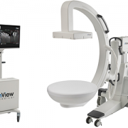
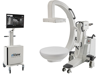
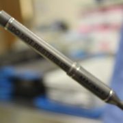
After implanting a MAGEC Spinal Growing Rod, doctors use an external remote control to lengthen the magnetically controlled rod as the child grows.

Children’s National Health System was among the first in the country to offer a novel spinal growing rod for children with scoliosis after it was approved by the FDA just three years ago – and has now treated 30 patients with this innovative technique. The MAGEC™ (MAGnetic Expansion Control) Spinal Growing Rod is a non-invasive treatment for children with early onset scoliosis.

After the initial procedure to implant the rod, doctors use an external remote control outside of the body to lengthen the magnetically controlled rod as the child grows. The adjustments are non-invasive, reducing the number of surgeries required during the course of treatment.

Growing rods have become effective tools for children whose spinal curvature is too significant to control with bracing or casting. The rods—which are surgically attached to the spine above and below the curve and then traditionally lengthened during follow-up surgical procedures—allow the spine to continue growing while managing the curve until the child is old enough for spinal fusion.

The problem: Children must bear the physical and psychological burden of undergoing lengthening procedures every six to 12 months until they are skeletally mature enough to have spinal fusion—typically around age 10 for girls and age 12 or 13 for boys.

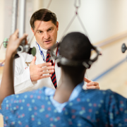
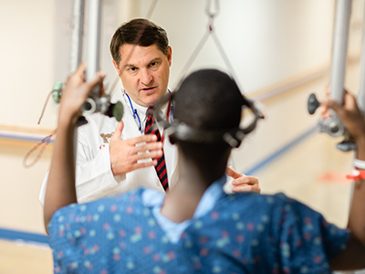
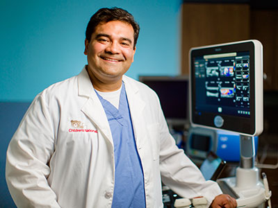
Now, instead of returning to the hospital for a major surgery to adjust growth rods twice a year, children with the MAGEC rod have adjustments in just a few minutes four times a year – minus invasive surgery and recovery time, says Matthew Oetgen, M.D., Division Chief of Orthopaedic Surgery and Sports Medicine and Director of Orthopaedic Research at Children’s National.

“Traditional growing rods work, but they require multiple surgeries that increase complication rates and time spent in the hospital,” he says. “We treat many children each year who have or are candidates for growing rods, so it’s important for us to embrace new technology to make the lengthening process easier and less painful for children while decreasing morbidity.”

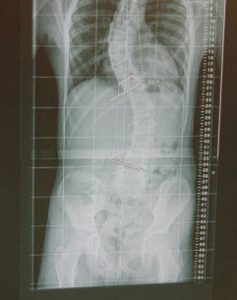
Children’s National orthopaedic surgeons lengthen the MAGEC rod every three to four months in the office using the electronic remote control. They then monitor the scoliosis and treatment progress with radiographs. Like traditional growing rods, MAGEC is a means, not an end—the system provides a bridge treatment spanning the years between the initial lengthening surgery and spinal fusion.

Dr. Oetgen says this game-changing technology may not be the right solution for every patient, but is the preferred choice because the patients can avoid some additional surgeries down the line. Patients in the 5-7 year age range at the time the rod is in place would potentially face 10 years of surgeries every six months with traditional growing rods.

“We’ve eliminated these regularly scheduled procedures, which is great if you’re a healthy kid,” Oetgen says. “But if you are a kid with other health challenges, such as a neuromuscular disease – it’s really life changing not to have to go into surgery every six months. It saves them a tremendous amount of intervention.”

Following MAGEC’s approval by the U.S. Food and Drug Administration in February 2014, surgeons at Children’s National performed two of the first 15 MAGEC implantations in the country, and the first in the greater Washington, DC, area. MAGEC rods are approved for children with scoliosis greater than 50 degrees in magnitude and under 10 years of age.

On the horizon for this new technology are some improvements Oetgen says would allow physicians using MAGEC rods to improve the patient experience even more. “Smarter” devices could potentially tell doctors how much lengthening has actually occurred after they’ve pushed the buttons on the remote control – instead of having to follow up the procedure with an x-ray to see how the rod interacted with soft tissue around the spine, he says.

And the next generations of MAGEC rods may be smaller devices, allowing younger, smaller kids to reap the benefits.

“In the future these improvements will allow us to treat more patients, and allow us to know what we’re doing and what kind of feedback we’re getting,” Oetgen says.