Tag Archive for: neuroprotection

Bone Marrow–Derived MSC Treatment Mitigates Structural Abnormalities Resulting From CPB

Cell therapy mitigates neurological impacts of cardiac surgery in pre-clinical model

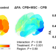
Differences of cortical fractional anisotropy between cardiopulmonary bypass and control (left), cardiopulmonary bypass + mesenchymal stromal cells and cardiopulmonary bypass (center), and 3 groups (right).

A pre-clinical study in the journal JACC: Basic to Translational Science shows that infusing bone marrow-derived mesenchymal stromal cells (BM-MSCs) during cardiac surgery provides both cellular-level neuroprotection for the developing brain and improvements in behavior alterations after (or resulting from) surgery.

What this means

According to lead author Nobuyuki Ishibashi, M.D., Oxidative and inflammatory stresses that are thought to be related to cardiopulmonary bypass cause prolonged microglia activation and cortical dysmaturation in the neonatal and infant brain. These issues are a known contributor to neurodevelopmental impairments in children with congenital heart disease.

This study found that, in a pre-clinical model, the innovative use of cardiopulmonary bypass to deliver these mesenchymal stromal cells minimizes microglial activation and neuronal apoptosis (cell death), with subsequent improvement of cortical dysmaturation and behavioral alteration after neonatal cardiac surgery.

Additionally, the authors note that further transcriptomic analyses provided a possible mechanism for the success: Exosome-derived miRNAs such as miR-21-5p, which may be key drivers of the suppressed apoptosis and STAT3-mediated microglial activation observed following BM-MSC infusion.

Why it matters

Significant neurological delay is emerging as one of the most important current challenges for children with congenital heart disease, yet few treatment options are currently available.

Applications of BM-MSC treatment will provide a new therapeutic paradigm for potential MSC-based therapies as a form of neuroprotection in children with congenital heart disease.

Children’s National Hospital leads the way

The Ishibashi lab is the first research team to demonstrate the safety, efficacy and utility of using cardiopulmonary bypass to deliver BM-MSCs with the goal of improving neurological impairments in children undergoing surgery for congenital heart disease. In addition to this pre-clinical research, a phase 1 clinical trial, MeDCaP, is underway at Children’s National.

Recent additional funding from the NIH will allow the team to identify molecular signatures of BM-MSC treatment and mine specific BM-MSC exosomes for unique cardiopulmonary bypass pathology to further increase understanding of precisely how and why this cell-based treatment shows success.

x-ray of child with congenital heart disease

Cell therapy research for neuroprotection in congenital heart disease receives another $3.3 million from NIH

x-ray of child with congenital heart disease

Significant neurological delay is emerging as one of the most important current challenges for children with congenital heart disease, yet few treatment options are currently available.

The research lab of Nobuyuki Ishibashi, M.D., at Children’s National Hospital, recently received $3.3 million in additional funding for research into cell therapy for neuroprotection in children with congenital heart disease. The new support comes from the National Heart, Lung and Blood Institute (NHLBI) of the National Institutes of Health.

The research goal

The overarching goal of the award is to establish detailed molecular signatures from critical cell populations for tissue repair and regeneration at single cell resolution after bone marrow-derived mesenchymal stromal cell (BM-MSC) delivery. The team has shown cellular, structural and behavioral improvements in pre-clinical models after delivery of BM-MSCs through cardiopulmonary bypass for children with congenital heart disease. However, the mechanisms underlying the therapeutic action of BM-MSCs still remain largely unknown. This R01 renewal will address the key knowledge gap.

Why it matters

Significant neurological delay is emerging as one of the most important current challenges for children with congenital heart disease, yet few treatment options are currently available.

The Ishibashi lab has demonstrated the efficacy and utility of using cardiopulmonary bypass to deliver BM-MSCs  to improve neurological impairments in children undergoing surgery for congenital heart disease. Most notably, this included development of a phase 1 clinical trial, MeDCaP, at Children’s National.

The big picture

Together with the ongoing clinical trial established from the previous award, identifying molecular signatures of BM-MSC treatment and mining specific BM-MSC exosomes for unique cardiopulmonary bypass pathology will significantly improve understanding of this cell-based treatment. This work will also provide a new therapeutic paradigm for potential cell-free MSC-based therapies for neuroprotection in children with congenital heart disease.

illustration of brain with stem cells

Innovative phase 1 trial to protect brains of infants with CHD during and after surgery

A novel phase 1 trial looking at how best to optimize brain development of babies with congenital heart disease (CHD) is currently underway at Children’s National Hospital.

Children with CHD sometimes demonstrate delay in the development of cognitive and motor skills. This can be a result of multiple factors including altered prenatal oxygen delivery, brain blood flow and genetic factors associated with surgery including exposure to cardiopulmonary bypass, also known as the heart lung machine.

This phase 1 trial is the first to deliver mesenchymal stromal cells from bone marrow manufactured in a lab (BM-MSC) into infants already undergoing cardiac surgery via cardiopulmonary bypass. The hypothesis is that by directly infusing the MSCs into the blood flow to the brain, more MSCs quickly and efficiently reach the subventricular zone and other areas of the brain that are prone to inflammation. The trial is open to eligible patients ages newborn to six months of age.


Learn more in this overview video.

The trial is part of a $2.5 million, three-year grant from the National Institutes of Health (NIH) led by Richard Jonas, M.D.Catherine Bollard, M.B.Ch.B., M.D., and Nobuyuki Ishibashi, M.D.. The project involves collaboration between the Prenatal Cardiology program of Children’s National Heart Institute, the Center for Cancer and Immunology Research, the Center for Neuroscience Research and the Sheikh Zayed Institute for Pediatric Surgical Innovation.

“NIH supported studies in our laboratory have shown that MSC therapy may be extremely helpful in improving brain development in animal models after cardiac surgery,” says Dr. Ishibashi. “MSC infusion can help reduce inflammation including prolonged microglia activation that can occur during surgery that involves the heart lung machine.”

Staff from the Cellular Therapy Laboratory, led by director Patrick Hanley, Ph.D., manufactured the BM-MSC at the Center for Cancer and Immunology Research, led by Dr. Bollard.

The phase 1 safety study will set the stage for a phase 2 effectiveness trial of this highly innovative MSC treatment aimed at reducing brain damage, minimizing neurodevelopmental disabilities and improving the postoperative course in children with CHD. The resulting improvement in developmental outcome and lessened behavioral impairment will be of enormous benefit to individuals with CHD.

For more information about this new treatment, contact the clinical research team: Gil Wernovsky, M.D., Shriprasad Deshpande, M.D., Maria Fortiz.

newborn in incubator

How EPO saves babies’ brains

newborn in incubator

Researchers have discovered that treating premature infants with erythropoietin can help protect and repair their vulnerable brains.

The drug erythropoietin (EPO) has a long history. First used more than three decades ago to treat anemia, it’s now a mainstay for treating several types of this blood-depleting disorder, including anemia caused by chronic kidney disease, myelodysplasia and cancer chemotherapy.

More recently, researchers discovered a new use for this old drug: Treating premature infants to protect and repair their vulnerable brains. However, how EPO accomplishes this feat has remained unknown. New genetic analyses presented at the Pediatric Academic Societies 2018 annual meeting that was conducted by a multi-institutional team that includes researchers from Children’s National show that this drug may work its neuroprotective magic by modifying genes essential for regulating growth and development of nervous tissue as well as genes that respond to inflammation and hypoxia.

“During the last trimester of pregnancy, the fetal brain undergoes tremendous growth. When infants are born weeks before their due dates, these newborns’ developing brains are vulnerable to many potential insults as they are supported in the neonatal intensive care unit during this critical time,” says An Massaro, M.D., an attending neonatologist at Children’s National Health System and lead author of the research. “EPO, a cytokine that protects and repairs neurons, is a very promising therapeutic approach to support the developing brains of extremely low gestational age neonates.”

The research team investigated whether micro-preemies treated with EPO had distinct DNA methylation profiles and related changes in expression of genes that regulate how the body responds to such environmental stressors as inflammation, hypoxia and oxidative stress.  They also investigated changes in genes involved in glial differentiation and myelination, production of an insulating layer essential for a properly functioning nervous system. The genetic analyses are an offshoot of a large, randomized clinical trial of EPO to treat preterm infants born between 24 and 27 gestational weeks.

The DNA of 18 newborns enrolled in the clinical trial was isolated from specimens drawn within 24 hours of birth and at day 14 of life. Eleven newborns were treated with EPO; a seven-infant control group received placebo.

DNA methylation and whole transcriptome analyses identified 240 candidate differentially methylated regions and more than 50 associated genes that were expressed differentially in infants treated with EPO compared with the control group. Gene ontology testing further narrowed the list to five candidate genes that are essential for normal neurodevelopment and for repairing brain injury:

“These findings suggest that EPO’s neuroprotective effect may be mediated by epigenetic regulation of genes involved in the development of the nervous system and that play pivotal roles in how the body responds to inflammation and hypoxia,” Dr. Massaro says.

In addition to Dr. Massaro, study co-authors include Theo K. Bammler, James W. MacDonald, biostatistician, Bryan Comstock, senior research scientist, and Sandra “Sunny” Juul, M.D., Ph.D., study principal investigator, all of University of Washington.