In a recent review article published in Circulation Research, Nobuyuki Ishibashi, M.D., and his colleagues at Children’s National Health System summarized what is currently known about how congenital heart disease affects brain maturation.
What’s known
Among all known birth defects, congenital heart disease (CHD) is the leading cause of death in infants. Fortunately, advances in surgical techniques and treatments are improving the outlook for these children, and more and more are reaching adulthood. However, because of this increased longevity, it has become increasingly clear that children born with CHD are at risk of developing life-long neurological deficits. Several risk factors for these neurodevelopmental abnormalities have been identified, but direct links between specific factors and neurological defects have yet to be established.
What’s new
In a recent review article published in Circulation Research, a team from Children’s National Health System summarized what is currently known about how CHD affects brain maturation. Drawing from studies conducted at Children’s National as well as other research institutions, Paul D. Morton, Ph.D., Nobuyuki Ishibashi, M.D., and Richard A. Jonas, M.D., write that clinical findings in patients, improvements in imaging analysis, advances in neuromonitoring techniques and the development of animal models have greatly contributed to our understanding of the neurodevelopmental changes that occur with CHD.
Findings from Children’s National include:
An assessment of the intraoperative effects of cardiopulmonary bypass surgery on white matter using neonatal piglets.
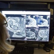
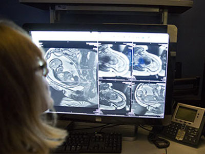
An arterial spin labeling MRI study that showed newborns with complex CHD have a significant reduction in global cerebral blood flow.
A rodent study that modeled diffuse white matter brain injury in premature birth and identified the cellular and molecular mechanisms underlying lineage-specific vulnerabilities of oligodendrocytes and their regenerative response after chronic neonatal hypoxia.
The authors conclude that although there is ample clinical evidence of neurological damage associated with CHD, there is limited knowledge of the cellular events associated with these abnormalities. They offer perspectives about what can be done to improve our understanding of neurological deficits in CHD, and emphasize that ultimately, a multidisciplinary approach combining multiple fields and myriad technology will be essential to improve or prevent adverse neurodevelopmental outcomes in individuals with CHD.
Questions for future research
Q: What are the cellular events associated with each factor involved in neurodevelopmental delays?
Q: How does the neurodevelopmental status of a patient with CHD change as they age?
Q: How do the genes involved in structural congenital cardiac anomalies affect brain development and function?
https://innovationdistrict.childrensnational.org/wp-content/uploads/2017/06/Nobuyuki-Ishibashi.jpg300400Innovation Districthttps://innovationdistrict.childrensnational.org/wp-content/uploads/2023/12/innovationdistrict_logo-1-1030x165.pngInnovation District2017-06-02 12:02:112024-09-06 15:02:26Congenital heart disease and the brain
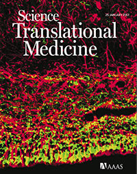
The cover of Science Translational Medicine features a new study of the cellular-level changes in the brain induced by congenital heart disease. Reprinted with permission from AAAS. Not for download
Disruptions in cerebral oxygen supply caused by congenital heart disease have significant impact on cortical growth, according to a research led by Children’s National Health System. The findings of the research team, which include co-authors from the National Institutes of Health, Boston Children’s Hospital and Johns Hopkins School of Medicine, appear on the cover of Science Translational Medicine. The subventricular zone (SVZ) in normal newborns’ brains is home to the largest stockpile of neural stem/progenitor cells, with newly generated neurons migrating from this zone to specific regions of the frontal cortex and differentiating into interneurons. When newborns experience disruptions in cerebral oxygen supply due to congenital heart disease, essential cellular processes go awry and this contributes to reduced cortical growth.
The preliminary findings point to the importance of restoring these cells’ neurogenic potential, possibly through therapeutics, to lessen children’s long-term neurological deficits.
“We know that congenital heart disease (CHD) reduces cerebral oxygen at a time when the developing fetal brain most needs oxygen. Now, we are beginning to understand the mechanisms of CHD-induced brain injuries at a cellular level, and we have identified a robust supply of cells that have the ability to travel directly to the site of injury and potentially provide help by replacing lost or damaged neurons,” says Nobuyuki Ishibashi, M.D., Director of the Cardiac Surgery Research Laboratory at Children’s National, and co-senior study author.
The third trimester of pregnancy is a time of dramatic growth for the fetal brain, which expands in volume and develops complex structures and network connections that growing children rely on throughout adulthood. According to the National Heart, Lung, and Blood Institute, congenital heart defects are the most common major birth defect, affecting 8 in 1,000 newborns. Infants born with CHD can experience myriad neurological deficits, including behavioral, cognitive, social, motor and attention disorders, the research team adds.
Cardiologists have tapped noninvasive imaging to monitor fetal hearts during gestation in high-risk pregnancies and can then perform corrective surgery in the first weeks of life to fix damaged hearts. Long term neurological deficits due to immature cortical development also have emerged as major challenges in pregnancies complicated by CHD.
“I think this is an enormously important paper for surgeons and for children and families who are affected by CHD. Surgeons have been worried for years that the things we do during corrective heart surgery have the potential to affect the development of the brain. And we’ve learned to improve how we do heart surgery so that the procedure causes minimal damage to the brain. But we still see some kids who have behavioral problems and learning delays,” says Richard A. Jonas, M.D., Chief of the Division of Cardiac Surgery at Children’s National, and co-senior study author. “We’re beginning to understand that there are things about CHD that affect the development of the brain before a baby is even born. What this paper shows is that the low oxygen level that sometimes results from a congenital heart problem might contribute to that and can slow down the growth of the brain. The good news is that it should be possible to reverse that problem using the cells that continue to develop in the neonate’s brain after birth.”
Among preclinical models, the spatiotemporal progression of brain growth in this particular model most closely parallels that of humans. Likewise, the SVZ cytoarchitecture of the neonatal preclinical model exposed to hypoxia mimics that of humans in utero and shortly after birth. The research team leveraged CellTracker Green to follow the path traveled by SVZ derived cells and to illuminate their fate, with postnatal SVZ supplying the developing cortex with newly generated neurons. SVZ derived cells were primarily neuroblasts. Superparamagnetic iron oxide nanoparticles supplied answers about long term SVZ migration, with SVZ derived cells making their way to the prefrontal cortex and the somatosensory cortex of the brain.
“We demonstrated that in the postnatal period, newly generated neurons migrate from the SVZ to specific cortices, with the majority migrating to the prefrontal cortex,” says Vittorio Gallo, Ph.D., Director of the Center for Neuroscience Research at Children’s National, and co-senior study author. “Of note, we revealed that the anterior SVZ is a critical source of newborn neurons destined to populate the upper layers of the cortex. We challenged this process through chronic hypoxia exposure, which severely impaired neurogenesis within the SVZ, depleting this critical source of interneurons.”
In the preclinical model of hypoxia as well as in humans, brains were smaller, weighed significantly less and had a significant reduction in cortical gray matter volume. In the prefrontal cortex, there was a significant reduction in white matter neuroblasts. Taken as a whole, according to the study authors, the findings suggest that impaired neurogenesis within the SVZ represents a cellular mechanism underlying hypoxia induced, region specific reduction in immature neurons in the cortex. The prefrontal cortex, the region of the brain that enables such functions as judgment, decision making and problem solving, is most impacted. Impairments in higher order cognitive functions involving the prefrontal cortex are common in patients with CHD.
https://innovationdistrict.childrensnational.org/wp-content/uploads/2017/01/CHD-Impaired-Cortical-Growth-HP.jpg248400Innovation Districthttps://innovationdistrict.childrensnational.org/wp-content/uploads/2023/12/innovationdistrict_logo-1-1030x165.pngInnovation District2017-02-07 14:08:072024-09-06 15:05:20Congenital heart disease and cortical growth
Although recent advances have greatly improved the survival of children with congenital heart disease, up to 55 percent will be left with injury to their brain’s white matter – an area that is critical for aiding connection and communication between various regions in the brain.
What’s known
Eight of every 1,000 children born each year have congenital heart disease (CHD). Although recent advances have greatly improved the survival of these children, up to 55 percent will be left with injury to their brain’s white matter – an area that is critical for aiding connection and communication between various regions in the brain. The resulting spectrum of neurological deficits can have significant costs for the individual, their family and society. Although studies have demonstrated that white matter injuries due to CHD have many contributing factors, including abnormal blood flow to the fetal brain, many questions remain about the mechanisms that cause these injuries and the best interventions.
What’s new
A Children’s National Health System research team combed existing literature, reviewing studies from Children’s as well as other research groups, to develop an article detailing the current state of knowledge on CHD and white matter injury. The scientists write that advances in neuroimaging – including magnetic resonance imaging, magnetic resonance spectroscopy, Doppler ultrasound and diffusion tensor imaging – have provided a wealth of knowledge about brain development in patients who have CHD. Unfortunately, these techniques alone are unable to provide pivotal insights into how CHD affects cells and molecules in the brain. However, by integrating animal models with findings in human subjects and in postmortem human tissue, the scientists believe that it will be possible to find novel therapeutic targets and new standards of care to prevent developmental delay associated with cardiac abnormalities.
For example, using a porcine model, the Children’s team was able to define a strategy for white matter protection in congenital heart surgery through cellular and developmental analysis of different white matter regions. Another study from Children’s combined rodent hypoxia with a brain slice model to replicate the unique brain conditions in neonates with severe and complex congenital heart disease. This innovative animal model provided novel insights into the possible additive effect of preoperative hypoxia on brain insults due to cardiopulmonary bypass and deep hypothermic circulatory arrest.
The Children’s research team also recently published an additional review article describing the key windows of development during which the immature brain is most vulnerable to CHD-related injury.
Questions for future research
Q: Can we create an animal model that recapitulates the morphogenic and developmental aspects of CHD without directly affecting other organs or developmental processes?
Q: What are the prenatal and neonatal cellular responses to CHD in the developing brain?
Q: What are the molecular mechanisms underlying white matter immaturity and vulnerability to CHD, and how can we intervene?
Q: How can we accurately assess the dynamic neurological outcomes of CHD and/or corrective surgery in animal models?
Q: Prenatal or postnatal insults to the developing brain: which is most devastating in regards to developmental and behavioral disabilities?
Q: How can we best extrapolate from, and integrate, neuroimaging findings/correlations in human patients with cellular/molecular approaches in animal models?
Source: Reprinted from Trends in Neurosciences, Vol. 38/Ed. 6, Paul D. Morton, Nobuyuki Ishibashi, Richard A. Jonas and Vittorio Gallo, “Congenital cardiac anomalies and white matter injury,” pp. 353-363, Copyright 2015, with permission from Elsevier.
https://innovationdistrict.childrensnational.org/wp-content/uploads/2017/02/CHD_White_Matter_article-1.jpg300400Innovation Districthttps://innovationdistrict.childrensnational.org/wp-content/uploads/2023/12/innovationdistrict_logo-1-1030x165.pngInnovation District2017-02-06 07:39:392017-06-07 14:12:46Congenital heart disease and white matter injury