Tag Archive for: Magge

Suresh Magge

Sudden blindness leads to unusual diagnosis

Suresh Magge

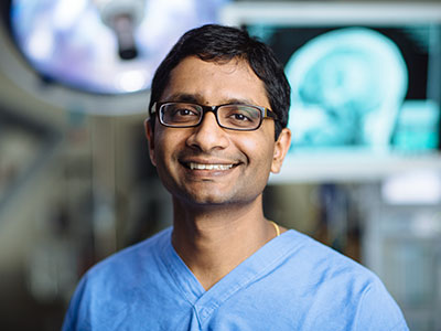
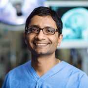
Suresh N. Magge, M.D., and his colleagues at Children’s National recently published the details of an unusual case of advanced moyamoya disease in the journal Stroke.

When Children’s National Health System Neurosurgeon Suresh N. Magge, M.D., met his new patient, the 16-year-old had suddenly lost her vision in both eyes.

To discover the reason for this abrupt loss of vision, her doctors ran a battery of tests. An ophthalmologist found no problems with her eyes.  Her optic nerves, which run signals generated from the eyes to the brain, also appeared to work normally. However, a computerized tomography scan and magnetic resonance imaging showed the unmistakable signs of a stroke in her occipital lobe, the portion of the brain responsible for interpreting signals relayed from the optic nerves.

“Her brain basically wasn’t seeing what her eyes saw,” Dr. Magge explains.

Delving deeper, her Children’s National care team found the reason why their young patient had suffered a stroke by using a cerebral angiogram, an imaging test that shows the blood vessels in and around the brain. The teen had moyamoya disease, a rare condition that causes blood vessels in the brain to narrow, often leading otherwise healthy adults and children to have strokes.

According to the National Institutes of Health, moyamoya is Japanese for “puff of smoke,” so named because of the telltale signs this condition presents on an angiogram. When arteries in the brain narrow, brain tissue becomes “thirsty” for more blood, Dr. Magge explains, leading its cells to produce chemicals that prompt new blood vessels to grow. These new collateral blood vessels often grow in a thin tangle that looks like smoke on an angiogram. Generally, however, they do not supply sufficient oxygenated blood to meet the brain’s needs, leaving it starved for oxygen. Eventually, the blood supply can get so low that patients suffer transient ischemic attacks, “mini-strokes” that temporarily deprive the brain tissue of oxygen, or full-blown strokes typically characterized by weakness, speech problems, facial paralysis or other problems.

Dr. Magge’s patient had little warning before her stroke occurred. The first major symptom that led her to seek medical attention was abrupt blindness, which Dr. Magge says is a highly unusual occurrence for a moyamoya diagnosis. That’s why he and colleagues decided to publish the details of her case as a teaching report April 14, 2017 in the journal Stroke.

The Children’s National co-authors wrote that once their patient was diagnosed with a stroke due to advanced moyamoya disease, with blood vessels severely narrowed throughout her brain, the first order of business was stabilizing her symptoms and making sure she did not have further strokes. Her blood pressure was stabilized, and she was started on aspirin therapy to decrease her risk of further strokes. She took time to recover as much as possible from her original stroke.

A few weeks later, Dr. Magge and his neurosurgery colleagues performed a type of surgery to revascularize – or restore blood flow – to areas of the brain that were still healthy but at risk of having subsequent strokes. The surgical procedure, known as pial synangiosis, reconfigures the brain’s blood vessels to make sure that these vulnerable areas of the brain have a sufficient blood supply.

Years later, Dr. Magge says, his patient is doing well, except for the original blindness, a permanent consequence of the stroke to her occipital lobe before her diagnosis. She has not had new strokes since the revascularization surgery. She will need aspirin therapy and periodic neurological checkups for the rest of her life, Dr. Magge explains, to make sure that the blood supply to her brain remains stable.

Children’s experts use a team approach to treat patients with complex care needs: Neurologists, neurosurgeons, intensivists, hematologists, anesthesiologists, neuroradiologists and nurses leverage their combined expertise with moyamoya disease to treat the complexities of this condition.

“A team approach is essential to deliver the best outcomes to children with life-changing diseases,” Dr. Magge says. “We try to help kids get back to living full and healthy lives.”

Teen Girl drawing a heart on an iPad

Illuminating cardiometabolic risk in Down syndrome

Teen Girl drawing a heart on an iPad

A leading researcher at Children’s National says researchers should look closely at the increased risks of obesity and thyroid disease common in patients with Down Syndrome, and determine how these long term comorbidities relate to cardiovascular and metabolic (cardiometabolic) risk, body image, and quality of life.

Over the last several decades, physicians’ improved ability to treat the common comorbidities of Down syndrome, such as congenital heart disease, has dramatically prolonged survival. Today, more than 400,000 people across the country are living with Down syndrome, and life expectancy has increased to 60 years.

New strategies to manage care for patients with Down syndrome must include preventive, evidence-based approaches to address the unique needs of these patients, according to Sheela N. Magge, M.D., M.S.C.E., Director of Research in the Division of Endocrinology and Diabetes at Children’s. She says that these efforts should include looking more closely at the increased risks of obesity and thyroid disease common in this population, and determining how these long term comorbidities relate to cardiovascular and metabolic (cardiometabolic) risk, body image, and quality of life.

An NIH-funded study from Children’s National and the Children’s Hospital of Philadelphia (CHOP), led by Dr. Magge and her colleague from CHOP, Dr. Andrea Kelly, seeks to better understand how the body composition of patients with Down syndrome impacts their likelihood for developing diabetes and obesity-related cardiovascular risks long term.

“We know that individuals with Down syndrome are at increased risk for obesity, but what hasn’t been clear is whether or not they also have the same cardiometabolic risk associated with obesity that we know holds true for other populations,” says Dr. Magge. “In this previously under-studied population, the common assumption based on very limited studies from the 1970’s was that individuals with Down syndrome were protected from the diabetes and cardiovascular risks that can develop in other overweight people. However, more recent epidemiologic studies contradict those early findings.”

The study has enrolled 150 Down syndrome patients and almost 100 controls to date, and the team is currently beginning to analyze the data. Dr. Magge believes that the findings from this study will help to provide new, research-driven evidence to inform the long term clinical management of obesity and cardiometabolic risk in adolescents with Down syndrome.

She concludes, “The goal is for our research to provide the foundation that will advance prevention and treatment strategies for this understudied group, so that individuals with Down syndrome not only have a longer life expectancy, but also a healthier and better quality of life.”

Dr. Keating and Abigail

Multidisciplinary approach to hydrocephalus care

Reflective of the myriad symptoms and complications that can accompany hydrocephalus, a multidisciplinary team at Children’s National works with patients and families for much of childhood.

The Doppler image on the oversized computer screen shows the path taken by blood as it flows through the newborn’s brain, with bright blue distinguishing blood moving through the middle cerebral artery toward the frontal lobe and bright red depicting blood coursing away. Pitch black zones indicate ventricles, cavities through which cerebrospinal fluid usually flows and where hydrocephalus can get its start.

The buildup of excess cerebrospinal fluid in the brain can begin in the womb and can be detected by fetal magnetic resonance imaging. Hydrocephalus also can crop up after birth due to trauma to the head, an infection, a brain tumor or bleeding in the brain, according to the National Institutes of Health. An estimated 1 to 2 per 1,000 newborns have hydrocephalus at birth.

When parents learn of the hydrocephalus diagnosis, their first question tends to be “Is my child going to be OK?” says Suresh Magge, M.D., a pediatric neurosurgeon at Children’s National Health System.

“We have a number of ways to treat hydrocephalus. It is one of the most common conditions that pediatric neurosurgeons treat,” Dr. Magge adds.

Unlike fluid build-up elsewhere in the body where there are escape routes, with hydrocephalus spinal fluid becomes trapped in the brain. To remove it, surgeons typically implant a flexible tube called a shunt that drains excess fluid into the abdomen, an interim stop before it is flushed away. Another surgical technique, called an endoscopic third ventriculostomy has the ability to drain excess fluid without inserting a shunt, but it only works for select types of hydrocephalus, Dr. Magge adds.

For the third year, Dr. Magge is helping to organize the Hydrocephalus Education Day on Feb. 25, a free event that offers parents an opportunity to learn more about the condition.

Reflective of the myriad symptoms and complications that can accompany hydrocephalus, such as epilepsy, cerebral palsy, cortical vision impairment and global delays, a multidisciplinary team at Children’s National works with patients and families for much of childhood.

Neuropsychologist Yael Granader, Ph.D., works with children ages 4 and older who have a variety of developmental and medical conditions. Granader is most likely to see children and adolescents with hydrocephalus once they become medically stable in order to assist in devising a plan for school support services and therapeutic interventions. Her assessments can last an entire day as she administers a variety of tasks that evaluate how the child thinks and learns, such as discerning patterns, assembling puzzles, defining words, and listening to and remembering information.

Neuropsychologists work with schools in order to help create the most successful academic environment for the child. For example, some children may struggle to visually track across a page accurately while reading; providing a bookmark to follow beneath the line is a helpful and simple accommodation to put in place. Support for physical limitations also are discussed with schools in order to incorporate adaptive physical education or to allow use of an elevator in school.

“Every child affected by hydrocephalus is so different. Every parent should know that their child can learn,” Granader says. “We’re going to find the best, most supportive environment for them. We are with them on their journey and, every few years, things will change. We want to be there to help with emerging concerns.”

Another team member, Justin Burton, M.D., a pediatric rehabilitation specialist, says rehabilitation medicine’s “piece of the puzzle is doing whatever I can to help the kids function better.” That means dressing, going to the bathroom, eating and walking independently. With babies who have stiff, tight muscles, that can mean helping them through stretches, braces and medicine management to move muscles smoothly in just the way their growing bodies want. Personalized care plans for toddlers can include maintaining a regular sleep-wake cycle, increasing attention span and strengthening such developmental skills as walking, running and climbing stairs. For kids 5 and older, the focus shifts more to academic readiness, since those youths’ “full-time job” is to become great students, Dr. Burton says.

The area of the hospital where children work on rehabilitation is an explosion of color and sounds, including oversized balance balls of varying dimensions in bright primary colors, portable basketball hoops with flexible rims at multiple heights, a set of foam stairs, parallel bars, a climbing device that looks like the entry to playground monkey bars and a chatterbox toy that lets a patient know when she has opened and closed the toy’s doors correctly.

“We end up taking care of these kids for years and years,” he adds. “I always love seeing the kids get back to walking and talking and getting back to school. If we can get them back out in the world and they’re doing things just like every other kid, that’s success.”

Meanwhile, Dr. Magge says research continues to expand the range of interventions and to improve outcomes for patients with hydrocephalus, including:

  • Fluid dynamics of cerebrospinal fluid
  • Optimal ways to drain excess fluid
  • Improving understanding of why shunts block
  • Definitively characterizing post-hemorrhagic ventricular dilation.

Unlike spina bifida, which sometimes can be corrected in utero at some health institutions, hydrocephalus cannot be corrected in the womb. “While we have come a long way in treating hydrocephalus, there is still a lot of work to be done. We continue to learn more about hydrocephalus with the aim of continually improving treatments,” Dr. Magge says.

During a recent office visit, 5-year-old Abagail’s head circumference had measured ¼ centimeter of growth, an encouraging trend, Robert Keating, M.D., Children’s Chief of Neurosurgery, tells the girl’s mother, Melissa J. Kopolow McCall. According to Kopolow McCall, who co-chairs the Hydrocephalus Association DC Community Network, it is “hugely” important that Children’s National infuses its clinical care with the latest research insights. “I have to have hope that she is not going to be facing a lifetime of brain surgery, and the research is what gives me the hope.”