It’s long been known that colds, flu and other respiratory illnesses are major triggers for asthma exacerbations, says asthma expert Stephen J. Teach, M.D., MPH. Consequently, a significant body of research has focused on trying to figure out what’s happening on the cellular or molecular level as these illnesses progress to exacerbations.
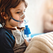
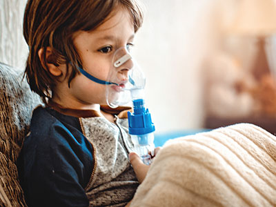
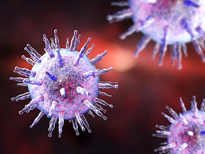
People with asthma can be indistinguishable from people who don’t have this chronic airway disease – until they have an asthma attack, also known as an exacerbation. During these events, their airways become inflamed and swollen and produce an abundance of mucus, causing dangerous narrowing of the bronchial tubes that leads to coughing, wheezing and trouble breathing. These events are a major cause of morbidity and mortality, leading to the deaths of 10 U.S. residents every day, according to the Centers for Disease Control and Prevention.
It’s long been known that colds, flu and other respiratory illnesses are major triggers for asthma exacerbations, says Children’s National in Washington, D.C., asthma expert Stephen J. Teach, M.D., MPH. Consequently, a significant body of research has focused on trying to figure out what’s happening on the cellular or molecular level as these illnesses progress to exacerbations. Targeted searches have identified several different molecular pathways that appear to be key players in this phenomenon. However, Dr. Teach says researchers have been missing a complete and unbiased snapshot of all the important pathways in illness-triggered exacerbations and how they interrelate.
To develop this big picture view, Dr. Teach and Inner-City Asthma Consortium colleagues recruited 208 children ages 6-17 years old with severe asthma – marked by the need for daily doses of inhaled corticosteroids, two hospitalizations or systemic corticosteroid treatments over the past year, and a high concentration of asthma-associated immune cells – from nine pediatric medical centers across the country, including Children’s National. (Inhaled corticosteroids are a class of medicine that calms inflamed airways.) The researchers collected samples of nasal secretions and blood from these patients at baseline, when all of them were healthy.
Then, they waited for these children to show symptoms of respiratory illnesses. Within six days of cold symptoms, the researchers took two more samples of nasal secretions and blood. They also administered breathing tests to determine whether these respiratory illnesses led to asthma exacerbations and recorded whether these patients were treated with systemic corticosteroids to stem the associated respiratory inflammation.
The researchers examined nasal fluid samples for evidence of viral infection during illness and used analytical methods to identify the causative virus. They analyzed all the samples they collected for changes in concentrations of various immune cells. They also looked globally in these samples for changes in gene expression compared with baseline and between the two collection periods during respiratory illness.
Together, this information told the molecular story about what took place after these children got sick and after some of them developed exacerbations. Of the 208 patients recruited, 106 got respiratory illnesses during the six-month study period, leading to a total of 154 illness events. Of those, 47 caused exacerbations, and 107 didn’t.
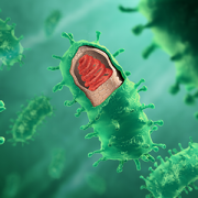
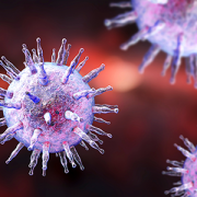
About half the exacerbations appeared to have been triggered by a rhinovirus, a cause of common colds, the research team reports in a study published online April 8, 2019, in Nature Immunology. The other children’s cold-like symptoms could have been triggered by pollution, allergens or other irritants.
In most exacerbations, virally triggered or not, the researchers saw early activation of a network of genes that appeared to be associated with SMAD3, a signaling molecule already known to be involved in airway inflammation. At the same time, genes that control a set of immune cells known as lymphocytes were turned down. However, as the exacerbation progressed and worsened, the researchers saw gene networks turned on that related to airway narrowing, mucus hypersecretion and activation of other immune cells.
Exacerbations triggered by viruses were associated with multiple inflammatory pathways, in contrast to those in which viruses weren’t found, which were associated with molecular pathways that affected cells in the airway lining.
The researchers validated these findings in 19 patients who each got respiratory illnesses at least twice during the study period but only developed an exacerbation during one of these episodes, finding the same upregulated and downregulated molecular pathways in these patients as in the study population as a whole. They also identified a set of molecular risk factors in patients at baseline – signatures of gene activation that appeared to put patients at risk for exacerbations when they got sick. When patients were treated with systemic corticosteroids during exacerbations, these medicines appeared to restore only some of the affected molecular pathways to normal, healthy levels. Other molecular pathways remained markedly changed.
Each finding could represent a new target for drugs that could prevent or more effectively treat exacerbations, keeping more patients with asthma healthy and out of the hospital.
“Our consortium study found increased gene expression of enzymes that produce molecules that contribute to narrowed airways and dilated blood vessels,” Dr. Teach adds. “This is especially intriguing because drugs that target kallikreins or bradykinin may help treat asthma attacks that aren’t caused by viruses.”
In addition to Dr. Teach, study co-authors include Lead Author Matthew C. Altman, University of Washington; Michelle A. Gill, Baomei Shao and Rebecca S. Gruchalla, all of University of Texas Southwestern Medical Center; Elizabeth Whalen and Scott Presnell of Benaroya Research Institute; Denise C. Babineau and Brett Jepson of Rho, Inc.; Andrew H. Liu, Children’s Hospital Colorado; George T. O’Connor, Boston University School of Medicine; Jacqueline A. Pongracic, Ann Robert H. Lurie Children’s Hospital of Chicago; Carolyn M. Kercsmar and Gurjit K. Khurana Hershey, , Cincinnati Children’s Hospital; Edward M. Zoratti and Christine C. Johnson, Henry Ford Health System; Meyer Kattan, Columbia University College of Physicians and Surgeons; Leonard B. Bacharier and Avraham Beigelman, Washington University, St. Louis; Steve M. Sigelman, Peter J. Gergen, Lisa M. Wheatley and Alkis Togias, National Institute of Allergy and Infectious Diseases; and James E. Gern, William W. Busse and Senior author Daniel J. Jackson, University of Wisconsin School of Medicine and Public Health.
Funding for research described in this post was provided by the National Institute of Allergy and Infectious Diseases under award numbers 1UM1AI114271 and UM2AI117870; CTSA under award numbers UL1TR000150, UL1TR001422 and 5UL1TR001425; the National Institutes of Health under award number UL1TR000451; CTSI under award number 1UL1TR001430; CCTSI under award numbers UL1TR001082 and 5UM1AI114271; and NCATS under award numbers UL1 TR001876 and UL1TR002345.
The Epstein-Barr virus (EBV) is best known as the cause of mononucleosis, the ubiquitous “kissing disease” that most people contract at some point in their life. But in rare instances, this virus plays a more sinister role as the impetus of lymphomas, cancers that affect the white blood cells known as lymphocytes.
The Epstein-Barr virus (EBV) is best known as the cause of mononucleosis, the ubiquitous “kissing disease” that most people contract at some point in their life. But in rare instances, this virus plays a more sinister role as the impetus of lymphomas, cancers that affect the white blood cells known as lymphocytes. EBV-associated lymphomas account for about 40% of Hodgkin lymphomas, 20% of diffuse large B-cell lymphomas, and more than 90% of natural killer/T-cell lymphomas. This latter type of lymphoma typically has a very poor prognosis even with the “standard of care” lymphoma treatments such as chemotherapy and/or radiation.
When these interventions fail, the only curative approach is an allogeneic hematopoietic stem cell transplant from a healthy donor, a treatment that’s tough on patients’ bodies and carries significant risks, says Lauren P. McLaughlin, M.D., a pediatrician specializing in hematology and oncology at Children’s National in Washington, D.C. Patients who receive these allogenic transplants are immune-compromised until the donor cells engraft; the grafts can attack patients’ healthy cells in a phenomenon called graft versus host disease; and if patients relapse or don’t respond to this treatment, few options remain.
To help improve outcomes, Dr. McLaughlin and colleagues tested an addition to the allogeneic hematopoietic stem cell transplant procedure for patients with EBV-associated lymphomas: infusion of a type of immune cell called T cells specifically trained to fight cells infected with EBV.
Dr. McLaughlin, along with Senior Author Catherine M. Bollard, M.D., M.B.Ch.B., director of the Center for Cancer and Immunology Research and the Program for Cell Enhancement and Technologies for Immunotherapy at Children’s National, and colleagues tested this therapy in 26 patients treated at Children’s National or Baylor College of Medicine. They published these results online on Sept. 27, 2018, in the journal Blood. The study was a Phase I clinical trial, meaning that the therapy was tested primarily for safety, with efficacy as a secondary aim.
Seven patients who received the therapy had active disease that had not responded to conventional therapies. The other 19 were patients deemed to be at high risk for relapse.
Before each patient received their stem cell transplant, their donors gave an additional blood sample to generate the cancer-fighting T cells. Over the next 8 to 10 weeks, the researchers painstakingly manufactured the immune cells known as T-cells that specifically targeted EBV, growing these cells into numbers large enough for clinical use. Then, as early as 30 days after transplant, the researchers infused these T-cells into patients administering at least two doses, spaced two weeks apart.
Over the next several weeks, the researchers at CNMC and Baylor College of Medicine monitored patients with comprehensive exams to see how they fared after these transplants. The results showed that adverse effects from the treatment were exceedingly rare. There were no immediate infusion-related toxicities to the T-cell therapy and only one incident of dose-limiting toxicity.
This therapy may be efficacious, depending on the individual patients’ circumstances, Dr McLaughlin adds. For those in complete remission but at high risk of relapsing, the two-year survival rate was 78%, suggesting that the administration of this novel T-cell therapy may give the immune system a boost to prevent the lymphoma from returning after transplant. For patients with active T-cell lymphomas, two-year survival rates were 60%. However, even these lower rates are better than the historical norm of 30-50%, suggesting that the targeted T-cell therapies could help fight disease in patients with this poor prognosis lymphoma.
Dr. McLaughlin, the study’s lead author and a Lymphoma Research Foundation grantee, notes that researchers have more work to do before this treatment becomes mainstream. For example, this treatment will need to be tested in larger populations of patients with EBV-related lymphoma to determine who would derive the most benefit, the ideal dose and dose timing. It also may be possible to extend targeted T-cell treatments like this to other types of cancers. In the future, Dr. McLaughlin adds, it may be possible to develop T-cells that could be used “off the shelf”—in other words, they wouldn’t need to come from a matched donor and would be ready to use whenever a recipient needs them. Another future goal is using this therapy as one of the first lines of treatment rather than as a last resort.
“Our ultimate goal is to find a way to avoid chemotherapy and/or radiation therapy while still effectively treating a patient’s cancer,” she says. “Can you use the immune system to do that job? We’re working to answer that question.”
In addition to Drs. McLaughlin and Bollard, study co-authors include Rayne Rouce, Stephen Gottschalk, Vicky Torrano, George Carrum, Andrea M. Marcogliese, Bambi Grilley, Adrian P. Gee, Malcolm K. Brenner, Cliona M. Rooney and Helen E. Heslop, all of Baylor College of Medicine; Meng-Fen Wu from the Dan L. Duncan Comprehensive Cancer Center; and Fahmida Hoq and Patrick J. Hanley, Ph.D. from Children’s National in Washington, D.C.

Research published online Dec. 13, 2017, by The Lancet Haematology and co-led by Kirsten M. Williams, M.D., suggests that a new imaging agent can safely show engraftment as early as days after transplant – giving a helpful and hopeful preview to patients and their doctors.
Leukemia can be a terrifying diagnosis for the more than 60,000 U.S. patients who are told they have this blood cancer every year. But the treatment for this disease can be just as frightening. For patients with certain forms of leukemia, the only chance they have for a cure is to receive a massive dose of radiation and chemotherapy that kills their hematopoietic stem cells (HSCs), the cells responsible for making new blood, and then receive new HSCs from a healthy donor.
While patients are waiting for these new cells to go to the bone marrow factory and begin churning out new blood cells, patients are left without an immune system. Devoid of working HSCs for two to four weeks – or longer, if a first transplant doesn’t take – patients are vulnerable to infections that can be just as deadly as their original cancer diagnosis.
As they wait in the protected confines of a hospital, patients who undergo HSC transplants receive blood tests every day to gauge successful engraftment, searching for the presence of immune cells called neutrophils, explains Kirsten M. Williams, M.D., blood and bone marrow transplant specialist at Children’s National Health System.
“As you head into week three post-transplant and a patient’s cell counts remain at zero, everyone starts to get nervous,” Dr. Williams says. The longer a patient goes without an immune system, the higher the chance that they’ll develop a life-threatening infection. Until recently, Dr. Williams says, there has been no way beyond those daily blood tests to assess whether the newly infused cells have survived and started to grow early healthy cells in the bone marrow, a process called engraftment.
A new study could change that paradigm. Research published online Dec. 13, 2017, by The Lancet Haematology and co-led by Dr. Williams suggests that a new imaging agent can safely show engraftment as early as days after transplant – giving a helpful and hopeful preview to patients and their doctors.
The study evaluated an investigational imaging test called 18F-fluorothymidine (18F-FLT). It’s a radio-labeled analogue of thymidine, a natural component of DNA. Studies have shown that this compound is incorporated into just three white blood cell types, including HSCs. Because it’s radioactive, it can be seen on various types of common clinical imaging exams, such as positron emission tomography (PET) and computed tomography (CT) scans. Thus, after infusion, the newly infused developing immune system and marrow is readily visible.
To see whether this compound can readily and safely visualize transplanted HSCs, Dr. Williams and colleagues tested it on 23 patients with various forms of high-risk leukemia.
After these patients received total-body irradiation to destroy their own HSCs, they received donor HSCs from relatives or strangers. One day before they were infused with these donor cells, and then at five or nine days, 28 days, and one year after transplantation, the patients underwent imaging with the novel PET/and CT scan imaging platform.
Each of these patients had successful engraftment, reflected in blood tests two to four weeks after their HSC transplants. However, the results of the imaging exams revealed a far more complicated and robust story.
With 18F-FLT clearly visible in the scans, the researchers saw that the cells took a complex journey as they engrafted. First, they migrated to the patients’ livers and spleens. Next, they went to the thoracic spine, the axial spine, the sternum, and the arms and legs. By one year, most of the new HSCs were concentrated in the bones that make up the trunk of the body, including the hip, where most biopsies to assess marrow function take place.
Interestingly, notes Dr. Williams, this pathway is the same one that HSCs take in the fetus when they first form. Although experimental model research had previously suggested that transplanted HSCs travel the same route, little was known about whether HSCs in human patients followed suit.
The study also demonstrated that the radiation in 18F-FLT did not adversely affect engraftment. Additionally, images could identify success of their engraftments potentially weeks faster than they would have through traditional blood tests – a definite advantage to this technique.
“Through the images we took, these patients could see the new cells growing in their bodies,” Dr. Williams says. “They loved that.”
Besides providing an early heads up about engraftment status, she adds, this technique also could help patients avoid painful bone marrow biopsies to make sure donor cells have taken residence in the bones or at the very least help target those biopsies. It also could be helpful for taking stock of HSCs in other conditions, such as aplastic anemia, in which the body’s own HSCs fade away. And importantly, if the new healthy cells don’t grow, this test could signal this failure to doctors, enabling rapid mobilization of new cells to avert life-threatening infections and help us save lives after transplants at high risk of graft failure.
“What happens with HSCs always has been a mystery,” Dr. Williams says. “Now we can start to open that black box.”
Dr. Williams’ co-authors include co-lead author Jennifer Holter-Chakrabarty, M.D., Quyen Duong, M.S., Sara K. Vesely, Ph.D., Chuong T. Nguyen, Ph.D., Joseph P. Havlicek, Ph.D., George Selby, M.D., Shibo Li, M.D., and Teresa Scordino, M.D., University of Oklahoma; Liza Lindenberg, M.D., Karen Kurdziel, M.D., Frank I. Lin, M.D., Daniele N. Avila, N.P., Christopher G. Kanakry, M.D., Stephen Adler, Ph.D., Peter Choyke, M.D., and senior author Ronald E. Gress, M.D., National Cancer Institute; Juan Gea-Banacloche, M.D., Mayo Clinic Arizona; and Catherine “Cath” M. Bollard, M.D., MB.Ch.B., Children’s National.
Research reported in this story was supported by the National Institutes of Health, Ben’s Run/Ben’s Gift, Albert and Elizabeth Tucker Foundation, Mex Frates Leukemia Fund, Jones Family fund and Oklahoma Center for Adult Stem Cell Research.
