$1M grant funds research on quantitative imaging for tumors
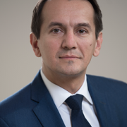
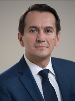
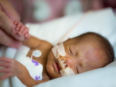
“For children who are at risk of losing their vision, this project will bring a window of opportunity for physicians to start treatment earlier and save their vision,” says Marius George Linguraru, DPhil, MA, MSc.
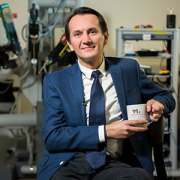
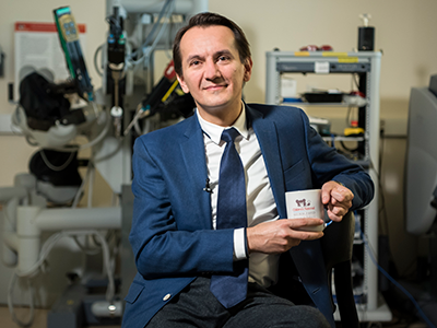
A team from Children’s National Hospital is part of a project receiving a two-year grant of nearly $1,000,000 from the National Institutes of Health (NIH) for the first pediatric project in the Quantitative Imaging Network (QIN) of the National Cancer Institute (NCI). Marius George Linguraru, DPhil, MA, MSc, principal investigator from the Sheikh Zayed Institute for Pediatric Surgical Innovation at Children’s National Hospital in Washington, D.C., is one of two principal investigators on the project, which focuses on developing quantitative imaging (QI) tools to improve pediatric tumor measurement, risk predictions and treatment response. Roger Packer, M.D., Senior Vice President of the Center for Neuroscience & Behavioral Health, Director of the Gilbert Neurofibromatosis Institute and Director of the Brain Tumor Institute, is co-investigator.
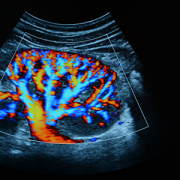
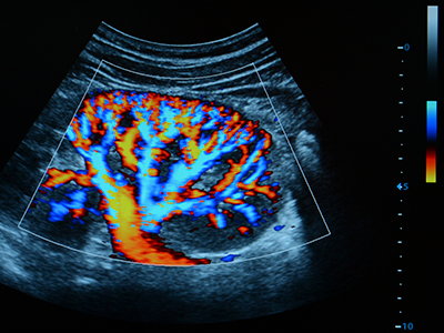
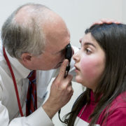
The project, in collaboration with Children’s Hospital of Philadelphia and Children’s Hospital Colorado, centers on the most common type of brain tumor in children, called a low-grade glioma. This project focuses on a clinically challenging group of children with neurofibromatosis type 1 (NF1), the most common inherited tumor predisposition syndrome. Nearly 20% of children with NF1 will develop a low-grade glioma called optic pathway glioma (OPG). In children with this type of brain tumor, the growth occurs around the optic nerve, chiasm and tracts, also called the optic pathway, which connects the eye to the brain. OPGs can cause vision loss and even blindness. Permanent vision loss usually occurs between one and eight years of age with doctors closely monitoring the tumor with magnetic resonance imaging (MRI) to assess the disease progression.
“Our traditional two-dimensional measures of tumor size are not appropriate to assess the changes in these amorphous tumors over time or how the tumor responds to treatment,” says Linguraru. “This means physicians have difficulty determining the size of the tumor as well as when treatment is working. Research such as this can lead to innovative medical technologies that can improve and possibly change the fate of children’s lives.”
Dr. Linguraru is leading the technical trials on this project, which take place in the first two years, or phase one, starting in June 2020. Phase one focuses on improving the often inaccurate human measurements of tumor size by developing QI tools to make precise and automated measures of tumor volume and shape using machine learning. In this phase, the project will use and homogenize MRI data from multiple centers to develop predictive models of the treatment response based on the tumor volume that are agnostic to the differences in imaging protocols. By doing this, it will allow physicians to make more informed decisions about the treatment’s success and whether the child will recover their vision.
When phase one is complete, Linguraru and the project’s other principal investigator Robert A. Avery, DO, MSCE, neuro-ophthalmologist in the Division of Ophthalmology at Children’s Hospital of Philadelphia, will initiate the second phase, which includes validating the QI application on data from the first ever phase III clinical trial comparing two treatments for NF1-OPGs. Phase two is scheduled to start in the Summer 2022 and continue through Summer 2025.
“For children who are at risk of losing their vision, this project will bring a window of opportunity for physicians to start treatment earlier and save their vision,” says Linguraru. “For those children who won’t benefit from chemotherapy because the tumor poses no threat to their sight, this project will save them from having to go through that difficult treatment unnecessarily. It will be life-changing for the children and their families, which is what excites me about this QI application.”
This project is a collaboration between Children’s Hospital of Philadelphia and Children’s National Hospital in Washington, D.C., in partnership with Children’s Hospital of Colorado and University of Pennsylvania. Upon project completion, the QI application will provide a precision-medicine approach for NF1-OPGs and improve clinical outcomes for pediatric tumors.