Tag Archive for: immune system

Hodgkin lymphoma cells

Clinical Trial Spotlight: Can Nivolumab make cellular therapy more effective for treating relapsed lymphomas?

Hodgkin lymphoma cells

Each year, about 9,000 new patients are diagnosed with Hodgkin lymphoma, 10-15% of them children.

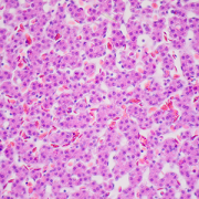
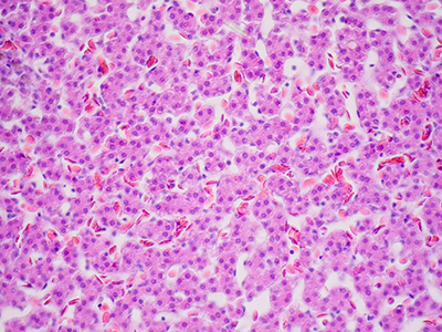
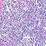
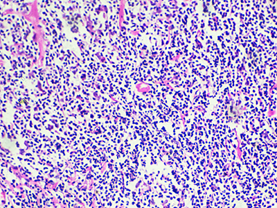
Each year, about 9,000 new patients are diagnosed with Hodgkin lymphoma, 10-15% of them children. Despite a relatively high cure rate for children with Hodgkin lymphoma, there are many debilitating long-term side effects of the treatments currently used. Additionally, 15-20% of children have a relapse and only half of them experience a long-term cure. Diffuse large B cell Lymphomas are another type of aggressive lymphoma that are difficult to cure, especially when they do not respond to upfront chemotherapy (refractory). Patients who experience relapse have to undergo more intensive chemotherapy followed by autologous stem cell transplantation and yet often times their lymphoma comes back.

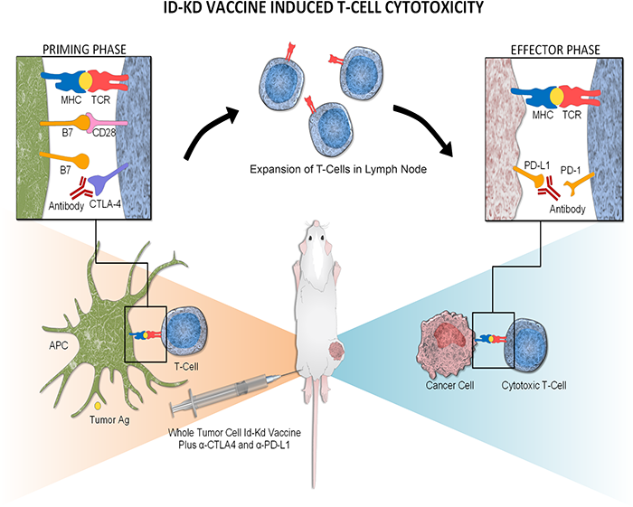
Physicians at Children’s National Hospital, in partnership with the Huntsman Cancer Institute at the University of Utah School of Medicine, are enrolling patients in a clinical trial to test the safety of administering PD-1 inhibitor Nivolumab given prior to and following the infusions of the patients’ own TAA-T cells which have been trained to target tumor cells in the laboratory. Nivolumab is currently approved by the FDA for relapsed Hodgkin lymphoma. Nivolumab acts by unleashing the brakes put on by the lymphoma cells, and by doing so, Nivolumab allows the immune system to overcome the tumor’s escape mechanism.

“We believe that if our T cells are deemed safe when given in combination with already approved drugs, we may be able to impact multiple lives and reduce long-term toxicities from conventional chemotherapies,” said Hema Dave, M.D., an oncologist at Children’s National. “We’re hopeful that combination immunotherapies will produce more durable responses than when immunotherapies are given alone as a single agent and, additionally, that they will reduce the use of cytotoxic chemotherapy.”

The investigators will collect blood from the patients to isolate peripheral blood mononuclear cells. They will then make special cells called dendritic cells to stimulate the T cells. Then they will add special mixtures of tumor proteins WT1, PRAME and Survivin and provide a cytokine milieu favorable to T cell expansion/activation, inducing selective expansion of T cells targeted to kill tumor cells. This process trains the T cells to recognize the tumor proteins and become specialized TAA-T cells. The cells will be grown and frozen until ready for use. While the T cells are growing, the patients will be given Nivolumab.

“We’re really trying to test if priming the patients with Nivolumab will make their T cells more effective when they get infused,” says Dr. Dave. “The Nivolumab will help prepare the immune system. Then, when we infuse the T cells, our hope is that the environment is primed for the T cells to expand, grow and work to attack the cancer. If we can prime the immune system and make it more conducive for the T cells, then maybe they will have a better chance to get to the lymphoma cells and thus have a more sustained response.”

Patients will then receive two infusions of the TAA-T cells and be monitored for side effects. The anticipated enrollment is 18 patients over the next 2-3 years. If there is a positive response in patients enrolled in this safety trial, it could expand to test for efficacy of the novel combination immunotherapy.

Phase 1 Study Utilizing Tumor Associated Antigen Specific T Cells (TAA-T) with PD1 Inhibitor Nivolumab for Relapsed/Refractory Lymphoma

  • PI: Hema Dave, M.D.
  • Status: Recruiting

For more information about this trial, contact:

Hema Dave, M.D.
202-476-6397
hkdave@childrensnational.org

Fahmida Hoq, MBBS, MS
202-476-3634
fhoq@childrensnational.org

Click here to view Open Phase 1 and 2 Cancer Clinical Trials at Children’s National.

The Children’s National Center for Cancer and Blood Disorders is committed to providing the best care for pediatric patients. Our experts play an active role in innovative clinical trials to advance pediatric cancer care. We offer access to novel trials and therapies, some of which are only available here at Children’s National. With research interests covering nearly every aspect of pediatric cancer care, our work is making great advancements in childhood cancer.

Vote for STAT Madness

It’s a three-peat! Children’s National again competes in STAT Madness

Vote for STAT Madness

Children’s National Hospital collects patients’ blood, extracts T-cells and replicates them in the presence of specific proteins found on cancer cells which, in essence, teaches the T-cells to target specific tumor markers. Training the T-cells, growing them to sufficient quantities and ensuring they are safe for administration takes weeks. But when patients return to the outpatient clinic, their T-cell infusion lasts just a few minutes.

For the third consecutive year, Children’s National was selected to compete in STAT Madness, an annual bracket-style competition that chooses the year’s most impactful biomedical innovation by popular vote. Children’s entry, “Immunotherapy of relapsed and refractory solid tumors with ex vivo expanded multi-tumor associated antigen specific cytotoxic T lymphocytes,” uses the body’s own immune system to attack and eliminate cancer cells in pediatric and adult patients with solid tumor malignancies.

In 2018, Children’s first-ever STAT Madness entry advanced through five brackets in the national competition and, in the championship round, finished second. That innovation, which enables more timely diagnoses of rare diseases and common genetic disorders, helping to improve kids’ health outcomes around the world, also was among four “Editor’s Pick” finalists, entries that spanned a diverse range of scientific disciplines.

An estimated 11,000 new cases of pediatric cancer were diagnosed in children 14 and younger in the U.S. in 2019. And, when it comes to disease, cancer remains the leading cause of death among children, according to the National Institutes of Health. An enterprising research team led by Children’s National faculty leveraged T-cells – essential players in the body’s immune system – to treat pediatric and adult patients with relapsed or refractory solid tumors who had exhausted all other therapeutic options.

“We’re using the patient’s own immune system to fight their cancer, rather than more traditional chemotherapy drugs,” says Catherine M. Bollard, M.D., director of the Center for Cancer & Immunology Research at Children’s National and co-senior author of the study. “It’s more targeted and less toxic to the patient. These T-cells home in on any cancer cells that might be in the body, allowing healthy cells to continue to grow,” Dr. Bollard adds.

That means patients treated in the Phase I, first-in-human trial didn’t lose their hair and weren’t hospitalized for the treatment. After a quick clinical visit for their treatment, they returned to normal activities, like school, with good energy levels.

“With our specially trained T-cell therapy, many patients who previously had rapidly progressing disease experienced prolonged disease stabilization,” says Holly J. Meany, M.D., a Children’s National oncologist and the study’s co-senior author. “Patients treated at the highest dose level showed the best clinical outcomes, with a six-month, progression-free survival of 73% after tumor-associated antigen cytotoxic T-cell (TAA-T) infusion, compared with 38% with their immediate prior therapy.”

The multi-institutional team published their findings from the study online July 29, 2019, in the Journal of Clinical Oncology.

“Our research team and our parents are delighted that some patients treated in our study continue to do well following T-cell therapy without additional treatment. In some cases, two years after treatment, patients do not appear to have active disease and are maintaining an excellent quality of life,” says Amy B. Hont, M.D., the study’s lead author. “One of these was a patient whose parents were told his only other option was palliative care. Our innovation gives these families new hope,” Dr. Hont adds.

The 2020 STAT Madness #Core64 bracket opened March 2, and the champion will be announced April 6.

In addition to Drs. Hont, Meany and Bollard, Children’s National co-authors include C. Russell Cruz, M.D., Ph.D., Robert Ulrey, MS, Barbara O’Brien, BS, Maja Stanojevic, M.D., Anushree Datar, MS, Shuroug Albihani, MS, Devin Saunders, BA, Ryo Hanajiri, M.D., Ph.D., Karuna Panchapakesan, MS, Payal Banerjee, MS, Maria Fernanda Fortiz, BS, Fahmida Hoq, MBBS, MS, Haili Lang, M.D., Yunfei Wang, DrPH, Patrick J. Hanley, Ph.D., and Jeffrey S. Dome, M.D., Ph.D.; and Sam Darko, MS, National Institute of Allergy and Infectious Diseases.

Financial support for the research described in this post was provided by the Children’s National Hospital Heroes Gala, Alex’s Army Foundation, the Children’s National Board of Visitors and Hyundai Hope on Wheels Young Investigator Grant to Support Pediatric Cancer Research, the Children’s National Research Institute Bioinformatics Unit, the Clinical and Translational Science Institute and the National Institutes of Health under award No. UL1-TR001876.

Hepatocytes

H-IPSE internalized by just a limited range of cells

Hepatocytes

A team led by Children’s National Hospital found that H-IPSE is internalized by just a limited range of cells, including hepatocytes.

Schistosoma mansoni is a parasite that hides out in snails, breaks free into waterways, and then infects humans, spending much of its life inside blood vessels, laying eggs and jeopardizing public health when those eggs are excreted in urine or feces. As parasitic diseases go, the ailment it causes, Schistosomiasis, is second only to malaria in global impact, according to the Centers for Disease Control and Prevention.

In order to elude the human host’s defenses, S. mansoni uses self-defense tactics that researchers are trying to better understand in order to outmaneuver the parasite. A research team led by Children’s National Hospital is trying to tease out the multiple steps that enable this parasite to reproduce and generate millions of eggs without killing its host.

The parasite’s eggs secrete a number of proteins, with IPSE as one of the most abundant, the team recently presented during the American Society of Tropical Medicine and Hygiene 2019 annual meeting. That protein binds immunoglobulin, which induces basophils and mast cells to release IL-4. After sequestering chemokines, H-IPSE infiltrates the cell nucleus (thus H-IPSE is called an infiltrin), modulating gene expression.

“H-IPSE tips the immune system balance, making it more likely to trigger a Th2 anti-inflammatory response,” says Michael Hsieh, M.D., Ph.D., director of transitional urology at Children’s National and the research project’s senior author. “It downregulates pro-inflammatory pathways, but we wanted to know more about which specific human cells it targets.”

Using Trypan Blue, a stain that selectively colors certain cells bright blue, they solved the mystery, finding that H-IPSE is internalized by just a limited range of cells. What’s more, some cell types, like urothelial cells and hepatocytes (the liver’s chief functioning cells, which activate innate immunity), are more susceptible than neurons, endothelial cells or immature dendritic cells.

In addition to Dr. Hsieh, presentation co-authors include Olivia Lamanna, Evaristus Mbanefo and Kenji Ishida, all of Children’s National; Franco Falcone, of University of Nottingham; and Theodore Jardetzky and Luke Pennington, of Stanford University.

t-cells

Tailored T-cell therapies neutralize viruses that threaten kids with PID

t-cells

Tailored T-cells specially designed to combat a half dozen viruses are safe and may be effective in preventing and treating multiple viral infections, according to research led by Children’s National Hospital faculty.

Catherine Bollard, M.B.Ch.B., M.D., director of the Center for Cancer and Immunology Research at Children’s National and the study’s senior author, presented the teams’ findings Nov. 8, 2019, during a second-annual symposium jointly held by Children’s National and the National Institute of Allergy and Infectious Diseases (NIAID), part of the National Institutes of Health (NIH). Children’s National and NIAID formed a research partnership in 2017 to develop and conduct collaborative clinical research studies focused on young children with allergic, immunologic, infectious and inflammatory diseases. Each year, they co-host a symposium to exchange their latest research findings.

According to the NIH, more than 200 forms of primary immune deficiency diseases impact about 500,000 people in the U.S. These rare, genetic diseases so impair the person’s immune system that they experience repeated and sometimes rare infections that can be life threatening. After a hematopoietic stem cell transplantation, brand new stem cells can rebuild the person’s missing or impaired immune system. However, during the window in which the immune system rebuilds, patients can be vulnerable to a host of viral infections.

Because viral infections can be controlled by T-cells, the body’s infection-fighting white blood cells, the Children’s National first-in-humans Phase 1 dose escalation trial aimed to determine the safety of T-cells with antiviral activity against a half dozen opportunistic viruses: adenovirus, BK virus, cytomegalovirus (CMV), Epstein-Barr virus (EBV), Human Herpesvirus 6 and human parainfluenza-3 (HPIV3).

Eight patients received the hexa-valent, virus-specific T-cells after their stem cell transplants:

  • Three patients were treated for active CMV, and the T-cells resolved their viremia.
  • Two patients treated for active BK virus had complete symptom resolution, while one had hemorrhagic cystitis resolved but had fluctuating viral loads in their blood and urine.
  • Of two patients treated prophylactically, one developed EBV viremia that was treated with rituximab.

Two additional patients received the T-cell treatments under expanded access for emergency treatment, one for disseminated adenoviremia and the other for HPIV3 pneumonia. While these critically ill patients had partial clinical improvement, they were being treated with steroids which may have dampened their antiviral responses.

“These preliminary results show that hexaviral-specific, virus-specific T-cells are safe and may be effective in preventing and treating multiple viral infections,” says Michael Keller, M.D., a pediatric immunologist at Children’s National and the lead study author. “Of note, enzyme-linked immune absorbent spot assays showed evidence of antiviral T-cell activity by three months post infusion in three of four patients who could be evaluated and expansion was detectable in two patients.”

In addition to Drs. Bollard and Keller, additional study authors include Katherine Harris M.D.; Patrick J. Hanley Ph.D., assistant research professor in the Center for Cancer and Immunology; Allistair Abraham, M.D., a blood and marrow transplantation specialist; Blachy J. Dávila Saldaña, M.D., Division of Blood and Marrow Transplantation; Nan Zhang Ph.D.; Gelina Sani BS; Haili Lang MS; Richard Childs M.D.; and Richard Jones M.D.

###

Children’s National-NIAID 2019 symposium presentations

“Welcome and introduction”
H. Clifford Lane, M.D., director of NIAID’s Division of Clinical Research

“Lessons and benefits from collaboration between the NIH and a free-standing children’s hospital”
Marshall L. Summar, M.D., director, Rare Disease Institute, Children’s National

“The hereditary disorders of PropionylCoA and Cobalamin Metabolism – past, present and future”
Charles P. Venditti, M.D., Ph.D., National Human Genome Research Institute Collaboration

“The road(s) to genetic precision therapeutics in pediatric neuromuscular disease: opportunities and challenges”
Carsten G. Bönnemann, M.D., National Institute of Neurological Disorders and Stroke

“Genomic diagnostics in immunologic diseases”
Helen Su, M.D., Ph.D., National Institute of Allergy and Infectious Diseases

“Update on outcomes of gene therapy clinical trials for X-SCID and X-CGD and plans for future trials”
Harry Malech, M.D., National Institute of Allergy and Infectious Diseases

“Virus-specific T-cell therapies: broadening applicability for PID patients”
Catherine Bollard, M.D., Children’s National 

“Using genetic testing to guide therapeutic decisions in Primary Immune Deficiency Disease”
Vanessa Bundy, M.D., Ph.D., Children’s National 

Panel discussion moderated by Lisa M. Guay-Woodford, M.D.
Drs. Su, Malech, Bollard and Bundy
Morgan Similuk, S.C.M., NIAID
Maren Chamorro, Parent Advocate

“Underlying mechanisms of pediatric food allergy: focus on B cells
Adora Lin, M.D., Ph.D., Children’s National 

“Pediatric Lyme outcomes study – interim update”
Roberta L. DeBiasi, M.D., MS, Children’s National 

“Molecular drivers and opportunities in neuroimmune conditions of pediatric onset”
Elizabeth Wells, M.D., Children’s National 


Epstein Barr virus

Fighting lymphoma with targeted T-cells

Epstein-Barr virus

The Epstein-Barr virus (EBV) is best known as the cause of mononucleosis, the ubiquitous “kissing disease” that most people contract at some point in their life. But in rare instances, this virus plays a more sinister role as the impetus of lymphomas, cancers that affect the white blood cells known as lymphocytes.

The Epstein-Barr virus (EBV) is best known as the cause of mononucleosis, the ubiquitous “kissing disease” that most people contract at some point in their life. But in rare instances, this virus plays a more sinister role as the impetus of lymphomas, cancers that affect the white blood cells known as lymphocytes. EBV-associated lymphomas account for about 40% of Hodgkin lymphomas, 20% of diffuse large B-cell lymphomas, and more than 90% of natural killer/T-cell lymphomas. This latter type of lymphoma typically has a very poor prognosis even with the “standard of care” lymphoma treatments such as chemotherapy and/or radiation.

When these interventions fail, the only curative approach is an allogeneic  hematopoietic stem cell transplant from a healthy donor, a treatment that’s tough on patients’ bodies and carries significant risks, says Lauren P. McLaughlin, M.D., a pediatrician specializing in hematology and oncology at Children’s National in Washington, D.C. Patients who receive these allogenic transplants are immune-compromised until the donor cells engraft; the grafts can attack patients’ healthy cells in a phenomenon called graft versus host disease; and if patients relapse or don’t respond to this treatment, few options remain.

To help improve outcomes, Dr. McLaughlin and colleagues tested an addition to the allogeneic hematopoietic stem cell transplant procedure for patients with EBV-associated lymphomas: infusion of a type of immune cell called T cells specifically trained to fight cells infected with EBV.

Dr. McLaughlin, along with Senior Author Catherine M. Bollard, M.D., M.B.Ch.B., director of the Center for Cancer and Immunology Research and the Program for Cell Enhancement and Technologies for Immunotherapy at Children’s National, and colleagues tested this therapy in 26 patients treated at Children’s National or Baylor College of Medicine. They published these results online on Sept. 27, 2018, in the journal Blood. The study was a Phase I clinical trial, meaning that the therapy was tested primarily for safety, with efficacy as a secondary aim.

Seven patients who received the therapy had active disease that had not responded to conventional therapies. The other 19 were patients deemed to be at high risk for relapse.

Before each patient received their stem cell transplant, their donors gave an additional blood sample to generate the cancer-fighting T cells. Over the next 8 to 10 weeks, the researchers painstakingly manufactured the immune cells known as T-cells that specifically targeted EBV, growing these cells into numbers large enough for clinical use. Then, as early as 30 days after transplant, the researchers infused these T-cells into patients administering at least two doses, spaced two weeks apart.

Over the next several weeks, the researchers at CNMC and Baylor College of Medicine monitored patients with comprehensive exams to see how they fared after these transplants. The results showed that adverse effects from the treatment were exceedingly rare. There were no immediate infusion-related toxicities to the T-cell therapy and only one incident of dose-limiting toxicity.

This therapy may be efficacious, depending on the individual patients’ circumstances, Dr McLaughlin adds. For those in complete remission but at high risk of relapsing, the two-year survival rate was 78%, suggesting that the administration of this novel T-cell therapy may give the immune system a boost to prevent the lymphoma from returning after transplant. For patients with active T-cell lymphomas, two-year survival rates were 60%. However, even these lower rates are better than the historical norm of 30-50%, suggesting that the targeted T-cell therapies could help fight disease in patients with this poor prognosis lymphoma.

Dr. McLaughlin, the study’s lead author and a Lymphoma Research Foundation grantee, notes that researchers have more work to do before this treatment becomes mainstream. For example, this treatment will need to be tested in larger populations of patients with EBV-related lymphoma to determine who would derive the most benefit, the ideal dose and dose timing. It also may be possible to extend targeted T-cell treatments like this to other types of cancers. In the future, Dr. McLaughlin adds, it may be possible to develop T-cells that could be used “off the shelf”—in other words, they wouldn’t need to come from a matched donor and would be ready to use whenever a recipient needs them. Another future goal is using this therapy as one of the first lines of treatment rather than as a last resort.

“Our ultimate goal is to find a way to avoid chemotherapy and/or radiation therapy while still effectively treating a patient’s cancer,” she says. “Can you use the immune system to do that job? We’re working to answer that question.”

In addition to Drs. McLaughlin and Bollard, study co-authors include Rayne Rouce, Stephen Gottschalk, Vicky Torrano, George Carrum, Andrea M. Marcogliese, Bambi Grilley, Adrian P. Gee, Malcolm K. Brenner, Cliona M. Rooney and Helen E. Heslop, all of Baylor College of Medicine; Meng-Fen Wu from the Dan L. Duncan Comprehensive Cancer Center; and Fahmida Hoq and Patrick J. Hanley, Ph.D. from Children’s National in Washington, D.C.

ID-KD vaccine induced T-cell cytotoxicity

Fighting lethal cancer with a one-two punch

The immune system is the ultimate yin and yang, explains Anthony D. Sandler, M.D., senior vice president and surgeon-in-chief of the Joseph E. Robert Jr. Center for Surgical Care at Children’s National in Washington, D.C. With an ineffective immune system, infections such as the flu or diarrheal illness can run unchecked, causing devastating destruction. But on the other hand, excess immune activity leads to autoimmune diseases, such as lupus or multiple sclerosis. Thus, the immune system has “checks and balances” to stay controlled.

Cancer takes advantage of “the checks and balances,” harnessing the natural brakes that the immune system puts in place to avoid overactivity. As the cancer advances, molecular signals from tumor cells themselves turn on these natural checkpoints, allowing cancers to evade immune attack.

Several years ago, a breakthrough in pharmaceutical science led to a new class of drugs called checkpoint inhibitors. These medicines take those proverbial brakes off the immune system, allowing it to vigorously attack malignancies. However, Dr. Sandler says, these drugs have not worked uniformly and in some cancers, they barely work at all against the cancer.

One of these non-responders is high risk neuroblastoma, a common solid tumor found outside the skull in children. About 800 U.S. children are diagnosed with this cancer every year. And kids who have the high-risk form of neuroblastoma have poor prognoses, regardless of which treatments doctors use.

However, new research could lead to promising ways to fight high-risk neuroblastoma by enabling the immune system to recognize these tumors and spark an immune response. Dr. Sandler and colleagues recently reported on these results in the Jan. 29, 2018, PLOS Medicine using an experimental model of the disease.

The researchers created this model by injecting the preclinical models with cancer cells from an experimental version of neuroblastoma. The researchers then waited several days for the tumors to grow. Samples of these tumors showed that they expressed a protein on their cell surfaces known as PD-L1, a protein that is also expressed in many other types of human cancers to evade immune system detection.

To thwart this protective feature, the researchers made a cancer vaccine by removing cells from the experimental model’s tumors and selectively turning off a gene known as Id2. Then, they irradiated them, a treatment that made these cells visible to the immune system but blocked the cells from dividing to avoid new tumors from developing.

They delivered these cells back to the experimental models, along with two different checkpoint inhibitor drugs – antibodies for proteins known as CLTA-4 and PD-L1 – over the course of three treatments, delivered every three days. Although most checkpoint inhibitors are administered over months to years, this treatment was short-term for the experimental models, Dr. Sandler explains. The preclinical models were completely finished with cancer treatment after just three doses.

Over the next few weeks, the researchers witnessed an astounding turnaround: While experimental models that hadn’t received any treatment uniformly died within 20 days, those that received the combined vaccine and checkpoint inhibitors were all cured of their disease. Furthermore, when the researchers challenged these preclinical models with new cancer cells six months later, no new tumors developed. In essence, Dr. Sandler says, the preclinical models had become immune to neuroblastoma.

Further studies on human patient tumors suggest that this could prove to be a promising treatment for children with high-risk neuroblastoma. The patient samples examined show that while tumors with a low risk profile are typically infiltrated with numerous immune cells, tumors that are high-risk are generally barren of immune cells. That means they’re unlikely to respond to checkpoint inhibiting drugs alone, which require a significant immune presence in the tumor microenvironment. However, Dr. Sandler says, activating an immune response with a custom-made vaccine from tumor cells could spur the immune response necessary to make these stubborn cancers respond to checkpoint inhibitors.

Dr. Sandler cautions that the exact vaccine treatment used in the study won’t be feasible for people. The protocol to make the tumor cells immunogenic is cumbersome and may not be applicable to gene targeting in human patients. However, he and his team are currently working on developing more feasible methods for crafting cancer vaccines for kids. They also have discovered a new immune checkpoint molecule that could make this approach even more effective.

“By letting immune cells do all the work we may eventually be able to provide hope for patients where there was little before,” Dr. Sandler says.

In addition to Dr. Sandler, study co-authors include Priya Srinivasan, Xiaofang Wu, Mousumi Basu and Christopher Rossi, all of the Joseph E. Robert Jr. Center for Surgical Care and The Sheikh Zayed Institute for Pediatric Surgical Innovation (SZI), at Children’s National in Washington, D.C.

Financial support for research described in this post was provided by the EVAN Foundation, the Catherine Blair foundation, the Michael Sandler Research Fund and SZI.

ID-KD vaccine induced T-cell cytotoxicity

Mechanism of Id2kd Neuro2a vaccination combined with α-CTLA-4 and α-PD-L1 immunotherapy in a neuroblastoma model. During a vaccine priming phase, CTLA-4 blockade enhances activation and proliferation of T-cells that express programmed cell death 1 (PD1) and migrate to the tumor. Programmed cell death-ligand 1 (PD-L1) is up-regulated on the tumor cells, inducing adaptive resistance. Blocking PD-L1 allows for enhanced cytotoxic effector function of the CD8+ tumor-infiltrating lymphocytes. Artist: Olivia Abbate

Schistosoma haematobium egg

For hemorrhagic cystitis, harnessing the power of a parasite

Schistosoma haematobium egg

“Urogenital Schistosoma infestation, which is caused by S. haematobium, also causes hemorrhagic cystitis, likely by triggering inflammation when the parasite’s eggs are deposited in the bladder wall or as eggs pass from the bladder into the urinary stream. S. haematobium eggs secrete proteins, including IPSE, that ensure human hosts are not so sickened that they succumb to hemorrhagic cystitis,” says Michael H. Hsieh, M.D., Ph.D.

Every year, hundreds of thousands of U.S. patients – and even more throughout the world – are prescribed cyclophosphamide or ifosfamide. These two chemotherapy drugs can be life-saving for a wide range of pediatric cancers, including leukemias and cancers of the eyes and nerves. However, these therapies come with a serious side effect: Both cause hemorrhagic cystitis in up to 40 percent of patients. This debilitating condition is characterized by severe inflammation in the bladder that can cause tremendous pain, life-threatening bleeding, and frequent and urgent urination.

Infection with a parasitic worm called Schistosoma haematobium also causes hemorrhagic cystitis, but this organism has a fail-safe: To keep its host alive, the parasite secretes a protein that suppresses inflammation and the associated pain and bleeding.

In a new study, a Children’s-led research team harnessed this protein to serve as a new therapy for chemotherapy-induced hemorrhagic cystitis.

“Urogenital Schistosoma infestation, which is caused by S. haematobium, also causes hemorrhagic cystitis, likely by triggering inflammation when the parasite’s eggs are deposited in the bladder wall or as eggs pass from the bladder into the urinary stream. S. haematobium eggs secrete proteins, including IPSE, that ensure human hosts are not so sickened that they succumb to hemorrhagic cystitis,” says Michael H. Hsieh, M.D., Ph.D., senior author of the study published April 3, 2018, by The FASEB Journal. “This work in an experimental model is the first published report of exploiting an uropathogen-derived host modulatory molecule in a clinically relevant model of bladder disease, and it points to the potential utility of this as an alternate treatment approach.”

S. mansoni IPSE binds to Immunoglobulin E (IgE), an antibody produced by the immune system that is expressed on the surface of basophils, a type of immune cell; and mast cells, another immune cell that mediates inflammation; and sequesters chemokines, signaling proteins that alert white cells to infection sites. The team produced an ortholog of the uropathogen-derived protein. A single IV dose proved superior to multiple doses of 2-Mercaptoethane sulfonate sodium (MESNA), the current standard of care, in suppressing chemotherapy-induced bladder hemorrhaging in an experimental model. It was equally potent as MESNA in dampening chemotherapy-induced pain, the research team finds.

“The current array of medicines we use to treat hemorrhagic cystitis all have shortcomings, so there is a definite need for novel therapeutic options,” says Dr. Hsieh, a Children’s National Health System urologist. “And other ongoing research projects have the potential to further expand patients’ treatment options by leveraging other urogenital parasite-derived, immune-modulating molecules to treat inflammatory bowel diseases and autoimmune disorders.”

Future research will aim to describe the precise molecular mechanisms of action, as well as to generate other orthologs that boost efficacy while reducing side effects.

In addition to Dr. Hsieh, Children’s study co-authors include Lead Author, Evaristus C. Mbanefo; Loc Le and Luke F. Pennington; Justin I. Odegaard and Theodore S. Jardetzky, Stanford University; Abdulaziz Alouffi, King Abdulaziz City for Science and Technology; and Franco H. Falcone, University of Nottingham.

Financial support for this research was provided by National Institutes of Health under award number RO1-DK113504.

Adora Lin

Funding will help uncover immune system differences that trigger food allergies

Adora Lin

“When it comes to food allergies, we really don’t know how they develop. We don’t know how to best differentiate between a child who can safely eat a potential allergen, like peanuts, compared with a child who cannot safely eat peanuts.” says Adora A. Lin, M.D., Ph.D.

Adora A. Lin, M.D., Ph.D., an attending physician in Children’s department of Allergy and Immunology, was awarded $240,000 to improve understanding of how children’s immune systems tolerate or react to certain food allergens – sometimes triggering a cascade of side effects that can be fatal.

The three-year American Academy of Allergy, Asthma & Immunology (AAAAI) Foundation award will underwrite Dr. Lin’s ongoing research into the regulation of the antibody Immunoglobulin E (IgE), which plays a pivotal role in these allergic responses.

“Our immune system maintains a delicate balance, working just enough to ward off potential invaders and pathogens, but not so much that it triggers problems of its own making,” Dr. Lin says. “When it comes to food allergies, we really don’t know how they develop. We don’t know how to best differentiate between a child who can safely eat a potential allergen, like peanuts, compared with a child who cannot safely eat peanuts.”

Food allergies have become a growing problem and affect about 1 in 13 U.S. children, or about two per classroom. Food items such as eggs, milk, peanuts, tree nuts, soy and wheat trigger allergic reactions that can include itching, swelling, hives and difficulty breathing. As children’s immune systems react to exposure to such allergens, their B-cells produce IgE antibodies.

Apart from avoiding these foods and carrying rescue medications, which must be used immediately after accidental exposure, there is no way to treat food allergies effectively. That makes it essential to better understand how the immune system works in order to innovate new and better food allergy treatments and diagnostics.

Dr. Lin’s work involves isolating immune cells from blood samples, culturing them and stimulating an immune response to known food allergy triggers. B-cells make IgE, but additional clarity is needed about what turns on the “make IgE” signal as well as which signals indicate it’s time to stop making IgE. Ultimately, the aim is to identify biomarkers that are akin to the “check engine” light that illuminates to warn of a potential problem long before a car stalls in traffic.

“I’m very excited about this funding,” Dr. Lin adds. “Our field has done an exceptional job with clinical work to help children with food allergies. This award recognizes the importance of the mechanistic side of the equation. I’m excited to help make that contribution to the research.”

As it stands now, blood tests are sensitive to food-related IgE, but are not specific. Only 30 to 55 percent of children who have IgE to common food allergens have an allergic reaction after eating the food, which means that 45 to 70 percent are merely sensitized and could tolerate eating the food. Current tests cannot distinguish between sensitized and allergic children.

“Our hope is to identify biomarkers that would serve as the ‘check engine’ light that tell us in advance which child’s immune system will react strongly to that food. Right now, there is no way to tell. This project will help uncover those differences,” she says.

Dr. Lin was one of three recipients of the AAAAI Foundation’s faculty development award, which was presented during a March 3, 2018, award ceremony held during the organization’s business meeting.