Tag Archive for: hsieh

pregnant woman at fertility consultant

Fertility preservation in sickle cell disease patients

pregnant woman at fertility consultant

Fertility is a long-standing concern for patients with sickle cell disease and their families.

In a recent review in the Journal of Clinical Medicine, researchers from Children’s National Hospital look at the current state of fertility preservation in patients with sickle cell disease and make recommendations for longitudinal post-treatment for these individuals.

Fertility is a long-standing concern for patients with sickle cell disease and their families. Current curative therapy for the disease requires gonadotoxic conditioning, which many patients resist because of the resulting risk of infertility. And, while standard fertility preserving interventions exist for pre- and postpubescent females and males, best practices for integrating these interventions into sickle cell disease care have not yet been established.

In their article, Children’s National hematologist Robert Sheppard Nickel, M.D., and co-authors review current fertility assessments, fertility considerations in pre- and post-transplant patients with sickle cell disease and fertility preserving interventions for patients. The authors conclude that in the future, less toxic curative approaches may make fertility preservation unnecessary, but at present, fertility preservation should be offered to patients with sickle cell disease pursing curative therapy.

Additional authors from Children’s National include Michael Hsieh, M.D., Ph.D., and Jacqueline Maher, M.D.

Read the full review article, Fertility after Curative Therapy for Sickle Cell Disease: a Comprehensive Review to Guide Care, in the Journal of Clinical Medicine.

kidneys with science images

PMA-based PCR amplifies DNA from only live bacteria in urine

kidneys with science images

The question of why urinary tract infection (UTI) symptoms can persist in some patients who have been seemingly appropriately treated with antibiotics and have negative post-antibiotic urine cultures is one that urologists have long sought to answer.

Experts at Children’s National Hospital have successfully developed propidium monoazide (PMA)-based polymerase chain reaction (PCR) methods that amplify DNA from only live bacteria in urine. The study’s results, published in Frontiers, suggest that non-PMA bound DNA from live bacteria can be present in urine, even after antibiotic treatment.

PMA has been shown to differentiate between non-viable and viable bacteria in various settings. However, its effectiveness in urine has not been previously studied.

The question of why urinary tract infection (UTI) symptoms can persist in some patients who have been seemingly appropriately treated with antibiotics and have negative post-antibiotic urine cultures is one that urologists have long sought to answer.

“One theory is that very low levels of bacteria that don’t show up on cultures may be the cause,” says Michael Hsieh, M.D., director of Transitional Urology at Children’s National and senior author of the study.

Although PCR has previously been used to try and confirm this theory, the use of this method has been criticized because PCR can amplify DNA from dead bacteria (which obviously don’t cause UTI).

The authors developed a PCR test that selectively detects DNA from live bacteria. In a preclinical setting, results show that with the use of antibiotics, cultures collected can be negative but urine can contain DNA from live bacteria, as detected using the PCR test.

“We think something similar can occur in patients and we show some data in the paper confirming the PCR test can work with patient samples,” Dr. Hsieh adds. “I’m excited that we might finally have an explanation as to why some patients have persistent UTI symptoms after antibiotic treatment.”

Namely, he adds, that these patients still have a UTI. This may lead to better follow-up management of patients with UTI. The next step is to confirm the theory in patients.

surgeon doing laparoscopic surgery

Autonomous robotic laparoscopic surgery for intestinal anastomosis

surgeon doing laparoscopic surgery

Children’s National Hospital in collaboration with the University of North Carolina Wilmington and Johns Hopkins University developed an enhanced autonomous strategy for laparoscopic soft tissue surgery.

A new approach to soft tissue surgery could simplify autonomous surgical planning and enable collaborative surgery between an autonomous robot and human, a new study published in Science Robotics finds. This is the first time a robot can complete an autonomous soft tissue surgical task under laparoscopic conditions, forming the foundation for future soft tissue surgeries.

Children’s National Hospital in collaboration with the University of North Carolina Wilmington and Johns Hopkins University developed an enhanced autonomous strategy for laparoscopic soft tissue surgery. The multi-institutional effort made it possible to perform a robotic laparoscopic small bowel anastomosis in phantom and in vivo intestinal tissues. The findings further suggest that autonomous robot-assisted surgery has the potential to provide more efficacy, safety and consistency independent of an individual surgeon’s skill and experience.

The hold-up in the field

Autonomous anastomosis is known to be a challenging soft tissue surgery task. And in the laparoscopic setting, surgeries like these prove to be more challenging because of the need for high maneuverability and repeatability under motion and vision constraints – especially in pediatric patients.

“This work represents the first time autonomous soft tissue surgery has been performed using a laparoscopic technique and is the first step in bridging the gap between human and machine towards completing autonomous surgical tasks in soft tissue surgeries,” says Hamed Saeidi, Ph.D., assistant professor at University of North Carolina Wilmington and lead author of the study.

To overcome the unpredictable motions of the tissue, the experts used machine learning based techniques to track the dynamic motions of the soft tissue during the surgery. These methods also pave the way for markerless methods for tracking the tissue motion in future surgeries.

“Until now, laparoscopic autonomous surgeries were not possible in soft tissue due to the unpredictable motions of the tissue and limitations on the size and capabilities of surgical tools,” says Justin Opfermann, M.S., Ph.D., student and Johns Hopkins University and co-author.

What’s unique

Performing autonomous surgery would require the development of novel suturing tools, imaging systems and robotic controls to visualize a surgical scene, generate an optimized surgical plan and then execute that surgical plan with the highest precision.

The autonomous robot takes its skill one step further when performing surgical tasks on soft tissues by enabling a robot-human collaboration to complete more complicated surgical tasks where preoperative planning is not possible.

Additionally, the robot used in this work uses a novel shared control scheme called “conditional autonomy,” whereby the robot performs the majority of the surgical task, which the surgeon oversees.

Bottom line

“Combining all of these features into a single system is non-trivial,” Opfermann adds. “In 2016, we were the first group to demonstrate feasibility of semi-autonomous small bowel anastomosis with a robot in soft tissue, and now we can perform autonomous laparoscopic anastomosis.”

The resulting anastomosis had more consistency and achieved higher burst strength than surgeons suturing with manual technique, resulting in less anastomotic leak.

In laparoscopic surgeries – and pediatric patients especially – these challenges are even more difficult due to the small size of the patient. Robotic anastomosis is one way to ensure that surgical tasks that require high precision and repeatability can be performed with more accuracy and precision in every patient independent of surgeon skill.

“As a surgeon, I can attest to the potential benefits of improving how we perform surgery on our patients,” says Michael Hsieh, M.D., Ph.D., director of Transitional Urology at Children’s National Hospital. “Working with my engineering colleagues at Johns Hopkins, we’ve been able to develop prototypes of supervised, autonomous suturing robots that may be a step towards such improvements.”

Micrograph of human parasite Schistosoma mansonii

Diagnosing and monitoring of urogenital schistosomiasis

Micrograph of human parasite Schistosoma mansonii

Urogenital schistosomiasis (UGS) is caused by egg-laying S. haematobium worms dwelling within the veins draining the main pelvic organs, including the bladder, uterus, and cervix.

Although urogenital schistosomiasis remains a major global challenge, Michael Hsieh, M.D., Ph.D., director of Transitional Urology at Children’s National Hospital, and other experts including Eglal Shalaby Rana, M.D., from Children’s National, show in a new study published in Advances in Parasitology that newer refinements in associated technologies may lead to improvements in patient care.

In addition, application of investigational imaging methods, such as confocal laser endomicroscopy and two-photon microscopy in urogenital schistosomiasis, are likely to contribute to the understanding of this infection’s pathogenesis.

Read the full study in Advances in Parasitology.

conceptual image of bladder cancer

Sensitivity to physical versus chemical factors in CAP

conceptual image of bladder cancer

To date, reactive oxygen species and reactive nitrogen species have been regarded as the key factors causing the observable cellular death of cold atmospheric plasma (CAP)-treated cancer cells. The chemical basis of the conventional CAP treatment highlights apoptosis as the main CAP-triggered cell death mechanism.

However, in a recent study published in the Journal of Physics, Michael Hsieh, M.D., Ph.D., director of Transitional Urology at Children’s National Hospital, and other experts demonstrated a strong anti-melanoma effect based on physically-based CAP treatment. The study, which also tested bladder cancer, compared the anti-cancer effect of chemically-based versus physically-based CAP treatment on four typical cancer cell lines in vitro.

Illustration of Bifidobacterium

Probiotic use in pediatric medicine

Illustration of Bifidobacterium

Probiotics have received significant attention within both the scientific and lay communities for their potential health-promoting properties, including the treatment or prevention of various conditions in children.

In a recent article published by Pediatric Research, Michael Hsieh, M.D., Ph.D., director of Transitional Urology at Children’s National Hospital, and other experts review the published data on use of specific probiotic strains for three common pediatric conditions: the prevention of urinary tract infections and antibiotic-associated diarrhea and the treatment of atopic dermatitis.

schistosome

Parasite-derived molecule could accelerate recovery from UTI

schistosome

Eggs from S. haematobium may produce the molecule IPSE to reduce the immune response against them, which happens to dampen UTI-induced bladder inflammation.

IPSE, a urogenital parasite-derived immunomodulatory molecule, can suppress bladder pathogenesis and anti-microbial peptide gene expression in bacterial urinary tract infection (UTI) according to a new study led by Michael Hsieh, Ph.D., director of Transitional Urology at Children’s National Hospital.

Half of all girls and women, and about 5% of boys and men, will have at least one urinary tract infection (UTI) in their lifetimes.

“Although antibiotics are very helpful for these infections, there are concerns that overuse of antibiotics may contribute to antibiotic-resistant infections,” Dr. Hsieh said. “There are also concerns that antibiotic therapy for UTI does not uniformly resolve infection-induced or inflammation-associated symptoms quickly.”

Parasitic infections are often associated with bacterial co-infections for unclear reasons. This may be true for urogenital schistosomiasis (caused by Schistosoma haematobium infection) and bacterial urinary tract co-infection (UTI), the study noted. Dr. Hsieh and other leading experts previously reported that this co-infection is facilitated by S. haematobium eggs triggering interleukin-4 (IL-4) production and sought to dissect the underlying mechanisms.

“Despite S. haematobium’s ability to make hosts more susceptible to UTI, we have identified IPSE, a bladder parasite protein, as a potential anti-inflammatory agent to accelerate recovery from UTI,” Dr. Hsieh explained. “S. haematobium eggs may produce IPSE to reduce the immune response against them, which happens to dampen UTI-induced bladder inflammation. It may be possible to develop IPSE as novel therapeutic to accelerate recovery from UTI.”

The study’s data showed that IPSE may play a major role in S. haematobium-associated urinary tract co-infection, although in an unexpected way. The study’s findings also indicated that IPSE either works in concert with other IL-4 -inducing factors to increase susceptibility of S. haematobium-infected hosts to bacterial co-infection or does not contribute to enchaining vulnerability to this co-infection.

You can find the full study published in Parasites and Vectors. Learn more about the Children’s National Department of Urology.

Schistosoma

Parasitic eggs trigger upregulation in genes associated with inflammation

Schistosoma

Of the 200 million people around the globe infected with Schistosomiasis, about 100 million of them were sickened by the parasite Schistosoma haematobium.

Of the 200 million people around the globe infected with Schistosomiasis, about 100 million of them were sickened by the parasite Schistosoma haematobium. As the body reacts to millions of eggs laid by the blood flukes, people can develop fever, cough and abdominal pain, according to the Centers for Disease Control and Prevention. Schistosomiasis triggered by S. haematobium can also include hematuria, bladder calcification and bladder cancer.

Despite the prevalence of this disease, there are few experimental models specifically designed to study it, and some tried-and-true preclinical models don’t display the full array of symptoms seen in humans. It’s also unclear how S. haematobium eggs deposited in the host bladder modulate local tissue gene expression.

To better understand the interplay between the parasite and its human host, a team led by Children’s National Hospital injected 6,000 S. haematobium eggs into the bladder wall of seven-week-old experimental models.

After four days, they isolated RNA for analysis, comparing differences in gene expression in various treatment groups, including those that had received the egg injection and experimental models whose bladders were not exposed to surgical intervention.

Using the Database for Annotation, Visualization and Integrated Discovery (DAVID) – a tool that helps researchers understand the biological meaning of a long list of genes – the team identified commonalities with other pathways, including malaria, rheumatoid arthritis and the p53 signaling pathway, the team recently presented during the American Society of Tropical Medicine and Hygiene 2019 annual meeting. Some 325 genes were differentially expressed, including 34 genes in common with previous microarray data.

“Of particular importance, we found upregulation in genes associated with inflammation and fibrosis. We also now know that the body may send it strongest response on the first day it encounters a bolus of eggs,” says Michael Hsieh, M.D., Ph.D., director of transitional urology at Children’s National, and the research project’s senior author. “Next, we need to repeat these experiments and further narrow the list of candidate genes to key genes associated with immunomodulation and bladder cancer.”

In addition to Dr. Hsieh, presentation co-authors include Lead Author Kenji Ishida, Children’s National; Evaristus Mbanefo and Nirad Banskota, National Institutes of Health; James Cody, Vigene Biosciences; Loc Le, Texas Tech University; and Neil Young, University of Melbourne.

Financial support for research described in this post was provided by the National Institutes of Health under award No. R01-DK113504.

clatharin cage viewed by electron microscopy

IPSE infiltrates nuclei through clathrin-mediated endocytosis

clatharin cage viewed by electron microscopy

IPSE, one of the important proteins excreted by the parasite Schistosoma mansoni, infiltrates human cellular nuclei through clathrin-coated vesicles, like this one.

IPSE, one of the important proteins excreted by the parasite Schistosoma mansoni infiltrates human cellular nuclei through clathrin-mediated endocytosis (a process by which cells absorb metabolites, hormones and proteins), a research team led by Children’s National Hospital reported during the American Society of Tropical Medicine and Hygiene 2019 annual meeting.

Because the public health toll from the disease this parasite causes, Schistosomiasis, is second only to malaria in global impact, research teams have been studying its inner workings to help create the next generation of therapies.

In susceptible host cells – like urothelial cells, which line the urinary tract – IPSE modulates gene expression, increasing cell proliferation and angiogenesis (formation of new blood vessels). On a positive note, neurons appear better able to fend off its nucleus-infiltrating ways.

“We know that IPSE contributes to the severity of symptoms in Schistosomiasis, which leads some patients to develop bladder cancer, which develops from the urothelial lining of the bladder. Our team’s carefully designed experiments reveal IPSE’s function in the urothelium and point to the potential of IPSE playing a therapeutic role outside of the bladder,” says Michael Hsieh, M.D., Ph.D., director of transitional urology at Children’s National and the research project’s senior author.

In addition to Dr. Hsieh, research co-authors include Evaristus Mbanefo, Ph.D.; Kenji Ishida, Ph.D.; Austin Hester, M.D.; Catherine Forster, M.D.; Rebecca Zee, M.D., Ph.D.; and Christina Ho, M.D., all of Children’s National; Franco Falcone, Ph.D., University of Nottingham; and Theodore Jardetzky, Ph.D., and Luke Pennington, M.D., Ph.D., candidate, both of Stanford University.

Financial support for research described in this post was provided by the National Institutes of Health under award No. R01-DK113504.

Hepatocytes

H-IPSE internalized by just a limited range of cells

Hepatocytes

A team led by Children’s National Hospital found that H-IPSE is internalized by just a limited range of cells, including hepatocytes.

Schistosoma mansoni is a parasite that hides out in snails, breaks free into waterways, and then infects humans, spending much of its life inside blood vessels, laying eggs and jeopardizing public health when those eggs are excreted in urine or feces. As parasitic diseases go, the ailment it causes, Schistosomiasis, is second only to malaria in global impact, according to the Centers for Disease Control and Prevention.

In order to elude the human host’s defenses, S. mansoni uses self-defense tactics that researchers are trying to better understand in order to outmaneuver the parasite. A research team led by Children’s National Hospital is trying to tease out the multiple steps that enable this parasite to reproduce and generate millions of eggs without killing its host.

The parasite’s eggs secrete a number of proteins, with IPSE as one of the most abundant, the team recently presented during the American Society of Tropical Medicine and Hygiene 2019 annual meeting. That protein binds immunoglobulin, which induces basophils and mast cells to release IL-4. After sequestering chemokines, H-IPSE infiltrates the cell nucleus (thus H-IPSE is called an infiltrin), modulating gene expression.

“H-IPSE tips the immune system balance, making it more likely to trigger a Th2 anti-inflammatory response,” says Michael Hsieh, M.D., Ph.D., director of transitional urology at Children’s National and the research project’s senior author. “It downregulates pro-inflammatory pathways, but we wanted to know more about which specific human cells it targets.”

Using Trypan Blue, a stain that selectively colors certain cells bright blue, they solved the mystery, finding that H-IPSE is internalized by just a limited range of cells. What’s more, some cell types, like urothelial cells and hepatocytes (the liver’s chief functioning cells, which activate innate immunity), are more susceptible than neurons, endothelial cells or immature dendritic cells.

In addition to Dr. Hsieh, presentation co-authors include Olivia Lamanna, Evaristus Mbanefo and Kenji Ishida, all of Children’s National; Franco Falcone, of University of Nottingham; and Theodore Jardetzky and Luke Pennington, of Stanford University.

germ cells in testicular tissues

Experimental fertility preservation provides hope for young men

germ cells in testicular tissues

Confirming the presence of germ cells in testicular tissues obtained from patients. Undifferentiated embryonic cell transcription factor 1 (UTF1) is an established marker of undifferentiated spermatogonia as well as the pan-germ cell marker DEAD-box helicase 4 (DDX4). UTF1 (green) and/or DDX4 (red) immunostaining was confirmed in 132 out of 137 patient tissues available for research, including patients who had received previous non-alkylating (B, E, H, K) or alkylating (C, F, I, L) chemotherapy treatment. © The Author(s) 2019. Published by Oxford University Press on behalf of the European Society of Human Reproduction and Embryology.

Testicular tissue samples obtained from 189 males who were facing procedures that could imperil fertility were cryopreserved at one university, proving the feasibility of centralized processing and freezing of testicular tissue obtained from academic medical centers, including Children’s National, scattered around the world.

“It’s not surprising that the University of Pittsburgh would record the highest number of samples over the eight-year period (51 patients), given its role as the central processing facility for our recruiting network of academic medical centers,” says Michael Hsieh, M.D., Ph.D., director of transitional urology at Children’s National. “Children’s National recruited the third-highest number of patients, which really speaks to the level of collaboration I have with Jeff Dome’s team and their commitment to thinking about the whole patient and longer-term issues like fertility.”

An estimated 2,000 U.S. boys and young men each year receive treatments or have cancers or blood disorders that place them at risk for infertility. While older youths who have undergone puberty can bank their sperm prior to undergoing sterilizing doses of chemotherapy or radiation, there have been scant fertility preservation options for younger boys. However, some older adolescents and young men are too sick or stressed to bank sperm. For patients with no sperm to bank or who are too sick or stressed to bank sperm, the experimental procedure of freezing testicular tissue in anticipation that future cell- or tissue-based therapies can generate sperm is the only option.

Recent research in experimental models indicates that such testicular tissue biopsies contain stem cells, blank slate cells, hinting at the potential of generating sperm from biopsied tissue.

“This study demonstrates that undifferentiated stem and progenitor spermatogonia may be recovered from the testicular tissues of patients who are in the early stages of their treatment and have not yet received an ablative dose of therapy. The function of these spermatogonia was not tested,” writes lead author Hanna Valli-Pulaski, Ph.D., research assistant professor at the University of Pittsburgh, and colleagues in a study published online May 21, 2019, in Human Reproduction.

Right now, hematologists and oncologists discuss future treatment options with patients and families, as well as possible long-term side effects, including infertility. At Children’s National, they also mention the ongoing fertility preservation study and encourage families to speak with Dr. Hsieh. He meets with families, explains the study goals – which include determining better ways to freeze and thaw tissue and separating malignant cells from normal cells – what’s known about experimental fertility preservation and what remains unknown. Roughly half of patients decide to enroll.

“This study is unique in that there is definitely a potential direct patient benefit,” Dr. Hsieh adds. “One of the reasons the study is compelling is that it presents a message of hope to the families. It’s a message of survivorship: We’re optimistic we can help your child get through this and think about long-term issues, like having their own families.”

In this phase of the study, testicular tissue was collected from centers in the U.S. and Israel from January 2011 to November 2018 and cryopreserved. Patients designated 25% of the tissue sample to be used for the research study; 75 percent remains stored in liquid nitrogen at temperatures close to absolute zero for the patient’s future use. The fertility preservation patients ranged from 5 months old to 34 years old, with an average age of 7.9 years.

Thirty-nine percent of patients had started medical treatment prior requesting fertility preservation. Sixteen percent received non-alkylating chemotherapy while 23% received alkylating chemotherapy, which directly damages the DNA of cancer cells.

The research team found that the number of undifferentiated spermatogonia per seminiferous tubule increase steadily with age until about age 11, then rise sharply.

“We recommend that all patients be counseled and referred for fertility preservation before beginning medical treatments known to cause infertility. Because the decision to participate may be delayed, it is encouraging that we were able to recover undifferentiated spermatogonia from the testes of patients already in the early stages of chemotherapy treatments,” Dr. Hsieh says.

In addition to Dr. Hsieh, study co-authors include lead author, H. Valli-Pulaski, K.A. Peters, K. Gassei, S.R. Steimer, M. Sukhwani, B.P. Hermann, L. Dwomor, S. David, A.P. Fayomi, S.K. Munyoki, T. Chu, R. Chaudhry, G.M. Cannon, P.J. Fox, T.M. Jaffe, J.S. Sanfilippo, M.N. Menke and senior author, K.E. Orwig, all of University of Pittsburgh; E. Lunenfeld, M. Abofoul-Azab and M. Huleihel, Ben-Gurion University of the Negev; L.S. Sender, J. Messina and L.M. Klimpel, CHOC Children’s Hospital;  Y. Gosiengfiao, and E.E. Rowell, Ann & Robert H. Lurie Children’s Hospital of Chicago; C.F. Granberg, Mayo Clinic; P.P. Reddy, Cincinnati Children’s Hospital Medical Center; and J.I. Sandlow, Medical College of Wisconsin.

Financial support for the research covered in this post was provided by Eunice Kennedy Shriver National Institute for Child Health and Human Development under awards HD061289 and HD092084; Scaife Foundation; Richard King Mellon Foundation; University of Pittsburgh Medical Center; United States-Israel Binational Science Foundation and Kahn Foundation.

schistosome blood fluke

Therapy derived from parasitic worms downregulates proinflammatory pathways

schistosome blood fluke

A therapy derived from the eggs of the parasitic Schistosoma helps to protect against one of chemotherapy’s debilitating side effects by significantly downregulating major proinflammatory pathways, reducing inflammation.

A therapy derived from the eggs of parasitic worms helps to protect against one of chemotherapy’s debilitating side effects by significantly downregulating major proinflammatory pathways and reducing inflammation, indicates the first transcriptome-wide profiling of the bladder during ifosfamide-induced hemorrhagic cystitis.

The experimental model study findings were published online Feb. 7, 2019, in Scientific Reports.

With hemorrhagic cystitis, a condition that can be triggered by anti-cancer therapies like the chemotherapy drug ifosfamide and other oxazaphosphorines, the lining of the bladder becomes inflamed and begins to bleed. Existing treatments on the market carry their own side effects, and the leading therapy does not treat established hemorrhagic cystitis.

Around the world, people can become exposed to parasitic Schistosoma eggs through contaminated freshwater. Once inside the body, the parasitic worms mate and produce eggs; these eggs are the trigger for symptoms like inflammation. To keep their human hosts alive, the parasitic worms tamp down excess inflammation by secreting a binding protein with anti-inflammatory properties.

With that biological knowledge in mind, a research team led by Michael H. Hsieh, M.D., Ph.D., tested a single dose of IPSE, an Interleukin-4 inducing, Schistosoma parasite-derived anti-inflammatory molecule and found that it reduced inflammation, bleeding and urothelial sloughing that occurs with ifosfamide-related hemorrhagic cystitis.

In this follow-up project, experimental models were treated with ifosfamide to learn more about IPSE’s protective powers.

The preclinical models were given either saline or IPSE before the ifosfamide challenge. The bladders of the experimental models treated with ifosfamide had classic symptoms, including marked swelling (edema), dysregulated contraction, bleeding and urothelial sloughing. In contrast, experimental models “pre-treated” with IPSE were shielded from urothelial sloughing and inflammation, the study team found.

Transcriptional profiling of the experimental models’ bladders found the IL-1-B TNFa-IL-6 proinflammatory cascade via NFkB and STAT3 pathways serving as the key driver of inflammation. Pretreatment with IPSE slashed the overexpression of Il-1b, Tnfa and Il6 by 50 percent. IPSE drove significant downregulation of major proinflammatory pathways, including the IL-1-B TNFa-IL-6 pathways, interferon signaling and reduced (but did not eliminate) oxidative stress.

“Taken together, we have identified signatures of acute-phase inflammation and oxidative stress in ifosfamide-injured bladder, which are reversed by pretreatment with IPSE,” says Dr. Hsieh, a urologist at Children’s National Health System and the study’s senior author. “These preliminary findings reveal several pathways that could be therapeutically targeted to prevent ifosfamide-induced hemorrhagic cystitis in humans.”

When certain chemotherapy drugs are metabolized by the body, the toxin acrolein is produced and builds up in urine. 2-mercaptoethane sulfonate Na (MESNA) binds to acrolein to prevent urotoxicity. By contrast, IPSE targets inflammation at the source, reversing inflammatory changes that damage the bladder.

“Our work demonstrates that there may be therapeutic potential for naturally occurring anti-inflammatory molecules, including pathogen-derived factors, as alternative or complementary therapies for ifosfamide-induced hemorrhagic cystitis,” Dr. Hsieh adds.

In addition to Dr. Hsieh, study co-authors include Lead Author Evaristus C. Mbanefo and Rebecca Zee, Children’s National; Loc Le, Nirad Banskota and Kenji Ishida, Biomedical Research Institute; Luke F. Pennington and Theodore S. Jardetzky, Stanford University; Justin I. Odegaard, Guardant Health; Abdulaziz Alouffi, King Abdulaziz City for Science & Technology; and Franco H. Falcone, University of Nottingham.

Financial support for the research described in this report was provided by the Margaret A. Stirewalt Endowment, the National Institute of Diabetes and Digestive and Kidney Diseases under award R01DK113504, the National Institute of Allergy and Infectious Diseases under award R56AI119168 and a Urology Care Foundation Research Scholar Award.

Dr. Michael Hsieh's clay shield

Innovative urologist Michael Hsieh takes unbeaten path

Dr. Michael Hsieh's clay shield

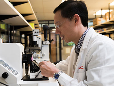
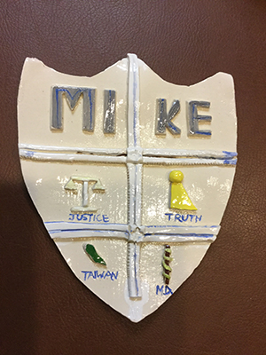
For an elementary school art project, Michael H. Hsieh, M.D., Ph.D., was instructed to fashion a coat of arms out of clay. In addition to panels for truth, justice and Taiwan, in the shield’s M.D. panel, a snake twists around a rod, like the staff for Asclepius, a Greek god associated with healing.

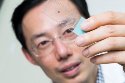
Children’s urologist Michael H. Hsieh, M.D., Ph.D., knew from age 10 that he would become a doctor. Proof is at his parents’ home. For an elementary school art project, students were instructed to fashion a coat of arms out of clay. In addition to panels for truth, justice and Taiwan, in the shield’s M.D. panel, a snake twists around a rod, like the staff for Asclepius, a Greek god associated with healing.

“I liked science. When I can use it to help patients, that is very rewarding,” says Dr. Hsieh, the first doctor in his family.

These days, Dr. Hsieh’s Twitter profile serves as a digital coat of arms, describing him as “tinker, tailor,” #UTI #biologist, epithelial #immunologist, helminthologist and #urologist.

Tinker/tailor is shorthand for the mystery drama, “Tinker Tailor Solider Spy,” he explains, adding that the “tinker” part also refers “to the fact that I am always questioning things, and science is about experimentation, trying to seek answers to questions.”

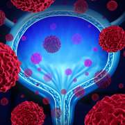
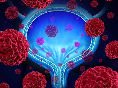
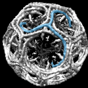
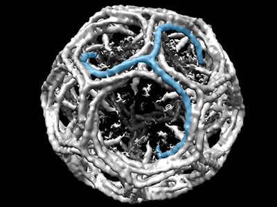
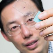
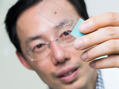
While still in medical school during a rotation Dr. Hsieh saw a bladder operation on a young child and thought it was “amazing.” That experience in part inspired Dr. Hsieh to become a urologist and bladder scientist. His training in immunology and study of the bladder naturally led him to study urinary tract infections and parasitic worms that affect the urinary tract. In addition, thanks to R01 funding from the National Institutes of Health (NIH), Dr. Hsieh is co-principal investigator with Axel Krieger, University of Maryland, and Jin U. Kang, Johns Hopkins, on a project to develop imaging robots for supervised autonomous surgery on soft tissue.

The $1 million in NIH funding pushes the boundaries on amazing by using multi-spectral imaging technology and improved techniques to reduce surgical complications.

Anastomosis is a technique used by surgeons to join one thing to another, whether it’s a vascular surgeon suturing blood vessels, an orthopedic surgeon joining muscles or a urologist stitching healthy parts of the urinary tract back together. Complications can set in if their stitching is too tight, prompting scar tissue to form, or too loose, letting fluid seep out.

“The human eye can see a narrow spectrum of electromagnetic radiation. These multi-spectral imaging cameras would see across greater set of wavelengths,” he says.

The project has three aims: figuring out the best way to place sutures using multi-spectral imaging, accurately tracking soft tissue as they model suturing and comparing the handicraft of a robot against anastomosis hand-sewn by surgeons.

“I like challenges, and I like new things. I am definitely not interested in doing permutations of other people’s work,” Dr. Hsieh explains. “I would much rather go on a path that hasn’t been tread. It is more difficult in some ways, but on a day-to-day basis, I know I am making a contribution.”

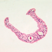
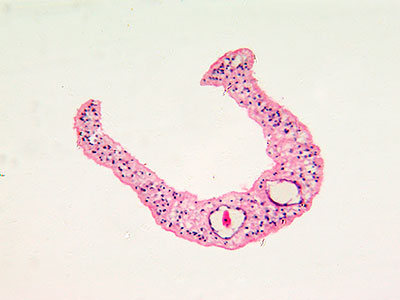
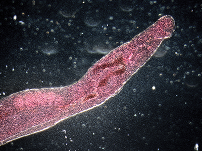
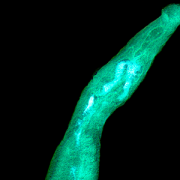
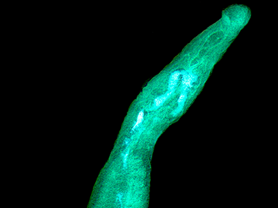
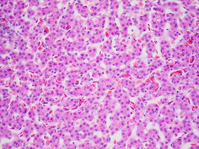
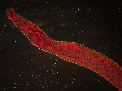
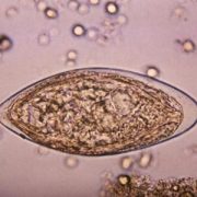
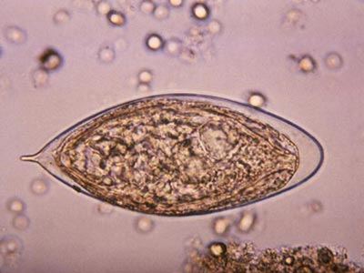
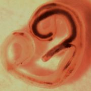
In another innovative research project, Dr. Hsieh leveraged a protein secreted by a parasitic worm, Schistosoma haematobium, that suppresses inflammation in hosts as a new therapeutic approach for chemotherapy-induced hemorrhagic cystitis, a form of inflammation of the bladder.

Watching his first surgery nearly 30 years ago, he had no idea robots might one day vie to take over some part of that complicated procedure, or that parasite proteins could be harnessed as drugs. However, he has a clear idea which innovations could be on the horizon for urology in the next three decades.

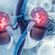
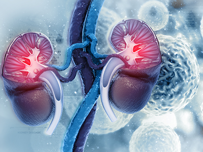
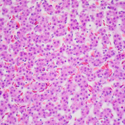
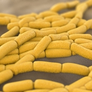
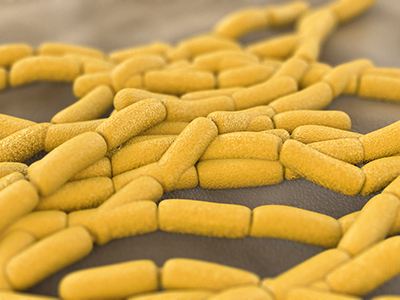
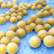
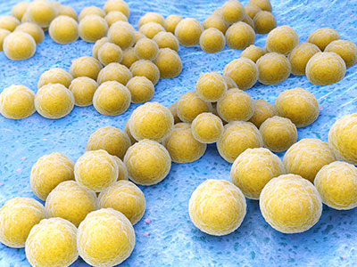
“My hope is 30 years from now, we will have a solid UTI vaccine and more non-antibiotic therapies. UTIs are the second-most common bacterial infection in childhood and, in severe cases, can contribute to kidney failure,” he says.

Globally, parasitic worms pose an ongoing challenge, affecting more than 1 billion worldwide – second only to malaria. People persistently infected by schistosome worms fail to reach their growth potential, struggle academically and lack sufficient energy for exercise or work.


“There is a feeling that the infection prevalence might be decreasing globally, but not as quickly as everyone hopes. In 30 years perhaps with more mass drug administration and additional drugs – including a vaccine – we’ll have it close to eliminated globally. It would become more like polio, casting a slim shadow with small pockets of infection here or there, rather than consigning millions to perpetual poverty.”

Lactobacillis-Bacteria

Does ZIP code factor into genitourinary system health?

Lactobacillis-Bacteria

Clinicians suspect that taking probiotics, such as lactobacillus supplements, and making changes to diet may prevent urinary diseases that occur commonly among pediatric patients. A research team led by Children’s faculty is exploring whether changes in the built environment also affect the urinary microbiome.

Emerging evidence suggests that the variety and volume of bacteria that reside in the bladder – the urinary microbiome – significantly impact whether people’s genitourinary systems remain healthy or become susceptible to disease.

Already, clinicians suspect that taking probiotics and making changes to diet may prevent urinary diseases that occur commonly among pediatric patients. A research team led by Children’s faculty is exploring whether changes in the built environment also affect the urinary microbiome.

Using experimental models, they looked at how stable the urinary microbiome was over time. Then, they measured the potential effect of changing the built environment on the urinary microbiome of preclinical models.

They did this by following six C57BL/6 experimental models for five months, starting from when they were nine weeks old. They collected urine specimens when the study began and repeated sample collections each month. The multidisciplinary team isolated microbial DNA from these specimens to determine the makeup of the bacterial community present in their urinary tracts.

All of the experimental models shared a single cage, drank the same water and ate the exact same chow. At four months, however, they moved the preclinical models to a different facility within the same county. Their chow and bedding remained unchanged, but the water source changed since they received tap water at both locations.

“There were no changes in the proportion of specific bacteria in the urinary microbiomes from month zero through month five, which means the urinary microbiomes of healthy experimental models remain stable over time,” says Michael Hsieh, M.D., Ph.D., a urologist at Children’s National Health System and senior author of the work presented during the Pediatric Urology Fall Conference. “However, the convergence of the Shannon Diversity Index, the clustering seen on Principal coordinate analyses and changes in functional analyses taken as a whole suggest that an overall shift of the urinary microbiome occurred due to a change in the physical environment.”

This work suggests that where patients live could influence which bacteria grow in the urinary tracts, including during urinary tract infections.

The Societies for Pediatric Urology’s Pediatric Urology Fall Conference

  • “Effects of time and the built environment on the stability of the mouse urinary microbiome: implications for clinical utility.”

Catherine S. Forster, M.D., MS, pediatric hospitalist, Children’s National; James Cody, Ph.D., Biomedical Research Institute; Nirad Banskota, MS, Biomedical Research Institute; Crystal Stroud, MS, Children’s National; Ljubica Caldovic, Ph.D., principal investigator, Children’s National; and Michael Hsieh, M.D., Ph.D., urologist, Children’s National.

Bladder cancer’s unique bacterial “fingerprint”

Michael H. Hsieh, M.D., Ph.D.

Michael H. Hsieh, M.D., Ph.D.

Decades ago, researchers thought that the native bacteria scattered throughout the human body—such as in the gut, the oral cavity and the skin—served little useful purpose. This microbiota, whose numbers at least match those of the cells in the body they live on and in, were considered mostly harmless hitchhikers.

More recently, research has revealed that these natural flora play key roles in maintaining and promoting health. In addition, studies have shown that understanding what a “typical” microbiome looks like and how it might change over time can provide an early warning system for some health conditions, including cancer.

Now, a small, multi-institutional study conducted in experimental models suggests that as bladder cancer progresses, it appears to be associated with a unique bacterial fingerprint within the bladder—a place thought to be bacteria-free except in the case of infection until just a few years ago. The finding opens the possibility of a new way to spot the disease earlier.

Bladder cancer is the fourth-most common malignancy among U.S. men but, despite its prevalence, mortality rates have remained stubbornly high. Patients often are diagnosed late, after bladder cancer has advanced. And, it remains difficult to discern which patients with non-invasive bladder cancer will go on to develop muscle-invasive disease.

Already, researchers know that patients with grade 4 oral squamous cell carcinoma, women with increasingly severe grades of cervical cancer and patients with cirrhosis who develop liver cancer have altered oral, vaginal and gut microbiomes, respectively.

New technological advances have led to identification of a diverse community of bacteria within the bladder, the urinary microbiome. Leveraging these tools, a research team that includes Children’s National Health System investigators studied whether an experimental model’s urinary bacterial community changed as bladder cancer progressed, evolving from a microbiome into a urinary “oncobiome.”

To test the hypothesis, the research team led by Michael H. Hsieh, M.D., Ph.D., a Children’s urologist, exposed an experimental model of bladder cancer to a bladder-specific cancer-causing agent, n-butyl-n-(4-hydroxybutyl) nitrosamine (BBN). Bladder cancers induced by BBN closely resemble human cancers in tissue structure at the microscopic level and by gene expression analyses. Ten of the preclinical models received a .05 percent concentration of BBN in their drinking water over five months and were housed together. Ten other experimental models received regular tap water and shared a separate, adjacent cage.

Researchers collected urine samples ranging from 10 to 100 microliters at the beginning of the longitudinal study, one week after it began, then once monthly. They isolated microbial DNA from the urine and quantified it to determine how much DNA was microbial. All of the bladders from experimental models exposed to BBN and two bladders from the control group were analyzed by a pathologist trained in bladder biology.

According to the study published online July 5, 2018, by the biology preprint server Biorxiv, they found a range of pathologies:

  • Five of the experimental models that received BBN did not develop cancer but had histology consistent with inflammation. Three had precancer on histology: urothelial dysplasia, hyperplasia or carcinoma in situ. Two developed cancer: invasive urothelial carcinomas, one of which had features of a squamous cell carcinoma.
  • The experimental model that developed invasive carcinoma had markedly different urinary bacteria at baseline, with Rubellimicrobium, a gram negative organism found in soil that has not been associated with disease previously, Escherichia and Kaistobacter, also found in soil, as the most prominent bacteria. By contrast, in the other experimental models the most common urinary bacteria were Escherichia, Prevotella, Veillonella, Streptococcus, Staphylococcus and Neisseria.
  • By month four, the majority of experimental models exposed to BBN had significantly higher proportion of Gardnerella and Bifidobacterium compared with their control group counterparts.

“Closely analyzing the urinary bacterial community among experimental models exposed to BBN, we saw distinct differences in microbial profiles by month four that were not present in earlier months,” Dr. Hsieh says. “While Gardnerella is associated with the development of cancer, Bifidobacterium has been shown to exert antitumor immunity, and its increasing abundance points to the need for additional research to understand its precise role in oncogenesis.”

Dr. Hsieh adds that although the study is small, its findings are of significance to children who are prone to developing urinary tract infections (UTIs), including children with spina bifida, due to the association between UTIs and bladder cancer. “This work is important because it not only suggests that the urinary microbiome could be used to diagnose bladder cancer, but that it could also perhaps predict cancer outcomes. If the urinary microbiome contributes to bladder carcinogenesis, it may be possible to favorably change the microbiome through antibiotics and/or probiotics in order to treat bladder cancer.”

In addition to Dr. Hsieh, co-authors include Catherine S. Forster, M.D., M.S., and Crystal Stroud, of Children’s National; James J. Cody, Nirad Banskota, Yi-Ju Hsieh and Olivia Lamanna, of the Biomedical Research Institute; Dannah Farah and Ljubica Caldovic, of The George Washington University; and Olfat Hammam, of Theodor Bilharz Research Institute.

Research reported in this news release was supported by the National Institutes of Health under award number R01 DK113504 and the Margaret A. Stirewalt Endowment.

Schistosoma haematobium egg

For hemorrhagic cystitis, harnessing the power of a parasite

Schistosoma haematobium egg

“Urogenital Schistosoma infestation, which is caused by S. haematobium, also causes hemorrhagic cystitis, likely by triggering inflammation when the parasite’s eggs are deposited in the bladder wall or as eggs pass from the bladder into the urinary stream. S. haematobium eggs secrete proteins, including IPSE, that ensure human hosts are not so sickened that they succumb to hemorrhagic cystitis,” says Michael H. Hsieh, M.D., Ph.D.

Every year, hundreds of thousands of U.S. patients – and even more throughout the world – are prescribed cyclophosphamide or ifosfamide. These two chemotherapy drugs can be life-saving for a wide range of pediatric cancers, including leukemias and cancers of the eyes and nerves. However, these therapies come with a serious side effect: Both cause hemorrhagic cystitis in up to 40 percent of patients. This debilitating condition is characterized by severe inflammation in the bladder that can cause tremendous pain, life-threatening bleeding, and frequent and urgent urination.

Infection with a parasitic worm called Schistosoma haematobium also causes hemorrhagic cystitis, but this organism has a fail-safe: To keep its host alive, the parasite secretes a protein that suppresses inflammation and the associated pain and bleeding.

In a new study, a Children’s-led research team harnessed this protein to serve as a new therapy for chemotherapy-induced hemorrhagic cystitis.

“Urogenital Schistosoma infestation, which is caused by S. haematobium, also causes hemorrhagic cystitis, likely by triggering inflammation when the parasite’s eggs are deposited in the bladder wall or as eggs pass from the bladder into the urinary stream. S. haematobium eggs secrete proteins, including IPSE, that ensure human hosts are not so sickened that they succumb to hemorrhagic cystitis,” says Michael H. Hsieh, M.D., Ph.D., senior author of the study published April 3, 2018, by The FASEB Journal. “This work in an experimental model is the first published report of exploiting an uropathogen-derived host modulatory molecule in a clinically relevant model of bladder disease, and it points to the potential utility of this as an alternate treatment approach.”

S. mansoni IPSE binds to Immunoglobulin E (IgE), an antibody produced by the immune system that is expressed on the surface of basophils, a type of immune cell; and mast cells, another immune cell that mediates inflammation; and sequesters chemokines, signaling proteins that alert white cells to infection sites. The team produced an ortholog of the uropathogen-derived protein. A single IV dose proved superior to multiple doses of 2-Mercaptoethane sulfonate sodium (MESNA), the current standard of care, in suppressing chemotherapy-induced bladder hemorrhaging in an experimental model. It was equally potent as MESNA in dampening chemotherapy-induced pain, the research team finds.

“The current array of medicines we use to treat hemorrhagic cystitis all have shortcomings, so there is a definite need for novel therapeutic options,” says Dr. Hsieh, a Children’s National Health System urologist. “And other ongoing research projects have the potential to further expand patients’ treatment options by leveraging other urogenital parasite-derived, immune-modulating molecules to treat inflammatory bowel diseases and autoimmune disorders.”

Future research will aim to describe the precise molecular mechanisms of action, as well as to generate other orthologs that boost efficacy while reducing side effects.

In addition to Dr. Hsieh, Children’s study co-authors include Lead Author, Evaristus C. Mbanefo; Loc Le and Luke F. Pennington; Justin I. Odegaard and Theodore S. Jardetzky, Stanford University; Abdulaziz Alouffi, King Abdulaziz City for Science and Technology; and Franco H. Falcone, University of Nottingham.

Financial support for this research was provided by National Institutes of Health under award number RO1-DK113504.

Staphylococcus

How our bladder’s microbiota affect health

Staphylococcus

The presence of bacteria such as Staphylococcus in the urine is linked to the incidence and severity of urge urinary incontinence as well as treatment success.

About half of the cells in our bodies aren’t really “ours” at all. They’re the microbiota: The vast array of microorganisms that live in our gut, skin, oral cavity and other places. Decades ago, researchers thought that these organisms simply happened to colonize these areas, playing only a tangential role in health, for example, helping to break down food in the intestines or causing cavities. More recent work has revealed the incredibly complex role they play in diseases ranging from diabetes and schizophrenia.

The bladder is no exception. Just a single decade ago, the bladder was thought to be a sterile environment. But that view has shifted radically, with more sensitive cultivation methods and precise 16S rRNA gene-sequencing techniques revealing a significant bladder microbiome that could have an enormous impact on pediatric urologic diseases. These findings have opened brand new fields of research aimed at clarifying the role that the bladder’s microbiome plays in common urological diseases that affect children, according to a review article published online Feb. 22, 2018, by Current Urology Reports.

“There is a growing appreciation for the role of diverse bacteria in contributing to improved health as well as triggering disease processes or exacerbating illness,” says Michael H. Hsieh, M.D., Ph.D., director of the Clinic for Adolescent and Adult Pediatric Onset Urology (CAPITUL) at Children’s National Health System and study senior author. “Already, we know that probiotics and dietary modifications have the potential to play powerful roles in preventing urinary diseases that commonly occur among pediatric patients,” Dr. Hsieh says. This underscores the importance of conducting even more studies to improve our understanding and to identify new therapies for health conditions that resist current treatment options.”

The review conducted by Dr. Hsieh and co-authors highlights the effects of the microbiome on a number of urologic diseases that affect children, including:

  • Urinary tract infection A number of studies point to the association between decreased microbial diversity and the incidence of what is commonly called urinary tract infection (UTI) or “dysbiosis.” This relationship suggests that using probiotics to replace or supplement antibiotics could favorably alter the urinary microbiome. Future research will focus on the pathophysiological role of the microbiome to determine whether it can be manipulated to prevent or treat UTIs.
  • Urge urinary incontinence While data vary by study, the presence of bacteria in the urine, especially certain bacterial species – such as Gardnerella, Staphylococcus, Streptococcus, Actinomyces, Aerococcus, Corynebacterium and Oligella – are linked to the incidence and severity of urge urinary incontinence (UUI) as well as treatment success. Most studies find an association between greater genitourinary biodiversity and reduced incidence and lessened severity of UUI as well as improved treatment response. Future research will focus on further clarifying this relationship.
  • Urolithiasis Calcium oxalate stones, the most common type of kidney stone, have a microbiome that differs from the urinary microbiome leading researchers to question whether the stone’s own bacterial makeup could help to predict recurrence of future kidney stones. What’s more, Oxalobacter formigenes, a gram-negative bacterium, lowers oxalate levels in the blood and are associated with a 70 percent reduction in the risk of kidney stones forming. In an experimental model, fecal transplants with the full microbiome represented had a pronounced and persistent effect on oxalate production. Patients who receive some antibiotics often have reduced rates of formigenes colonization. However, the bacteria are resistant to amoxicillin, augmentin, ceftriaxone and vancomycin, which could point to preferential use of these antibiotics to stave off disease and ward off kidney stone formation.

Additional authors include Daniel Gerber, study lead author, The Georgetown University School of Medicine and Health Sciences; and Catherine Forster, M.D., study co-author, Children’s National.

Love is in the air and, for parasites, inside our bodies

Michael H. Hsieh

As featured in a PBS video, schistosome worms form lifelong bonds and females produce thousands of eggs daily only when they live inside human hosts, says Michael H. Hsieh, M.D., Ph.D.

“Love is in the air, the sea, the earth and all over and inside our bodies,” the PBS Valentine’s Day-themed video begins. As the public television station notes, what humans consider romance can look vastly different for creatures big and small, including serenading mice, spiders who wrap their gifts in silk and necking giraffes.

The “spooning” parasites segment of the video is where viewers see research conducted by Michael H. Hsieh, M.D., Ph.D., director of the Clinic for Adolescent and Adult PedIatric OnseT UroLogy at Children’s National Health System, and video filmed in his lab.

Schistosomiasis, a chronic infection with schistosome worms, is a distinctly one-sided love affair. As shown in Dr. Hsieh’s video clips, the male worm is shorter and fatter and equipped with a groove, a love canal where the longer, thinner female lodges, enabling the pair to mate for decades. This lifelong bond and the thousands of eggs it produces daily can only occur when the worms are inside the human host, Dr. Hsieh says.

While the video stresses Valentine’s Day romance, there are few rosy outcomes for humans who are the subject of the schistosome worms’ attention.

“Heavily and chronically infected individuals can have lots of problems,” Dr. Hsieh says. “This is a stunting and wasting health condition that prevents people from reaching their growth potential, impairs their academic performance and leaves them sapped of the energy needed to exercise or work. It truly perpetuates a cycle of poverty, particularly for affected children.”

Even the potential bright spot in this sobering story, the ability of the body’s immune system to fend off the parasitic worms, is only partly good news.

Schistosome worms have co-evolved with their human hosts, learning to take advantage of human vulnerabilities. Take the immune system. If it kicks too far into overdrive in trying to wall off the eggs from the rest of the body, it can interfere with organ function and trigger liver failure, kidney failure and early onset of bladder cancer, he says.

However, Dr. Hsieh and other schistosomiasis researchers are working on ways to positively harness the human immune response to schistosome worms, including developing diagnostics, drugs and vaccines. He says he and his colleagues would “love” to eliminate schistosomiasis as a global scourge.

Twitter Pediatric Urology Journal Club @pedurojc

Journal club, with a 140-character limit

Twitter Pediatric Urology Journal Club @pedurojc

@perforin & @chrbayne have launched a new journal club focused on pediatric urology via Twitter, a platform that democratizes and distills the academic discussion.

Journal club is a rite of passage for nearly everyone who works in an academic laboratory. What might sound like an exclusive group of readers and authors united by a secret handshake is actually a regular meeting of scientists – faculty members and young trainees alike – who gather to discuss a highlighted paper in their field of expertise.

Some of these gatherings might involve a handful of people from the same lab; others might include a larger group from the same institutional department or division. Typically, one person presents a paper, sharing all the relevant details about a study’s methodology and conclusions. Afterward, everyone has the chance to pose questions, make comments and thoroughly discuss conclusions.

“It’s an excellent academic opportunity in terms of teaching and training of early career scientists and clinicians, and it remains useful no matter what stage you are in your career,” says Michael Hsieh, M.D., Ph.D., a urologist who directs the Clinic for Adolescent and Adult PedIatric OnseT UroLogy (CAPITUL) at Children’s National Health System who has participated in a heavy share of journal club meetings over the years.

But, what if journal club didn’t have to adhere to this traditional format? What if this academic discussion could move to a venue more fitting for the 21st century, more inclusive of scientists in different geographic locations, with varying viewpoints and expertise?

That’s what Dr. Hsieh and others are trying to accomplish with a new pediatric urology-focused journal club on Twitter. When Christopher Bayne, a second-year fellow training in pediatric urology at Children’s National under Dr. Hsieh’s mentorship, approached him with the idea, Dr. Hsieh said that he jumped at the chance.

Traditional journal clubs, the two explain, can be hindered by several factors. One is a tendency toward “group think,” Dr. Hsieh says – members of the same lab, or even the same institution, tend to have the same training and practices, so they’re less likely to feel comfortable introducing new ideas about these areas into the discussion. Journal club discussions also are limited by uncertainties about what a study author might have had in mind with their methodology and conclusions. Study authors are rarely included in the discussion, Dr. Hsieh adds.

Michael Hsieh

“It’s an excellent academic opportunity in terms of teaching and training of early career scientists and clinicians, and it remains useful no matter what stage you are in your career,” says Michael Hsieh, M.D., Ph.D., a urologist who directs the Clinic for Adolescent and Adult PedIatric OnseT UroLogy (CAPITUL) at Children’s National Health System.

Twitter, Bayne says, offers an easy way around these barriers. Rather than including just members of the same lab, their Pediatric Urology Journal Club (PUJC) can accommodate any registered Twitter user in their discussions. That means that any interested person around the world – researchers, clinician-scientists, other health care providers, as well as patients and their families, for example – can participate in the monthly discussions.

Participation also isn’t dictated by geography. During recent PUJC meetings, individuals joined the thread from Brazil, Ireland and Turkey. The meetings, sponsored by the Journal of Pediatric Urology, take place in the first days to weeks after the selected paper has been available under “open access,” giving anyone a chance to read it – even if they lack a journal subscription. This format enables all participants to join threads, erasing the restrictions of geography or busy clinical and research schedules.

Thus far, the meetings have included papers on:

  • A comparison of the cost and complications of performing a surgery either robotically or through an open procedure to fix the tubes that connect the kidneys to the bladder in patients with a condition known as vesicouretal reflux, in which urine flows in the wrong direction.
  • The pros and cons of treating varicoceles, enlarged veins inside the scrotum that potentially cause fertility problems. The condition is asymptomatic in adolescents.
  • The importance of the diameter of the ureter, the part of the tube closest to the outside of the body that carries urine to be expelled, for resolving vesicouretal reflux, an abnormal flow of urine.

This new platform has attracted a core group of relatively young and young-at-heart devotees, Bayne says. He and other organizers have included study authors in every meeting thus far, often guiding older and Twitter-naive scientists through the process of creating an account.

And the typical 140-character limit Twitter imposes on comments known as tweets? “It might be counterintuitive,” Bayne says, “but I see the character limit as one of this journal club’s biggest strengths.” This cutoff encourages discussion members to distill their thoughts, often including two or three distinct points, into concise and deeply meaningful statements. “Participants have really latched on to the efficiency of this approach to learning about a topic and having a lively discussion.”

Thus far, their approach has been increasing in popularity. Their very first PUJC meeting in February 2017 attracted a modest number of just 24 active participants who sent 310 tweets, but generated nearly 136,000 impressions, or views.

The researchers plan to continue the monthly PUJC meetings through the Twitter handle @pedurojc. You can follow updates from Dr. Hsieh on his handle: @perforin and updates from Bayne’s on his: @chrbayne.

Michael Hsieh

Michael Hsieh receives grant to explore parasite proteins for pain relief

Michael Hsieh

Michael Hsieh, M.D., hopes to use parasite proteins to alleviate pain in multiple types of bladder inflammation.

Children’s National Health System Urologist Michael Hsieh, M.D., was awarded a National Institutes of Health (NIH) grant to optimize a set of parasite proteins that could alleviate pain in multiple types of bladder inflammation.

The $1 million R01 grant will fund a five-year study to exploit a parasite-derived protein, IPSE, as a candidate therapeutic. Dr. Hsieh hypothesizes that IPSE may have the ability to modulate host immune and non-immune responses to bladder injury. IPSE could be optimized for therapeutic potential, while minimizing toxicity, by generating forms that have the ability to modulate host responses via three distinct mechanisms: IL-4-binding, chemokine-binding and nuclear localization.