Tag Archive for: focal segmental glomerulosclerosis

High magnification micrograph of focal segmental glomerulosclerosis

Reducing urinary protein for patients with FSGS slows kidney decline

High magnification micrograph of focal segmental glomerulosclerosis

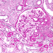
High magnification micrograph of focal segmental glomerulosclerosis (FSGS).

Reducing the amount of protein in the urine of patients with focal segmental glomerulosclerosis (FSGS), a rare disease in which scar tissue forms on the parts of the kidneys that filter waste from the blood, can significantly slow declines in kidney function and extend time before patients’ kidneys fail, a new analysis by a Children’s National Hospital researcher and her colleagues shows. These findings, published online Aug. 10, 2020, in the American Journal of Kidney Disease, could provide hope for patients who are able to lower their urinary protein with available treatments but aren’t able to achieve complete remission, the researchers say.

FSGS affects about seven per every million people in the general population. However, in the United States, it’s responsible for between 5 and 20% of all cases of end stage kidney disease (ESKD), a condition in which the kidney function declines enough that patients can’t survive without dialysis or a kidney transplant. There are no proven treatments specifically targeting FSGS, but steroids and other immunosuppressants have shown promise in clinical trials.

One characteristic sign of FSGS is proteinuria, in which too much protein is present in patients’ urine. Most clinical trials of FSGS treatments have focused on complete remission of proteinuria as a sign that the intervention is working. However, says Marva Moxey-Mims, M.D., researcher and chief of the Children’s National Division of Nephrology, only a fraction of patients meet that end goal. Instead, many patients achieve some reduction in proteinuria, but it’s been unclear whether those reductions lead to significant benefits for kidney health.

To investigate this question, Dr. Moxey-Mims and her colleagues used data from the National Institutes of Health-funded FSGS clinical trial that took place between November 2004 and May 2008. Participants in this study — 138 patients who developed proteinuria due to FSGS between the ages of 2 and 40 and didn’t respond to steroids — received one of two different immunosuppressant regimens. They received frequent checkups including urinary protein tests during the duration of the study and were followed for a maximum of 54 months.

Results showed that about 49% of the study participants’ proteinuria improved by 26 weeks of treatment on either regimen. More importantly, says Dr. Moxey-Mims, these patients retained significantly better kidney function over time, determined by a test called estimated glomerular filtration rate (eGFR), compared to those whose urinary protein remained high. The greater the reduction in proteinuria, the better their kidney function remained, and the longer their kidneys remained active before they developed ESKD.

“Even a modest reduction in proteinuria, as small as 20 or 30%, had an impact on these patients’ kidney health,” Dr. Moxey-Mims says.

Dr. Moxey-Mims notes that the finding could impact the design of clinical trials for FSGS treatments. Currently, these trials typically must include large numbers of patients to show a benefit if complete remission of proteinuria — which only occurred in about 20% of patients in the National Institute of Diabetes and Digestive and Kidney Diseases trial — is used as the end point.

If researchers use a range of proteinuria reduction as end points, she says, it could be easier to see if a drug or other intervention is working.

Similarly, she says, patients with FSGS and their doctors should view any proteinuria reduction as a positive.

“They shouldn’t be discouraged if they can’t reach full remission,” Dr. Moxey-Mims says. “Doctors and patients alike can feel reassured that if they’re reducing protein in the urine to some degree, then patients are getting some benefit.”


mitochondria

Treating nephrotic-range proteinuria with tacrolimus in MTP

mitochondria

Mitochondria are the cell’s powerplants and inside them the MTP enzymatic complex catalyzes three steps in beta-oxidation of long-chain fatty acids.

In one family, genetic lightning struck twice. Two sisters were diagnosed with mitochondrial trifunctional protein (MTP) deficiency. This is a rare condition that stops the body from converting fats to energy, which can lead to lactic acidosis, recurrent breakdown of muscle tissue and release into the bloodstream (rhabdomyolysis), enlarged heart (cardiomyopathy) and liver failure.

Mitochondria are the cell’s powerplants and inside them the MTP enzymatic complex catalyzes three steps in beta-oxidation of long-chain fatty acids. MTP deficiency is so rare that fewer than 100 cases have been reported in the literature says Hostensia Beng, M.D., who presented an MTP case study during the American Society of Nephrology’s Kidney Week.

The 7-month-old girl with known MTP deficiency arrived at Children’s National lethargic with poor appetite. Her laboratory results showed a low corrected serum calcium level, elevated CK level and protein in the urine (proteinuria) at a nephrotic range. The infant was treated for primary hypoparathyroidism and rhabdomyolysis.

Even though the rhabdomyolysis got better, the excess protein in the girl’s urine remained at worrisome levels. A renal biopsy showed minimal change disease and foot process fusion. And electron microscopy revealed shrunken, dense mitochondria in visceral epithelial cells and endothelium.

“We gave her tacrolimus, a calcineurin inhibitor that we are well familiar with because we use it after transplants to ensure patient’s bodies don’t reject the donated organ. By eight months after treatment, the girl’s urine protein-to-creatinine (uPCR) ratio was back to normal. At 35 months, that key uPCR measure rose again when tacrolimus was discontinued. When treatment began again, uPCR was restored to normal levels one month later,” Dr. Beng says.

The girl’s older sister also shares the heterozygous deletion in the HADHB gene, which provides instructions for making MTP. That missing section of the genetic how-to guide was predicted to cause truncation and loss of long-chain-3-hydroxyacl CoA dehydrogenase function leading to MTP deficiency.

The older sister was diagnosed with nephrotic syndrome and having scar tissue in the kidney’s filtering unit (focal segmental glomerulosclerosis) when she was 18 months old. By contrast, she developed renal failure and progressed to end stage renal disease at 20 months of age.

“Renal involvement has been reported in only one patient with MTP deficiency to date, the older sister of our patient,” Dr. Beng adds.

Podocytes are specialized cells in the kidneys that provide a barrier, preventing plasma proteins from leaking into the urine. Podocytes, however, need energy to function and are rich in mitochondria.

“The proteinuria in these two sisters may be related to their mitochondrial dysfunction. Calcineurin inhibitors like tacrolimus have been reported to reduce proteinuria by stabilizing the podocyte actin cytoskeleton. Tacrolimus was an effective treatment for our patient, who has maintained normal renal function, unlike her sister,” Dr. Beng says.

American Society of Nephrology’s Kidney Week presentation

  • “Treatment of nephrotic-range proteinuria with tacrolimus in mitochondrial trifunctional protein deficiency

Hostensia Beng, M.D., lead author; Asha Moudgil, M.D., medical director, transplant, and co-author; Sun-Young Ahn, M.D., MS, medical director, nephrology inpatient services, and senior author, all of Children’s National Health System.

little girl in hosptial corridor

A growing list of factors that impact CKD severity for kids

little girl in hosptial corridor

Myriad biological and societal factors can impact the occurrence and accelerate progression of chronic kidney disease for children of African descent – including preterm birth, exposure to toxins during gestation and lower socioeconomic status – and can complicate these children’s access to effective treatments.

Myriad biological and societal factors can impact the occurrence and accelerate progression of chronic kidney disease (CKD) for children of African descent – including preterm birth, exposure to toxins during gestation and lower socioeconomic status – and can complicate these children’s access to effective treatments, according to an invited commentary published in the November 2018 edition of American Journal of Kidney Diseases.

Clinicians caring for “these vulnerable children should be mindful of these multiple competing and compounding issues as treatment options are being considered along the continuum from CKD to kidney failure to transplantation,” writes Marva Moxey-Mims, M.D., chief of the Division of Nephrology at Children’s National Health System.

The supplemental article was informed by lessons learned from The Chronic Kidney Disease in Children (CKiD) longitudinal study and conversations that occurred during the Frank M. Norfleet Forum for Advancement of Health, “African Americans and Kidney Disease in the 21st Century.”

African American children represent 23 percent of the overall population of kids with CKD in the CKiD study. While acquired kidney diseases can get their start during childhood when the diseases betray few symptoms, the full impact of illness may not be felt until adulthood. A number of factors can uniquely affect children of African descent, heightening risk for some kids who already are predisposed to suffering more severe symptoms. These include:

  • Preterm birth. African American children make up 36 percent of patients in CKiD with glomerular disease, which tends to have faster progression to end-stage renal disease. These diseases impair kidney function by weakening glomeruli, which impairs the kidneys’ ability to clean blood. Patients with a high-risk apolipoprotein L1 (APOL1) genotype already are at higher risk for focal segmental glomerulosclerosis (FSGS) and CKD. Researchers hypothesize that preterm birth may represent “a second hit that facilitates the development of glomerular damage resulting from the high-risk genotype.” According to the Centers for Disease Control and Prevention, 1 in 10 U.S. infants in 2016 was born preterm, e.g., prior to 37 weeks gestation.
  • APOL1 genotype. Compared with children who had a low-risk genotype and FSGS, children with a high-risk genotype had higher rates of uncontrolled hypertension, left ventricular hypertrophy, elevated C-reactive protein levels and obesity.
  • Human immunodeficiency viral (HIV) status. About 65 percent of U.S. children with HIV-1/AIDS are African American. In a recent nested case-control study of children infected with HIV in the womb, infants with high-risk APOL1 genotypes were 3.5 times more likely to develop CKD with viral infection serving as “a likely second hit.”
  • Access to kidney transplant. African American adults experience a faster transition to end-stage renal disease and are less likely to receive kidney transplants. African American children with CKD from nonglomerular diseases begin renal replacement therapy 1.6 years earlier than children of other races, after adjusting for socioeconomic status. Their wait for dialysis therapy was 37.5 percent shorter. However, these African American children waited 53.7 percent longer for transplants. Although donor blood types, genetic characteristics and other biological factors each play contributing roles, “these findings may reflect sociocultural and institutional differences not captured by socioeconomic status,” Dr. Moxey-Mims writes.

To alleviate future health care disparities, she suggests that additional research explore the impact of expanding services to pregnant women to lower their chances of giving birth prematurely; early childhood interventions to help boost children’s educational outcomes, future job prospects and income levels; expanded studies about the impact of environmental toxicities on prenatal and postnatal development; and heightened surveillance of preterm infants as they grow older to spot signs of kidney disease earlier to slow or prevent disease progression.

“Clinicians can now begin to take into account genetics, socioeconomic status and the impact of the built environment, rather than blaming people and assuming that their behavior alone brought on kidney disease,” Dr. Moxey-Mims adds. “Smoking, not eating properly and not exercising can certainly make people vulnerable to disease. However, there are so many factors that go into developing a disease that patients cannot control: You don’t control to whom you’re born, where you live or available resources where you live. These research projects will be useful to help us really get to the bottom of which factors we can impact and which things can’t we prevent but can strive to mitigate.”

The article covered in this post is part of a supplement that arose from the Frank M. Norfleet Forum for Advancement of Health: African Americans and Kidney Disease in the 21st Century, held March 24, 2017, in Memphis, Tennessee. The Forum and the publication of this supplement were funded by the Frank M. Norfleet Forum for Advancement of Health, the Community Foundation of Greater Memphis and the University of Tennessee Health Science Center.

Coenzyme Q10

Supplement might help kidney disease

Coenzyme Q10

A research team was able to “rescue” phenotypes caused by silencing the fly CoQ2 gene by providing nephrocytes with a normal human CoQ2 gene, as well as by providing flies with Q10, a popular supplement.

A new study led by Children’s National research scientists shows that coenzyme Q10 (CoQ10), a popular over-the-counter supplement sold for pennies a dose, could alleviate genetic problems that affect kidney function. The work, done in genetically modified fruit flies — a common model for human genetic diseases since people and fruit flies share a majority of genes — could give hope to human patients with problems in the same genetic pathway.

The new study, published April 20 by Journal of the American Society of Nephrology, focuses on genes the fly uses to create CoQ10.

“Transgenic Drosophila that carry mutations in this critical pathway are a clinically relevant model to shed light on the genetic mutations that underlie severe kidney disease in humans, and they could be instrumental for testing novel therapies for rare diseases, such as focal segmental glomerulosclerosis (FSGS), that currently lack treatment options,” says Zhe Han, Ph.D., principal investigator and associate professor in the Center for Cancer & Immunology Research at Children’s National and senior study author.

Nephrotic syndrome (NS) is a cluster of symptoms that signal kidney damage, including excess protein in the urine, low protein levels in blood, swelling and elevated cholesterol. The version of NS that is resistant to steroids is a major cause of end stage renal disease. Of the more than 40 genes that cause genetic kidney disease, the research team concentrated on mutations in genes involved in the biosynthesis of CoQ10, an important antioxidant that protects the cell against damage from reactive oxygen.

Drosophila pericardial nephrocytes perform renal cell functions including filtering of hemolymph (the fly’s version of blood), recycling of low molecular weight proteins and sequestration of filtered toxins. Nephrocytes closely resemble, in structure and function, the podocytes of the human kidney.  The research team tailor-made a Drosophila model to perform the first systematic in vivo study to assess the roles of CoQ10 pathway genes in renal cell health and kidney function.

One by one, they silenced the function of all CoQ genes in nephrocytes. If any individual gene’s function was silenced, fruit flies died prematurely. But silencing three specific genes in the pathway associated with NS in humans – Coq2, Coq6 and Coq8 – resulted in abnormal localization of slit diaphragm structures, the most important of the kidney’s three filtration layers; collapse of membrane channel networks surrounding the cell; and increased numbers of abnormal mitochondria with deformed inner membrane structure.

Journal of the American Society of Nephrology September 2017 cover

The flies also experienced a nearly three-fold increase in levels of reactive oxygen, which the study authors say is a sufficient degree of oxidative stress to cause cellular injury and to impair function – especially to the mitochondrial inner membrane. Cells rely on properly functioning mitochondria, the cell’s powerhouse, to convert energy from food into a useful form. Impaired mitochondrial structure is linked to pathogenic kidney disease.

The research team was able to “rescue” phenotypes caused by silencing the fly CoQ2 gene by providing nephrocytes with a normal human CoQ2 gene, as well as by providing flies with Q10, a readily available dietary supplement. Conversely, a mutant human CoQ2 gene from an patient with FSGS failed to rescue, providing evidence in support of that particular CoQ2 gene mutation causing the FSGS. The finding also indicated that the patient could benefit from Q10 supplementation.

“This represents a benchmark for precision medicine,” Han adds. “Our gene-replacement approach silenced the fly homolog in the tissue of interest – here, the kidney cells – and provided a human gene to supply the silenced function. When we use a human gene carrying a mutation from a patient for this assay, we can discover precisely how a specific mutation – in many cases only a single amino acid change – might lead to severe disease. We can then use this personalized fly model, carrying a patient-derived mutation, to perform drug testing and screening to find and test potential treatments. This is how I envision using the fruit fly to facilitate precision medicine.”

Related resources:
News release: Drosophila effectively models human genes responsible for genetic kidney diseases
Video: Using the Drosophila model to learn more about disease in humans