Tag Archive for: fetal

newborn in incubator

A bronchopulmonary dysplasia primer to guide clinicians and researchers

newborn in incubator

Six months in the writing, the “Bronchopulmonary Dysplasia Primer” published recently by Nature Reviews will be the gold standard review on this topic for years to come.

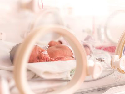
The term bronchopulmonary dysplasia, or BPD, was first coined in 1967 to describe a chronic lung disease of preterm newborns after treatment with supplemental oxygen via mechanical ventilation in an effort to save their lives. Back then, infants had 50-50 odds of surviving.

In the intervening years, survival has improved and the characteristics of BPD have evolved. Now, BPD is the most common complication of preterm birth for infants born at fewer than 28 weeks’ gestation, as more and more newborns survive premature birth. Hence, the primer.

“The contributing authors are some of the biggest thinkers on this topic,” says Robin H. Steinhorn, M.D., senior vice president, Center for Hospital-Based Specialties, at Children’s National Hospital and author of the section about BPD diagnosis, screening and prevention. “This document will guide clinical education and investigators in the field of BPD. I anticipate this will be the definitive review article on the subject for the next several years.”

Gestational age and low birth weight remain the most potent predictors of BPD. Some 50,000 extremely low gestational age newborns are born each year in the U.S. About 35% will develop some degree of BPD, according to the primer authors.

These newborns are introduced to life outside the womb well before their lungs are ready. Indeed, the pulmonary surfactants needed for normal lung function – a complex mixture of phospholipids that reduce surface tension within the lungs – don’t differentiate until late in pregnancy. Infants who persistently need respiratory support after the 14th day of life are at the highest risk of being diagnosed with BPD at 36 weeks, the coauthors note.

A number of complicating factors can come into play, including maternal diet; fetal exposure to maternal smoking and infection; structural issues such as pre-eclampsia; acute injury from mechanical ventilation and supplemental oxygen; as well as the body’s halting efforts to repair injured, inflamed lung tissue.

“The good news is the number of the smallest and youngest preterm infants who survive extreme preterm birth has steadily increased. Neonatal intensive care units, like our award-winning NICU, now routinely care for babies born at 22 weeks’ gestation,” Dr. Steinhorn says.

Treatment strategies include:

  • Reducing exposure to intubation and ventilation.
  • Leveraging respiratory stimulants, like caffeine.
  • Postnatal steroid therapy.

“Children’s National Hospital is the only center in our immediate region that provides comprehensive care for infants and children with severe BPD,” Dr. Steinhorn adds. “As the population of vulnerable and fragile infants has grown, we have invested in the equipment and the personnel – including at the Hospital for Sick Children Pediatric Center (HSC) – to create a very safe and supportive environment that improves survival and quality of life.”

Some preterm infants spend their first 9 to 10 months of life at Children’s National, and their days are filled with concentrated physical, occupational and speech therapy, as well as music and play therapy to hasten their rehabilitation.

Once their medical condition stabilizes, they transition to HSC to focus more intently on rehabilitation.

“We see HSC as filling a very important role in their care. When our children graduate to HSC, they are going for ongoing care of their lung disease, but also their ongoing rehabilitation. At HSC, they focus on creating the most normal life that we can possibly create and, over time, that is a life free of ventilators and tracheostomy tubes.”

In addition to Dr. Steinhorn, BPD Primer co-authors include Bernard Thébaud, Children’s Hospital of Eastern Ontario; Kara N. Goss, University of Wisconsin-Madison; Matthew Laughon, The University of North Carolina at Chapel Hill; Jeffrey A. Whitsett and Alan H. Jobe, Cincinnati Children’s Hospital Medical Center; Steven H. Abman, Children’s Hospital Colorado;  Judy L. Aschner, Joseph M. Sanzari Children’s Hospital; Peter G. Davis, The Royal Women’s Hospital; Sharon A. McGrath- Morrow, Johns Hopkins University School of Medicine; and Roger F. Soll, University of Vermont.

Financial support for the research described in this post was provided by the National Institutes of Health under grant Nos. U01HL122642, U01HL134745, RO1HL68702, R01HL145679, U01HL12118-01 and K24 HL143283; the Australian National Health and Medical Research Council; the Canadian Institute for Health Research; Stem Cell Network and the Ontario Institute for Regenerative Medicine.

T2-Weighted Magnetic Resonance (MR) Imaging Brain Segmentation

Maternal mental health alters structure and biochemistry of developing fetal brain

Even when pregnant women have uncomplicated pregnancies and high socioeconomic status, when they experience elevated anxiety, stress or depression these prenatal stressors can alter the structure of the developing fetal brain and disrupt its biochemistry, according to Children’s National Hospital research published online Jan. 29, 2020, in JAMA Network Open.

The Children’s National research findings “have enormous scientific, clinical and public health implications,” Charles A. Nelson III, Ph.D.,  Boston Children’s Hospital, writes in a companion editorial.

“Previously we found that 65% of pregnant women who received a diagnosis of fetal congenital heart disease had elevated levels of stress. It’s concerning but not surprising that pregnant women who wonder if their baby will need open heart surgery would feel stress,” says Catherine Limperopoulos, Ph.D., director of the Center for the Developing Brain at Children’s National and the study’s senior author. “In this latest study, we ran the same panel of questionnaires and were surprised to find a high proportion of otherwise healthy pregnant women whose unborn babies are doing well also report high levels of stress.”

Anxiety and depression are the most common mental health problems during pregnancy. To learn more about the implications for the developing fetal brain, the Children’s National research team recruited 119 healthy volunteers with low-risk pregnancies from obstetric clinics in Washington, D.C., from Jan. 1, 2016, to April 17, 2019. The women’s mean age was 34.4 years old. All were high school graduates, 83% were college graduates, and 84% reported professional employment.

T2-Weighted Magnetic Resonance (MR) Imaging Brain Segmentation.

T2-Weighted Magnetic Resonance (MR) Imaging Brain Segmentation. Segmentation results of total brain (orange), cortical gray matter (green), white matter (blue), deep gray matter (brown), brainstem (yellow), cerebellum (light blue), left hippocampus (purple) and right hippocampus (red) on a 3-Dimensional reconstructed T2-weighted MR image of a fetus at 26.4 gestational weeks. The hippocampus plays a central role in memory and behavioral inhibition and contains high concentrations of corticosteroid receptors and, thus, this brain region is sensitive to stress. Credit: JAMA Network Open.

The team performed 193 fetal brain magnetic resonance imaging (MRI) sessions between 24-40 weeks gestation and measured the volume of the total fetal brain as well as the cortical gray matter, white matter, deep gray matter, cerebellum, brainstem and hippocampus volumes. On the same day as their MRI visit, the pregnant women completed validated questionnaires to measure maternal stress, anxiety and depression, answering questions such as “how do you feel right now,” “how do you generally feel” as well as the degree of stressful feelings they experienced the month prior.

Of the pregnant women in the study:

  • 27% tested positive for stress
  • 26% tested positive for anxiety
  • 11% tested positive for depression
  • Maternal anxiety and stress were associated with increased fetal cortical gyrification
  • Elevated maternal depression was associated with decreased creatine and choline levels in the fetal brain
  • Maternal stress scores decreased with increasing gestational age, while anxiety and depression did not

“We report for the first time that maternal psychological distress may be associated with increased fetal local gyrification index in the frontal and temporal lobes,” says Yao Wu, Ph.D., a research associate working with Limperopoulos at Children’s National and the study’s lead author. “We also found an association with left fetal hippocampal volume, with maternal psychological distress selectively stunting the left hippocampal volumetric growth more than the right. And elevated maternal depression was associated with decreased creatine and choline levels in the fetal brain,” Wu adds.

Late in pregnancy – at the time these women were recruited into the cohort study – the fetal brain grows exponentially and key metabolite levels also rise. Creatine facilitates recycling of adenosine triphosphate, the cell’s energy currency. Typically, levels of this metabolite rise, denoting rapid changes and higher cellular maturation; creatine also is known to support cognitive function. Choline levels also typically rise, marking cell membrane turnover as new cells are generated and support memory, mental focus and concentration.

“These women were healthy, and of high socioeconomic status and educational level, leading us to conclude that the prevalence of prenatal maternal psychological distress may be underestimated,” Limperopoulos adds. “While stress is an everyday reality for most of us, this is different because elevated stress during pregnancy can alter fetal brain programming. Our findings underscore the critical need to universally screen all pregnant women for prenatal psychological distress, even young mothers whose pregnancies wouldn’t otherwise raise red flags.”

In addition to Limperopoulos and Wu, Children’s National study co-authors include Yuan-Chiao Lu, Ph.D., research associate; Marni Jacobs, Ph.D., biostatistician; Subechhya Pradhan, Ph.D., research faculty; Kushal Kapse, MS, staff engineer; Li Zhao, Ph.D., research faculty; Nickie Niforatos-Andescavage, M.D., neonatologist; Gilbert Vezina, M.D., director of the neuroradiology program; and Adré  J. du Plessis, M.B.Ch.B., director, Fetal Medicine Institute. Research coordinators Catherine Lopez, MS, Kathryn Lee Bannantine, BSN, and Jessica Lynn Quistorff, MPH, assisted with subject recruitment.

Financial support for the research described in this post was provided by the National Institutes of Health under grant No. RO1 HL116585-01 and the Thrasher Research Fund under Early Career award No. 14764.

Journal Reference:
Yao Wu, Yuan-Chiao Lu, Marni Jacobs, Subechhya Pradhan, Kushal Kapse, Li Zhao, Nickie Niforatos-Andescavage, Gilbert Vezina, Adré J. du Plessis, Catherine Limperopoulos. “Association of prenatal maternal psychological distress with fetal brain growth, metabolism and cortical maturation,” JAMA Network Open, 3(1): e1919940, 2020

Catherine Limperopoulos

Stressful pregnancies can leave fingerprint on fetal brain

Catherine Limperopoulos

“We were alarmed by the high percentage of pregnant women with a diagnosis of a major fetal heart problem who tested positive for stress, anxiety and depression,” says Catherine Limperopoulos, Ph.D., director of the Center for the Developing Brain at Children’s National and the study’s corresponding author.

When a diagnosis of fetal congenital heart disease causes pregnant moms to test positive for stress, anxiety and depression, powerful imaging can detect impaired development in key fetal brain regions, according to Children’s National Hospital research published online Jan. 13, 2020, in JAMA Pediatrics.

While additional research is needed, the Children’s National study authors say their unprecedented findings underscore the need for universal screening for psychological distress as a routine part of prenatal care and taking other steps to support stressed-out pregnant women and safeguard their newborns’ developing brains.

“We were alarmed by the high percentage of pregnant women with a diagnosis of a major fetal heart problem who tested positive for stress, anxiety and depression,” says Catherine Limperopoulos, Ph.D., director of the Center for the Developing Brain at Children’s National and the study’s corresponding author. “Equally concerning is how prevalent psychological distress is among pregnant women generally. We report for the first time that this challenging prenatal environment impairs regions of the fetal brain that play a major role in learning, memory, coordination, and social and behavioral development, making it all the more important for us to identify these women early during pregnancy to intervene,” Limperopoulos adds.

Congenital heart disease (CHD), structural problems with the heart, is the most common birth defect. Still, it remains unclear how exposure to maternal stress impacts brain development in fetuses with CHD.

The multidisciplinary study team enrolled 48 women whose unborn fetuses had been diagnosed with CHD and 92 healthy women with uncomplicated pregnancies. Using validated screening tools, they found:

  • 65% of pregnant women expecting a baby with CHD tested positive for stress
  • 27% of women with uncomplicated pregnancies tested positive for stress
  • 44% of pregnant women expecting a baby with CHD tested positive for anxiety
  • 26% of women with uncomplicated pregnancies tested positive for anxiety
  • 29% of pregnant women expecting a baby with CHD tested positive for depression and
  • 9% women with uncomplicated pregnancies tested positive for depression

All told, they performed 223 fetal magnetic resonance imaging sessions for these 140 fetuses between 21 and 40 weeks of gestation. They measured brain volume in cubic centimeters for the total brain as well as volumetric measurements for key regions such as the cerebrum, cerebellum, brainstem, and left and right hippocampus.

Maternal stress and anxiety in the second trimester were associated with smaller left hippocampi and smaller cerebellums only in pregnancies affected by fetal CHD. What’s more, specific regions — the hippocampus head and body and the left cerebellar lobe – were more susceptible to stunted growth. The hippocampus is key to memory and learning, while the cerebellum controls motor coordination and plays a role in social and behavioral development.

The hippocampus is a brain structure that is known to be very sensitive to stress. The timing of the CHD diagnosis may have occurred at a particularly vulnerable time for the developing fetal cerebellum, which grows faster than any other brain structure in the second half of gestation, particularly in the third trimester.

“None of these women had been screened for prenatal depression or anxiety. None of them were taking medications. And none of them had received mental health interventions. In the group of women contending with fetal CHD, 81% had attended college and 75% had professional educations, so this does not appear to be an issue of insufficient resources,” Limperopoulos adds. “It’s critical that we routinely to do these screenings and provide pregnant women with access to interventions to lower their stress levels. Working with our community partners, Children’s National is doing just that to help reduce toxic prenatal stress for both the health of the mother and for the future newborns. We hope this becomes standard practice elsewhere.”

Adds Yao Wu, Ph.D., a research associate working with Limperopoulos at Children’s National and the study’s lead author: “Our next goal is exploring effective prenatal cognitive behavioral interventions to reduce psychological distress felt by pregnant women and improve neurodevelopment in babies with CHD.”

In addition to Limperopoulos and Wu , Children’s National study co-authors include Kushal Kapse, MS, staff engineer; Marni Jacobs, Ph.D., biostatistician; Nickie Niforatos-Andescavage, M.D., neonatologist; Mary T. Donofrio, M.D., director, Fetal Heart Program; Anita Krishnan, M.D., associate director, echocardiography; Gilbert Vezina, M.D., director, Neuroradiology Program; David Wessel, M.D., Executive Vice President and Chief Medical Officer; and Adré  J. du Plessis, M.B.Ch.B., director, Fetal Medicine Institute. Jessica Lynn Quistorff, MPH, Catherine Lopez, MS, and Kathryn Lee Bannantine, BSN, assisted with subject recruitment and study coordination.

Financial support for the research described in this post was provided by the National Institutes of Health under grant No. R01 HL116585-01 and the Thrasher Research Fund under Early Career award No. 14764.

allopregnanolone molecule

Autism spectrum disorder risk linked to insufficient placental steroid

allopregnanolone molecule

A study led by Children’s National Hospital and presented during Neuroscience 2019 finds that loss of allopregnanolone, a key hormone supplied by the placenta, leads to long-term structural alterations of the cerebellum – a brain region essential for smooth motor coordination, balance and social cognition – and increases the risk of developing autism.

An experimental model study suggests that allopregnanolone, one of many hormones produced by the placenta during pregnancy, is so essential to normal fetal brain development that when provision of that hormone decreases – as occurs with premature birth – offspring are more likely to develop autism-like behaviors, a Children’s National Hospital research team reports at the Neuroscience 2019 annual meeting.

“To our knowledge, no other research team has studied how placental allopregnanolone (ALLO) contributes to brain development and long-term behaviors,” says Claire-Marie Vacher, Ph.D., lead author. “Our study finds that targeted loss of ALLO in the womb leads to long-term structural alterations of the cerebellum – a brain region that is essential for motor coordination, balance and social cognition ­– and increases the risk of developing autism,” Vacher says.

According to the Centers for Disease Control and Prevention, about 1 in 10 infants is born preterm, before 37 weeks gestation; and 1 in 59 children has autism spectrum disorder.

In addition to presenting the abstract, on Monday, Oct. 21, Anna Penn, M.D., Ph.D., the abstract’s senior author, will discuss the research with reporters during a Neuroscience 2019 news conference. This Children’s National abstract is among 14,000 abstracts submitted for the meeting, the world’s largest source of emerging news about brain science and health.

ALLO production by the placenta rises in the second trimester of pregnancy, and levels of the neurosteroid peak as fetuses approach full term.

To investigate what happens when ALLO supplies are disrupted, a research team led by Children’s National created a novel transgenic preclinical model in which they deleted a gene essential in ALLO synthesis. When production of ALLO in the placentas of these experimental models declines, offspring had permanent neurodevelopmental changes in a sex- and region-specific manner.

“From a structural perspective, the most pronounced cerebellar abnormalities appeared in the cerebellum’s white matter,” Vacher adds. “We found increased thickness of the myelin, a lipid-rich insulating layer that protects nerve fibers. From a behavioral perspective, male offspring whose ALLO supply was abruptly reduced exhibited increased repetitive behavior and sociability deficits – two hallmarks in humans of autism spectrum disorder.”

On a positive note, providing a single ALLO injection during pregnancy was enough to avert both the cerebellar abnormalities and the aberrant social behaviors.

The research team is now launching a new area of research focus they call “neuroplacentology” to better understand the role of placenta function on fetal and newborn brain development.

“Our team’s data provide exciting new evidence that underscores the importance of placental hormones on shaping and programming the developing fetal brain,” Vacher notes.

  • Neuroscience 2019 presentation
    Sunday, Oct. 20, 9:30 a.m. (CDT)
    “Preterm ASD risk linked to cerebellar white matter changes”
    Claire-Marie Vacher, lead author; Sonia Sebaoui, co-author; Helene Lacaille, co-author; Jackie Salzbank, co-author; Jiaqi O’Reilly, co-author; Diana Bakalar, co-author; Panagiotis Kratimenos, M.D., neonatologist and co-author; and Anna Penn, M.D., clinical neonatologist and developmental neuroscientist and senior author.
Billie Lou Short and Kurt Newman at Research and Education Week

Research and Education Week honors innovative science

Billie Lou Short and Kurt Newman at Research and Education Week

Billie Lou Short, M.D., received the Ninth Annual Mentorship Award in Clinical Science.

People joke that Billie Lou Short, M.D., chief of Children’s Division of Neonatology, invented extracorporeal membrane oxygenation, known as ECMO for short. While Dr. Short did not invent ECMO, under her leadership Children’s National was the first pediatric hospital to use it. And over decades Children’s staff have perfected its use to save the lives of tiny, vulnerable newborns by temporarily taking over for their struggling hearts and lungs. For two consecutive years, Children’s neonatal intensive care unit has been named the nation’s No. 1 for newborns by U.S. News & World Report. “Despite all of these accomplishments, Dr. Short’s best legacy is what she has done as a mentor to countless trainees, nurses and faculty she’s touched during their careers. She touches every type of clinical staff member who has come through our neonatal intensive care unit,” says An Massaro, M.D., director of residency research.

For these achievements, Dr. Short received the Ninth Annual Mentorship Award in Clinical Science.

Anna Penn, M.D., Ph.D., has provided new insights into the central role that the placental hormone allopregnanolone plays in orderly fetal brain development, and her research team has created novel experimental models that mimic some of the brain injuries often seen in very preterm babies – an essential step that informs future neuroprotective strategies. Dr. Penn, a clinical neonatologist and developmental neuroscientist, “has been a primary adviser for 40 mentees throughout their careers and embodies Children’s core values of Compassion, Commitment and Connection,” says Claire-Marie Vacher, Ph.D.

For these achievements, Dr. Penn was selected to receive the Ninth Annual Mentorship Award in Basic and Translational Science.

The mentorship awards for Drs. Short and Penn were among dozens of honors given in conjunction with “Frontiers in Innovation,” the Ninth Annual Research and Education Week (REW) at Children’s National. In addition to seven keynote lectures, more than 350 posters were submitted from researchers – from high-school students to full-time faculty – about basic and translational science, clinical research, community-based research, education, training and quality improvement; five poster presenters were showcased via Facebook Live events hosted by Children’s Hospital Foundation.

Two faculty members won twice: Vicki Freedenberg, Ph.D., APRN, for research about mindfulness-based stress reduction and Adeline (Wei Li) Koay, MBBS, MSc, for research related to HIV. So many women at every stage of their research careers took to the stage to accept honors that Naomi L.C. Luban, M.D., Vice Chair of Academic Affairs, quipped that “this day is power to women.”

Here are the 2019 REW award winners:

2019 Elda Y. Arce Teaching Scholars Award
Barbara Jantausch, M.D.
Lowell Frank, M.D.

Suzanne Feetham, Ph.D., FAA, Nursing Research Support Award
Vicki Freedenberg, Ph.D., APRN, for “Psychosocial and biological effects of mindfulness-based stress reduction intervention in adolescents with CHD/CIEDs: a randomized control trial”
Renee’ Roberts Turner for “Peak and nadir experiences of mid-level nurse leaders”

2019-2020 Global Health Initiative Exploration in Global Health Awards
Nathalie Quion, M.D., for “Latino youth and families need assessment,” conducted in Washington
Sonia Voleti for “Handheld ultrasound machine task shifting,” conducted in Micronesia
Tania Ahluwalia, M.D., for “Simulation curriculum for emergency medicine,” conducted in India
Yvonne Yui for “Designated resuscitation teams in NICUs,” conducted in Ghana
Xiaoyan Song, Ph.D., MBBS, MSc, “Prevention of hospital-onset infections in PICUs,” conducted in China

Ninth Annual Research and Education Week Poster Session Awards

Basic and Translational Science
Faculty:
Adeline (Wei Li) Koay, MBBS, MSc, for “Differences in the gut microbiome of HIV-infected versus HIV-exposed, uninfected infants”
Faculty: Hayk Barseghyan, Ph.D., for “Composite de novo Armenian human genome assembly and haplotyping via optical mapping and ultra-long read sequencing”
Staff: Damon K. McCullough, BS, for “Brain slicer: 3D-printed tissue processing tool for pediatric neuroscience research”
Staff: Antonio R. Porras, Ph.D., for “Integrated deep-learning method for genetic syndrome screening using facial photographs”
Post docs/fellows/residents: Lung Lau, M.D., for “A novel, sprayable and bio-absorbable sealant for wound dressings”
Post docs/fellows/residents:
Kelsey F. Sugrue, Ph.D., for “HECTD1 is required for growth of the myocardium secondary to placental insufficiency”
Graduate students:
Erin R. Bonner, BA, for “Comprehensive mutation profiling of pediatric diffuse midline gliomas using liquid biopsy”
High school/undergraduate students: Ali Sarhan for “Parental somato-gonadal mosaic genetic variants are a source of recurrent risk for de novo disorders and parental health concerns: a systematic review of the literature and meta-analysis”

Clinical Research
Faculty:
Amy Hont, M.D., for “Ex vivo expanded multi-tumor antigen specific T-cells for the treatment of solid tumors”
Faculty: Lauren McLaughlin, M.D., for “EBV/LMP-specific T-cells maintain remissions of T- and B-cell EBV lymphomas after allogeneic bone marrow transplantation”

Staff: Iman A. Abdikarim, BA, for “Timing of allergenic food introduction among African American and Caucasian children with food allergy in the FORWARD study”
Staff: Gelina M. Sani, BS, for “Quantifying hematopoietic stem cells towards in utero gene therapy for treatment of sickle cell disease in fetal cord blood”
Post docs/fellows/residents: Amy H. Jones, M.D., for “To trach or not trach: exploration of parental conflict, regret and impacts on quality of life in tracheostomy decision-making”
Graduate students: Alyssa Dewyer, BS, for “Telemedicine support of cardiac care in Northern Uganda: leveraging hand-held echocardiography and task-shifting”
Graduate students: Natalie Pudalov, BA, “Cortical thickness asymmetries in MRI-abnormal pediatric epilepsy patients: a potential metric for surgery outcome”
High school/undergraduate students:
Kia Yoshinaga for “Time to rhythm detection during pediatric cardiac arrest in a pediatric emergency department”

Community-Based Research
Faculty:
Adeline (Wei Li) Koay, MBBS, MSc, for “Recent trends in the prevention of mother-to-child transmission (PMTCT) of HIV in the Washington, D.C., metropolitan area”
Staff: Gia M. Badolato, MPH, for “STI screening in an urban ED based on chief complaint”
Post docs/fellows/residents:
Christina P. Ho, M.D., for “Pediatric urinary tract infection resistance patterns in the Washington, D.C., metropolitan area”
Graduate students:
Noushine Sadeghi, BS, “Racial/ethnic disparities in receipt of sexual health services among adolescent females”

Education, Training and Program Development
Faculty:
Cara Lichtenstein, M.D., MPH, for “Using a community bus trip to increase knowledge of health disparities”
Staff:
Iana Y. Clarence, MPH, for “TEACHing residents to address child poverty: an innovative multimodal curriculum”
Post docs/fellows/residents:
Johanna Kaufman, M.D., for “Inpatient consultation in pediatrics: a learning tool to improve communication”
High school/undergraduate students:
Brett E. Pearson for “Analysis of unanticipated problems in CNMC human subjects research studies and implications for process improvement”

Quality and Performance Improvement
Faculty:
Vicki Freedenberg, Ph.D., APRN, for “Implementing a mindfulness-based stress reduction curriculum in a congenital heart disease program”
Staff:
Caleb Griffith, MPH, for “Assessing the sustainability of point-of-care HIV screening of adolescents in pediatric emergency departments”
Post docs/fellows/residents:
Rebecca S. Zee, M.D., Ph.D., for “Implementation of the Accelerated Care of Torsion (ACT) pathway: a quality improvement initiative for testicular torsion”
Graduate students:
Alysia Wiener, BS, for “Latency period in image-guided needle bone biopsy in children: a single center experience”

View images from the REW2019 award ceremony.

Nickie Andescavage

To understand the preterm brain, start with the fetal brain

Nickie Andescavage

“My best advice to future clinician-scientists is to stay curious and open-minded; I doubt I could have predicted my current research interest or described the path between the study of early oligodendrocyte maturation to in vivo placental development, but each experience along the way – both academic and clinical – has led me to where I am today,” Nickie Andescavage, M.D., writes.

Too often, medical institutions erect an artificial boundary between caring for the developing fetus inside the womb and caring for the newborn whose critical brain development continues outside the womb.

“To improve neonatal outcomes, we must transform our current clinical paradigms to begin treatment in the intrauterine period and continue care through the perinatal transition through strong collaborations with obstetricians and fetal-medicine specialists,” writes Nickie Andescavage, M.D., an attending in Neonatal-Perinatal Medicine at Children’s National.

Dr. Andescavage’s commentary was published online March 25, 2019, in Pediatrics Research and accompanies recently published Children’s research about differences in placental development in the setting of placental insufficiency. Her commentary is part of a new effort by Nature Publishing Group to spotlight research contributions from early career investigators.

The placenta, an organ shared by a pregnant woman and the developing fetus, plays a critical but underappreciated role in the infant’s overall health. Under the mentorship of Catherine Limperopoulos, Ph.D., director of MRI Research of the Developing Brain, and Adré J. du Plessis, M.B.Ch.B., MPH, chief of the Division of Fetal and Transitional Medicine, Dr. Andescavage works with interdisciplinary research teams at Children’s National to help expand that evidence base. She has contributed to myriad published works, including:

While attending Cornell University as an undergraduate, Dr. Andescavage had an early interest in neuroscience and neurobehavior. As she continued her education by attending medical school at Columbia University, she corroborated an early instinct to work in pediatrics.

It wasn’t until the New Jersey native began pediatric residency at Children’s National that those complementary interests coalesced into a focus on brain autoregulation and autonomic function in full-term and preterm infants and imaging the brains of both groups. In normal, healthy babies the autonomic nervous system regulates heart rate, blood pressure, digestion, breathing and other involuntary activities. When these essential controls go awry, babies can struggle to survive and thrive.

“My best advice to future clinician-scientists is to stay curious and open-minded; I doubt I could have predicted my current research interest or described the path between the study of early oligodendrocyte maturation to in vivo placental development, but each experience along the way – both academic and clinical – has led me to where I am today,” Dr. Andescavage writes in the commentary.