Tag Archive for: fetal development

pregnant woman talking to doctor

Prenatal COVID exposure associated with changes in newborn brain

pregnant woman talking to doctor

The team found differences in the brains of both infants whose mothers were infected with COVID while pregnant, as well as those born to mothers who did not test positive for the virus.

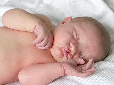
Babies born during the COVID-19 pandemic have differences in the size of certain structures in the brain, compared to infants born before the pandemic, according to a new study led by researchers at Children’s National Hospital.

The team found differences in the brains of both infants whose mothers were infected with COVID while pregnant, as well as those born to mothers who did not test positive for the virus, according to the study published in Cerebral Cortex.

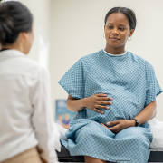
The findings suggest that exposure to the coronavirus and being pregnant during the pandemic could play a role in shaping infant brain development, said Nickie Andescavage, M.D., the first author of the paper and associate chief for the Developing Brain Institute at Children’s National.

The fine print

The study’s authors looked at three groups of infants: 108 born before the pandemic; 47 exposed to COVID before birth; and 55 unexposed infants. In all cases, researchers performed magnetic resonance imaging (MRI) scans of the newborns’ brains during the first few weeks of life. The MRI scans, which are non-invasive and do not expose patients to radiation, provided 3D images of the brain, allowing doctors to calculate the volume of different areas.

Researchers found several differences in the brains of babies exposed to COVID. They had larger volumes of the gray matter that makes up the brain’s outermost layer, compared to the two other groups. In contrast, an inner area of the brain, known as deep gray matter, was smaller than in unexposed babies. These are areas that contain large numbers of neurons that generate and process signals throughout the brain. “Their brains formed differently if they were exposed to COVID,” said Dr. Andescavage, adding that “those exposed to COVID had unique signatures” in the brain.

Doctors also measured the depths of the folds in the babies’ brains – a way to determine how the brain is maturing during early development. Babies born to mothers who had COVID in pregnancy had deeper grooves in the frontal lobe, while babies born during the pandemic – even without being exposed to COVID – had increased folds and grooves throughout the brain, compared to babies born before the pandemic. “There was something about being born during the pandemic that changed how the brain developed,” Dr. Andescavage said.

What’s ahead

The study authors can’t fully explain what caused the differences in brain development in these babies, Dr. Andescavage said. But other studies have linked maternal stress and depression to changes in the newborn brain. In a future study, Dr. Andescavage and her colleagues will examine the relationship between infant brain development and how stress and anxiety during the pandemic may have played a role in early development.

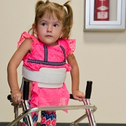
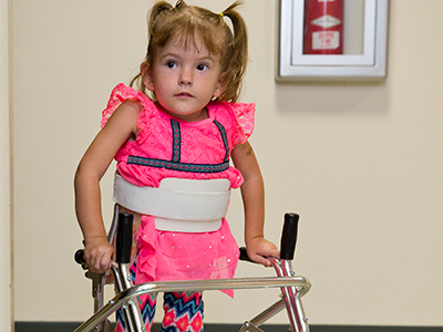
Because the babies in the study were just a few weeks old, researchers don’t know if their altered brain development will affect how they learn or behave. Researchers plan to follow the children until age 6, allowing them to observe whether pandemic-era babies hit key developmental milestones on time, such as walking, talking, holding a crayon and learning the alphabet.

Researchers have been worried about the effect of COVID on the fetus since the beginning of the pandemic. Studies show that babies exposed to COVID in the womb may experience developmental impacts, and research is underway to better understand long-term outcomes.

Although the coronavirus rarely crosses the placenta to infect the fetus directly, there are other ways maternal infection can influence the developing baby. Dr. Andescavage said inflammation is one potential harm to a developing baby. In addition, if a pregnant woman becomes so sick that the levels of oxygen in her blood fall significantly, that can deprive the fetus of oxygen, she added.

In recent decades, studies of large populations have found that maternal infections with influenza and other viruses increased the risk of serious problems in children even years later, including autism, attention deficit hyperactivity disorder and schizophrenia, although the reasons behind the association are not well understood. Technology may allow doctors to answer a number of questions about COVID and the infant brain.

“With advanced imaging and MRI, we’re in a position now to be able to understand how the babies are developing in ways we never previously could,” Dr. Andescavage said. “That will better allow us to identify the exposures that may be harmful, and at what times babies may be especially vulnerable, to better position us to promote maternal wellness. This, in turn, helps infant wellness.”

Mark Batshaw

40 years, 8 editions: Writing “Children With Disabilities”

Mark Batshaw

Forty years ago, Mark L. Batshaw, M.D., almost singlehandedly wrote a 23-chapter first edition that ran about 300 pages. Now Dr. Batshaw’s tome, “Children With Disabilities,” is in its eighth edition, and this new volume is almost 1,000 pages, with 42 chapters, two co-editors and over 35 authors from Children’s National.

Back in 1978, Mark L. Batshaw, M.D., was a junior faculty member at John’s Hopkins University School of Medicine. In the evenings he taught a course in the university’s School of Education  titled “The Medical and Physical Aspects of the Handicapped Child,” for Master’s level special education students. Because no textbook at that time focused on that specific topic, Batshaw developed his own slide set.

“At the end of the first year of teaching the course my students said ‘You really ought to consider writing a text book based on your slides to help us move forward,’ ” Dr. Batshaw recalls. The father of three carved out time by writing on weekends and at night, cutting back on sleep.

His first goal was to create a textbook that would serve as a curriculum for a series of courses that would be taught at universities to specialists who work with children with disabilities, including social workers, physical and occupational therapists, speech and language pathologists, special education teachers, nurses, doctors and dentists.

“I wanted to cover the whole range of disabilities and divided the book initially into a series of sections, including embryology, to help students understand what can go wrong in fetal development to lead to a developmental disability; and chapters on each developmental disability, including autism, attention-deficit/hyperactivity disorder (ADHD), cerebral palsy, learning disabilities and traumatic brain injury,” he says. “The third section was devoted to available treatments, including occupational and physical therapy, speech language therapy, nutrition and medications. The final section focused on outcomes.”

His second aim was for the book to serve as a reference text for professionals in the field. The 33-year-old contacted a brand-new new publisher, Paul H. Brookes Publishing Co., that focused on special education. “They took a chance on me, and I took a chance on them,” he says.

Forty years ago, he almost singlehandedly produced a 23-chapter first edition that ran about 300 pages. Now Dr. Batshaw’s tome is in its eighth edition, and this new volume is almost 1,000 pages. And, rather than being its sole author, Dr. Batshaw enlisted two co-editors and at least five dozen authors who contributed specialty expertise in genetic counseling, social work, physical and occupational therapy, medicine and nursing. His daughter, Elissa, a special education teacher and school psychologist, authored a chapter about special education services, and his son, Drew, an executive at a start-up company, contributed autobiographical letters about the effect ADHD has had on his life.

The book, “Children With Disabilities,” also includes:

  • A glossary of medical terms so that as the reader reviews patient reports they can easily look up an unfamiliar term
  • An appendix on commonly used drugs to treat children with disabilities in order to look up the medicine by name and see the range of doses
  • An appendix devoted to different syndromes children might have
  • A reference section with organizations and foundations that help children with disabilities
  • A web site with sections designed for students and other content designed for teachers with thought questions to guide practical use of information in each chapter and more than 450 customizable PowerPoint slides for download
  • Call-out boxes for interdisciplinary team members, such as genetic counselors, explaining the roles they serve and their educational background, and
  • Excerpts of recent research articles.

“The students say they don’t sell the book. Usually when students have a textbook, they try to sell it second hand after the course ends,” explains Dr. Batshaw, now Executive Vice President, Physician-in-Chief and Chief Academic Officer at Children’s National. “Instead, students keep it and use it as a practical reference as they become professionals in their field. It has had the impact I had hoped for both as a textbook and a reference book: They say they refer to it when they have patients with a particular disorder they’re not used to treating to read up on it.”

Now a bestseller, there are more than 200,000 copies in print, including Portuguese and Ukrainian translations. “It didn’t start that way. It grew organically,” he says.

In addition to Dr. Batshaw, Children’s contributors to “Children With Disabilities” include Nicholas Ah Mew, M.D., pediatric geneticist; Nickie N. Andescavage, M.D., neonatologist; Mackenzie E. Brown, D.O., fellow in Pediatric Rehabilitation Medicine; Justin M. Burton, M.D., chief, Division of Pediatric Rehabilitation Medicine; Gabrielle Sky Cardwell, BA, clinical research assistant; Catherine Larsen Coley, PT, DPT, PCS, physical therapist; Laurie S. Conklin, M.D., pediatric gastroenterologist; Denice Cora-Bramble, M.D., MBA, executive vice president and chief medical officer; Heather de Beaufort, M.D., pediatric ophthalmologist; Dewi Frances T. Depositario-Cabacar, M.D., pediatric neurologist; Lina Diaz-Calderon, M.D., fellow in Pediatric Gastroenterology; Olanrewaju O. Falusi, M.D., associate medical director of municipal and regional affairs, Child Health Advocacy Institute; Melissa Fleming, M.D., pediatric rehabilitation specialist; William Davis Gaillard, M.D., chief Division of Epilepsy, Neurophysiology and Critical Care; Satvika Garg, Ph.D., occupational therapist; Virginia C. Gebus, R.N., MSN, APN, CNSC, nutritionist; Monika K. Goyal, M.D., MSCE, assistant chief, Division of Emergency Medicine; Andrea Gropman, M.D., chief, Division of Neurodevelopmental Pediatrics and Neurogenetics, geneticist and Neurodevelopmental pediatrician; Mary A. Hadley, BS, senior executive assistant; Susan Keller, MLS., MS-HIT, research librarian; Lauren Kenworthy, Ph.D., director, Center for Autism Spectrum Disorders; Monisha S. Kisling, MS, CGC, genetic counselor; Eyby Leon, M.D., pediatric geneticist; Erin MacLeod, Ph.D., RD, LD, director, Metabolic Nutrition; Margaret B. Menzel, MS, CGC, genetic counselor; Shogo John Miyagi, Ph.D., PharmD, BCPPS, Pediatric Clinical Pharmacology fellow; Mitali Y. Patel, DDS, program director, Pediatric Dentistry; Deborah Potvin, Ph.D., neuropsychologist; Cara E. Pugliese, Ph.D., clinical psychologist; Khodayar Rais-Bahrami, M.D., neonatologist and director, Neonatal-Perinatal Medicine Fellowship Program; Allison B. Ratto, Ph.D., clinical psychologist; Adelaide S. Robb, M.D., chief, Division of Psychiatry and Behavioral Sciences; Joseph Scafidi, D.O., neonatal neurologist; Erik Scheifele, D.M.D., chief, Division of Oral Health; Rhonda L. Schonberg, MS, CGC, genetic counselor; Billie Lou Short, M.D., chief, Division of Neonatology; Kara L. Simpson, MS, CGC, genetic counselor; Anupama Rao Tate, D.M.D., MPH, pediatric dentist; Lisa Tuchman, M.D., MPH, chief, Division of Adolescent and Young Adult Medicine; Johannes N. van den Anker, M.D., Ph.D., FCP, chief, Division of Clinical Pharmacology, Vice Chair of Experimental Therapeutics; Miriam Weiss, CPNP-PC, nurse practitioner; and Tesfaye Getaneh Zelleke, M.D., pediatric neurologist.

little girl with spina bifida

Oral clefts may stem from a shared genetic cause as neural tube defects

little girl with spina bifida

Research by an international team that includes Children’s National faculty, published online Jan. 25, 2019 in Human Molecular Genetics, suggests that genetic mutations that cause cleft lip and palate also may contribute to neural tube defects, such as spina bifida.

Oral clefts are some of the most common birth defects worldwide, affecting about one in every 700 births. In the U.S., more than 4,000 babies are born each year with cleft lip, with or without cleft palate.

This defect isn’t simply a cosmetic manner: Oral clefts can severely affect feeding, speech and hearing, and they cause about 3,300 deaths annually worldwide.

To better understand these conditions, researchers have isolated a number of genetic mutations that appear to play contributing roles. These include those in a gene known as Interferon Regulatory Factor 6. New research by an international team that includes Children’s National faculty, published online Jan. 25, 2019 in Human Molecular Genetics, suggests that these mutations also may contribute to neural tube defects such as spina bifida.

In the first weeks of fetal development, the neural plate curves, creating a neural tube that, once fused shut, becomes the fetal brain and fetal spinal cord. Neural tube defects, which can range from mild to severe, are characterized by incomplete development of the brain, spinal cord or meninges. These defects can potentially result in paralysis or even fetal or neonatal demise. According to the National Institutes of Health, spina bifida, which affects the spinal cord, is the most common neural tube defect in the U.S., affecting up to 2,000 infants each year.

“Despite its high frequency, spina bifida remains among the least understood structural birth defects,” says Brian C. Schutte, an associate professor of Microbiology and Molecular Genetics, Pediatrics and Human Development at Michigan State University and the study’s senior author. “There is strong evidence that genetic factors are a leading cause of such structural birth defects, but in most cases, the cause is unknown. Our team’s study is the first published research to demonstrate that DNA variants in the gene IRF6 can cause spina bifida,” Schutte says.

What’s more, the research team identified a mechanism to explain how altering IRF6 leads to neural tube defects. This mechanism links IRF6 function to two other genes – known as transcription Factor AP2A (TFAP2A) and Grainyhead Like 3 (GRHL3) – that are also known to be required for the development of the neural tube, lip and palate.

“We’re all on the hunt for the reasons when, how and why birth defects happen,” adds Youssef A. Kousa, MS, D.O., Ph.D., a clinical fellow in the Division of Child Neurology at Children’s National Health System and the study’s lead author. “Our main goal is prevention. This paper is a significant development because our team has identified a group of genes that can potentially contribute to very common types of birth defects: craniofacial as well as neural tube defects.”

The scientific odyssey is a wonderful example of serendipity. Kousa, then working in Schutte’s lab, was studying the effects of a new mutant experimental model strain on development of the palate. But one day, he walked into Schutte’s office holding a deformed preclinical embryo and said: “Brian, look at this!”

“Weird things happen in biology,” Schutte replied and counseled him to return if it happened again. Less than two weeks later, Kousa was back with several more of the deformed preclinical embryos, saying: “OK, Brian. It happened again.”

Within hours Kousa had unearthed recently published research that included an image of a similarly affected preclinical embryo. The pair then sketched out possible intersecting genetic pathways, as they brainstormed the myriad ways to end up with that specific phenotype. Initially, they tested their hypotheses in experimental models and eventually corroborated findings through human genetic studies.

The human studies could only be performed by collaborations. Schutte shared their initial observations with human genetics researchers scattered across the country. Those labs then generously agreed to test whether DNA variants in IRF6 were associated with neural tube defects in samples from patients that they had collected over decades of research.

The team found that Tfap2aIrf6 and Grhl3 are components of a gene regulatory network required for neurulation, a folding process that results in the neural tube bending and then fusing to become the basis of the embryo’s nervous system, from brain to spinal cord.

“Since this network is also required for formation of the lip, palate, limbs and epidermis, which develop at different times and places during embryogenesis, we suggest that the Tfap2aIrf6Grhl3 network is a fundamental pathway for multiple morphogenetic processes,” the researchers write.

Interferon Regulatory Factor 6 functions best when there is neither too much expression nor too little. Overexpression of Irf6 suppresses Transcription Factor Activation Protein 2A and Grainyhead Like 3, causing exencephaly, a neural tube defect characterized by the brain being located outside of the skull. Counterintuitively, experimental models that had too little Irf6 also ended up with reduced levels of Tfap2a and Grhl3 that led to a structural birth defect, but at the opposite end of the neural tube.

To test whether the experimental model findings held true in humans, they sequenced samples from people who had spina bifida and anencephaly – the rare birth defect that Kousa spotted in the experimental models – and found IRF6 function was conserved in people. Because of the genetic complexity of these birth defects, and the challenges inherent in collecting samples from cases of severe birth defects, many research teams were invited to participate in the study.

As testament to their collegiality, researchers from Stanford University, University of Texas at Austin, University of Iowa, University of Texas at Houston and Duke University agreed to share precious samples from the California Birth Defects Monitoring Program, from the Hereditary Basis of Neural Tube Defects study and from their own institutional sample collections.

“As we get better at personalized medicine, we could use this information to one day help to counsel families about their own risk and protective factors,” Kousa adds. “If we can identify the genetic pathway, we might also be able to modify it to prevent a birth defect. For example, prenatal supplementation with folic acid has led to a decrease in babies born with neural tube defects, but not all neural tube defects are sensitive to folic acid. This knowledge will help us develop individual-based interventions.”

Financial support for the research covered in this post was provided by the National Institutes of Health under grants DE13513, F31DE022696, DE025060, P01HD067244 and GM072859; startup funding from Michigan State University and the UT-Health School of Dentistry in Houston; and the Centers for Disease Control and Prevention under award number 5U01DD001033.

In addition to Kousa and Schutte, study co-authors include Huiping Zhu, Yunping Lei and Richard H. Finnell, University of Texas at Austin; Walid D. Fakhouri, University of Texas Health Science Center at Houston; Akira Kinoshita, Nagasaki University; Raeuf R. Roushangar, Nicole K. Patel, Tamer Mansour, Arianna L. Smith, and Dhruv B. Sharma, Michigan State University; A.J. Agopian and Laura E. Mitchell, University of Texas School of Public Health; Wei Yang and Gary M. Shaw, Stanford University School of Medicine; Elizabeth J. Leslie, Emory University; Xiao Li, Tamara D. Busch, Alexander G. Bassuk and Brad A. Amendt, University of Iowa; Edward B. Li and Eric C. Liao, Massachusetts General Hospital; Trevor J. Williams, University of Colorado Denver at Anschutz Medical Campus; Yang Chai, University of Southern California; and Simon Gregory and Allison Ashley-Koch, Duke University Medical Center.

Baby with Cleft Palate

Understanding genetic synergy in cleft palate

Baby with Cleft Palate

Like mechanics fixing a faulty engine, Youssef A. Kousa, M.S., D.O., Ph.D., says researchers will not be able to remedy problems related to IRF6, a gene implicated in cleft palate, until they better understand how the gene works.

Like all of the individual elements of fetal development, palate growth is a marvel of nature. In part of this process, ledges of tissue on the sides of the face grow downwards on each side of the tongue, then upward, fusing at the midline at the top of the mouth. The vast majority of the time, this process goes correctly. However, some part of it goes awry for the 2,650 babies born in the United States each year with cleft palates and the thousands more born worldwide with the defect.

For nearly two decades, researchers have known that a gene known as IRF6 is involved in palate formation. Studies have shown that this gene contributes about 12 percent to 18 percent of the risk of cleft palate, more than any other gene identified thus far. IRF6 is active in epithelial tissues – those that line cavities and surfaces throughout the body – including the periderm, a tissue that lines the mouth cavity and plays an important role during development.

According to Youssef A. Kousa, M.S., D.O., Ph.D., a child neurology fellow at Children’s National Health System, the periderm acts like a nonstick layer, preventing the tongue or other structures from adhering to the growing palate and preventing it from sealing at the midline. While researchers have long suspected that IRF6 plays a strong role in promoting this nonstick quality, exactly how it exerts its influence has not been clear.

“Gaining a better understanding of this gene might help us to eventually address deficits or perturbations in the system that creates the palate,” Dr. Kousa says. “Like a mechanic fixing a faulty engine, we will not be able to remedy problems related to this gene until we know how the gene works.”

Youssef Kousa

“Gaining a better understanding of this gene might help us to eventually address deficits or perturbations in the system that creates the palate,” Dr. Kousa says. “Like a mechanic fixing a faulty engine, we will not be able to remedy problems related to this gene until we know how the gene works.”

In a study published July 19, 2017 by the Journal of Dental Research, Dr. Kousa and colleagues seek to decipher one piece of this puzzle by investigating how this key gene might interact with others that are active during fetal development. The researchers were particularly interested in genes that work together in a cascade of activity known as the tyrosine kinase receptor signaling pathway.

Because this pathway includes a large group of genes, Dr. Kousa and colleagues reasoned that they could answer whether IRF6 interacts with this pathway by looking at whether the gene interacts with the last member of the cascade, a gene called SPRY4. To do this, the researchers worked with experimental models that had mutations in IRF6, SPRY4 or both. If these two genes interact, the scientists hypothesized, carrying mutations in both genes at the same time should result in a dramatically different outcome compared with animals that carried mutations in just one gene.

Using selective breeding techniques, the researchers created animals that had mutations in either of these genes or in both. Their results suggest that IRF6 and SPRY4 indeed do interact: Significantly more of the oral surface was adhered to the tongue during fetal development in experimental models that had mutations in both genes compared with those that had just one single gene mutated. Examining the gene activity in the periderm cells of these affected animals, the researchers found that doubly mutated experimental models also had decreased activity in a third gene known as GRHL3, which also has been linked with cleft lip and palate.

Dr. Kousa says the research team plans to continue exploring this interaction to better understand the flow of events that lead from perturbations in these genes to formation of cleft palate. Some of the questions they would like to answer include exactly which gene or genes in the tyrosine kinase receptor signaling pathway specifically interact with IRF6 – since SPRY4 represents just the end of that pathway, others genes earlier in the pathway are probably the real culprits responsible for driving problems in palate formation. They also will need to verify if these interactions take place in humans in the same way they occur in preclinical models.

Eventually, Dr. Kousa adds, the findings could aid in personalized prenatal counseling, diagnosis and screening related to cleft palate, as well as preventing this condition during pregnancy. Someday, doctors might be able to advise couples who carry mutations in these genes about whether they are more likely to have a baby with a cleft palate or determine which select group of pregnancies need closer monitoring. Additionally, because research suggests that GRHL3 might interact with nutrients, including inositol, it might be possible to prevent some cases of cleft palate by taking additional supplements during pregnancy.

“The more we know about how these genes behave,” Dr. Kousa says, “the more we can potentially avoid fetal palate development going down the wrong path.”