Tag Archive for: fetal brain development

Neighborhood disadvantage alters brain networks in unborn babies

Illustration of high and low brain efficiencyGrowing up in a disadvantaged neighborhood changes a child’s brain even before birth. These new findings, in the latest edition of The Journal of Pediatrics, underscore the need to support young families from a baby’s earliest days.

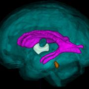
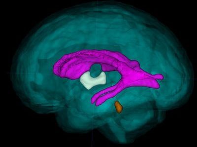
According to this new research from the Center for Prenatal, Neonatal & Maternal Health Research at Children’s National Hospital, exposure to neighborhood disadvantage lessens the functional integration of neural networks in the fetal brain, as seen on functional MRIs of healthy babies. The center compared the brains of 68 healthy babies on 79 scans. Researchers then mapped details about neural activity to a “social vulnerability index” from the Centers for Disease Control and Prevention, which indicates proximity to environmental stressors.

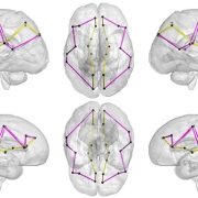
“We specifically looked at brain architecture to see how easily information flows between different regions,” said Kevin Cook, Ph.D., research faculty at the center and the manuscript’s first author. “To do this, we used graph theory, which borrows concepts from social network theory. It’s widely applied in computer science to understand how information flows within groups, and neuroscience has adapted it to study how information travels within the brain.”

What we found

Dr. Cook said researchers focused on the three metrics:

  • Path length, which measures how many stops information needs to make along its way through the brain.
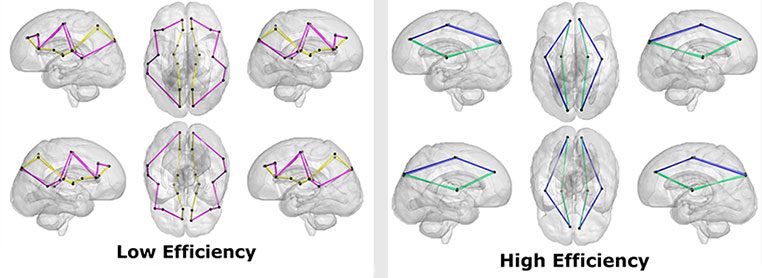
  • Global efficiency, which measures the overall efficiency of the entire brain’s network.
  • Small-world propensity, which describes how the brain’s network is organized and indicates how well the brain is organized into smaller, efficient networks.

As social vulnerability increased, the research team found global efficiency decreased, meaning the brain’s neural network was less efficient. The path lengths were also longer in children with greater neighborhood disadvantage, reinforcing the global efficiency findings.

The fine print

The under-development and over-development of fetal brains may contribute to neurological disorders, such as autism, epilepsy and other conditions of interest to researchers. Yet science’s understanding of how the brain matures in utero is still limited.

In this study, researchers found a notable difference related to age. At the youngest gestational ages, path lengths are longer, and both global efficiency and small world propensity are lower. As the fetus gets closer to term, path length and global efficiency show rapid maturation and less advantaged fetuses catch up to their peers who have greater advantages.

Researchers saw the same findings for small-world propensity, but the maturation didn’t stop. These unborn babies overshot their peers and had greater small-world propensity, suggesting their brains are divided into a greater number of smaller networks than their advantaged peers.

“We believe that length and global efficiency are on a trend to overshoot,” Dr. Cook said. “These findings are notable because they agree with what we know about older children and adolescents. Greater disadvantage is associated with hyper- or over-maturation of the brain. Our findings suggest that this may be starting even before birth.”

What’s ahead

While still early, this research improves the understanding of how environmental complexities can impact an unborn baby. Catherine Limperopoulos, Ph.D., director of the research center, which opened in 2023, said this work will be foundational as they continue to study the impact of a child’s environment on development.

“These findings have important implications for understanding how status and disadvantage may have a cumulative effect on fetal brain development,” Dr. Limperopoulos said. “We must study and consider how to conceptualize the impact of socioenvironmental disadvantage in communities to better care for children and work to improve outcomes.”

Read the full study – “Greater Neighborhood Disadvantage Is Associated with Alterations in Fetal Functional Brain Network Structure” – in The Journal of Pediatrics.

Prenatal COVID exposure associated with changes in newborn brain

pregnant woman talking to doctor

The team found differences in the brains of both infants whose mothers were infected with COVID while pregnant, as well as those born to mothers who did not test positive for the virus.

Babies born during the COVID-19 pandemic have differences in the size of certain structures in the brain, compared to infants born before the pandemic, according to a new study led by researchers at Children’s National Hospital.

The team found differences in the brains of both infants whose mothers were infected with COVID while pregnant, as well as those born to mothers who did not test positive for the virus, according to the study published in Cerebral Cortex.

The findings suggest that exposure to the coronavirus and being pregnant during the pandemic could play a role in shaping infant brain development, said Nickie Andescavage, M.D., the first author of the paper and associate chief for the Developing Brain Institute at Children’s National.

The fine print

The study’s authors looked at three groups of infants: 108 born before the pandemic; 47 exposed to COVID before birth; and 55 unexposed infants. In all cases, researchers performed magnetic resonance imaging (MRI) scans of the newborns’ brains during the first few weeks of life. The MRI scans, which are non-invasive and do not expose patients to radiation, provided 3D images of the brain, allowing doctors to calculate the volume of different areas.

Researchers found several differences in the brains of babies exposed to COVID. They had larger volumes of the gray matter that makes up the brain’s outermost layer, compared to the two other groups. In contrast, an inner area of the brain, known as deep gray matter, was smaller than in unexposed babies. These are areas that contain large numbers of neurons that generate and process signals throughout the brain. “Their brains formed differently if they were exposed to COVID,” said Dr. Andescavage, adding that “those exposed to COVID had unique signatures” in the brain.

Doctors also measured the depths of the folds in the babies’ brains – a way to determine how the brain is maturing during early development. Babies born to mothers who had COVID in pregnancy had deeper grooves in the frontal lobe, while babies born during the pandemic – even without being exposed to COVID – had increased folds and grooves throughout the brain, compared to babies born before the pandemic. “There was something about being born during the pandemic that changed how the brain developed,” Dr. Andescavage said.

What’s ahead

The study authors can’t fully explain what caused the differences in brain development in these babies, Dr. Andescavage said. But other studies have linked maternal stress and depression to changes in the newborn brain. In a future study, Dr. Andescavage and her colleagues will examine the relationship between infant brain development and how stress and anxiety during the pandemic may have played a role in early development.

Because the babies in the study were just a few weeks old, researchers don’t know if their altered brain development will affect how they learn or behave. Researchers plan to follow the children until age 6, allowing them to observe whether pandemic-era babies hit key developmental milestones on time, such as walking, talking, holding a crayon and learning the alphabet.

Researchers have been worried about the effect of COVID on the fetus since the beginning of the pandemic. Studies show that babies exposed to COVID in the womb may experience developmental impacts, and research is underway to better understand long-term outcomes.

Although the coronavirus rarely crosses the placenta to infect the fetus directly, there are other ways maternal infection can influence the developing baby. Dr. Andescavage said inflammation is one potential harm to a developing baby. In addition, if a pregnant woman becomes so sick that the levels of oxygen in her blood fall significantly, that can deprive the fetus of oxygen, she added.

In recent decades, studies of large populations have found that maternal infections with influenza and other viruses increased the risk of serious problems in children even years later, including autism, attention deficit hyperactivity disorder and schizophrenia, although the reasons behind the association are not well understood. Technology may allow doctors to answer a number of questions about COVID and the infant brain.

“With advanced imaging and MRI, we’re in a position now to be able to understand how the babies are developing in ways we never previously could,” Dr. Andescavage said. “That will better allow us to identify the exposures that may be harmful, and at what times babies may be especially vulnerable, to better position us to promote maternal wellness. This, in turn, helps infant wellness.”

Pandemic-related stressors in pregnant women affect fetal brain development

Dr. Limperopoulos talks to a mom

Dr. Catherine Limperopoulos walking with a mom.

Prolonged levels of stress and depression during the COVID-19 pandemic contributed to altering key features of fetal brain development — even if the mother was not infected by the virus. This is what a study published in Communications Medicine suggests after following more than 200 pregnant women. The study, led by Children’s National Hospital experts, emphasized the need for more scientific inquiry to shed light on the long-term neurodevelopmental consequences of their findings and COVID-19 exposures on fetal brain development.

“Understanding how contemporary stressors may influence fetal brain development during pregnancy has major implications for basic science and informing public policy initiatives,” said Catherine Limperopoulos, Ph.D., chief and director of the Developing Brain Institute at Children’s National and senior author of the study. “With this work, we are able to show there’s a problem, it’s happening prenatally, and we can use this model to start exploring how we can reduce stress in moms and support unborn babies.”

To better understand the effects of environmental exposures on the fetus during pregnancy, further confirmation of the team’s latest findings is needed by ruling out other possibilities, such as maternal nutrition, financial security and genetic factors.

The psychosocial impact of COVID-19 on fetal brain development remains vastly understudied. The neurologic underpinnings of fetal development that turn into psycho-behavioral disorders later in life, including bipolar disorder, mood disorder or anxiety disorder, remain complex and difficult to explain.

Among the 202 participants from the Washington D.C. metropolitan area, 137 were part of the pre-pandemic cohort and 65 were part of the pandemic cohort.

Through advanced MRI imaging techniques and reconstruction of high-resolution 3D brain models, the researchers found a reduction of fetal white matter, hippocampal and cerebellar volumes and delayed brain gyrification in COVID-19 pandemic-era pregnancies. Validated maternal stress, anxiety and depression scales were also used to compare the scores between the two cohorts.

This study builds upon previous work from the Developing Brain Institute led by Limperopoulos, which discovered that anxiety in pregnant women appears to affect the brain development of their babies. Her team also found that maternal mental health, even in high socioeconomic status, alters the structure and biochemistry of the developing fetal brain, emphasizing the importance of mental health support for pregnant women.

“We’re looking at modifiable conditions,” said Limperopoulos. “What’s clear is the next frontier is intervening early to see how we can prevent or reduce stress in the mom’s current setting.”

Low parental socioeconomic status alters brain development in unborn babies

doctor examining pregnant woman

A first-of-its-kind study with 144 pregnant women finds that socioeconomic status (SES) has an impact in the womb, altering several key regions in the developing fetal brain as well as cortical features.

Maternal socioeconomic status impacts babies even before birth, emphasizing the need for policy interventions to support the wellbeing of pregnant women, according to newly published research from Children’s National Hospital.

A first-of-its-kind study with 144 pregnant women finds that socioeconomic status (SES) has an impact in the womb, altering several key regions in the developing fetal brain as well as cortical features. Parental occupation and education levels encompassing populations with lower SES hinder early brain development, potentially affecting neural, social-emotional and cognitive function later in the infant’s life.

Having a clear understanding of early brain development can also help policymakers identify intervention approaches such as educational assistance and occupational training to support and optimize the well-being of people with low SES since they face multiple psychological and physical stressors that can influence childhood brain development, Lu et al. note in the study published in JAMA Network Open.

“While there has been extensive research about the interplay between socioeconomic status and brain development, until now little has been known about the exact time when brain development is altered in people at high-risk for poor developmental outcomes,” said Catherine Limperopoulos, Ph.D., director of the Developing Brain Institute and senior author. “There are many reasons why these children can be vulnerable, including high rates of maternal prenatal depression and anxiety. Later in life, these children may experience conduct disorders and impaired neurocognitive functions needed to acquire knowledge, which is the base to thrive in school, work or life.”

The findings suggest that fetuses carried by women with low socioeconomic backgrounds had decreased regional brain growth and accelerated brain gyrification and surface folding patterns on the brain. This observation in lower SES populations may in part be explained by elevated parental stress and may be associated with neuropsychiatric disorders and mental illness later in life.

In contrast, fetuses carried by women with higher education levels, occupation and SES scores showed an increased white matter, cerebellar and brainstem volume during the prenatal period, and lower gyrification index and sulcal depth in the parietal, temporal and occipital lobes of the brain. These critical prenatal brain growth and development processes lay the foundation for normal brain function, which ready the infant for life outside the womb, enabling them to attain key developmental milestones after birth, including walking, talking, learning and social skills.

There is also a knowledge gap in the association between socioeconomic status and fetal cortical folding — when the brain undergoes structural changes to create sulcal and gyral regions. The study’s findings of accelerated gyrification in low SES adds to the scientific record, helping inform future research, Limperopoulos added.

The Children’s National research team gathered data from 144 healthy women at 24 to 40 weeks gestation with uncomplicated pregnancies. To establish the parameters for socioeconomic status, which included occupation and education in lieu of family income, parents completed a questionnaire at the time of each brain magnetic resonance imaging (MRI) visit. The researchers used MRI to measure fetal brain volumes, including cortical gray matter, white matter, deep gray matter, cerebellum and brain stem. Out of the 144 participants, the scientists scanned 40 brain fetuses twice during the pregnancy, and the rest were scanned once. The 3-dimensional computational brain models among healthy fetuses helped determine fetal brain cortical folding.

Potential proximal risk factors like maternal distress were also measured in the study using a questionnaire accounting for 60% of the participants but, according to the limited data available, there was no significant association with low and high socioeconomic status nor brain volume and cortical features.

Authors in the study from Children’s National include: Yuan-Chiao Lu, Ph.D., Kushal Kapse, M.S., Nicole Andersen, B.A., Jessica Quistorff, M.P.H., Catherine Lopez, M.S., Andrea Fry, B.S., Jenhao Cheng, Ph.D., Nickie Andescavage, M.D., Yao Wu, Ph.D., Kristina Espinosa, Psy.D., Gilbert Vezina, M.D., Adre du Plessis, M.D., and Catherine Limperopoulos, Ph.D.

Protecting the fetal brain from harm

Anna Penn

Ongoing placental dysfunction and allopregnanolone loss, not the increase that was expected due to stress, may alter cortical development in complicated pregnancies and put babies at risk, says Anna Penn, M.D., Ph.D.

Researchers long have known that allopregnanolone (ALLO), a derivative of the hormone progesterone, is produced in adults’ brains during times of acute stress and modulates how easily the brain’s neurons fire. ALLO also is produced in the placenta during fetal development, one of more than 200 different hormones that each uniquely contribute to fostering a smooth pregnancy and maintaining a fetus’ overall health. Although ALLO is thought to protect the developing brain in pregnancies complicated by conditions that might harm it, such as high blood pressure, how its levels evolve during pregnancy and in newborns shortly after birth has remained unknown.

Now, a new study presented during the Pediatric Academic Societies (PAS) 2018 annual meeting suggests that the placenta ramps up ALLO production over the second trimester, peaking just as fetuses approach full term.

To investigate this phenomenon, Anna Penn, M.D., Ph.D., a neonatologist/neuroscientist at Children’s National Health System, and colleagues created a designer experimental model to study how premature loss of ALLO alters orderly brain development. Knowing more about the interplay between ALLO and normal development of the cortex, the outer layer of the cerebrum, is a first step that could lead to strategies to rescue this vital brain region.

“The cortex is basically the brain’s command-and-control center for higher functions. In our experimental model, it develops from the middle of gestation through to the end of gestation. If ALLO levels are disrupted just as these cells are being born, neurons migrating to the cortex are altered and the developing neural network is compromised,” says Dr. Penn, senior author of the research presented at PAS 2018. “We’re concerned this same phenomenon occurs in human infants whose preterm birth disrupts their supply of this essential hormone.”

To better understand the human placental hormone pattern, the research team analyzed cord blood or serum samples collected within the first 36 hours of life for 61 preterm newborns born between 24 to 36 gestational weeks. They compared those preemie samples with samples drawn from 61 newborns carried to term who were matched by race, gender, size for gestational age, delivery method and maternal demographics.

They used liquid-chromatography-tandem mass spectrometry, a technique that can precisely analyze trace levels of compounds, to compare levels of 27 different steroids, including ALLO and its precursors as well as better-known adrenal gland hormones, such as cortisol and 17-Hydroxyprogesterone.

“Pregnancies complicated by hypertension tended to correlate with lower ALLO levels, though this finding did not reach statistical significance. This suggests that ongoing placental dysfunction and ALLO loss, not the increase that we expected to be caused by stress, may alter cortical development in these pregnancies and put babies at risk,” Dr. Penn adds. “In addition, having the largest neonatal sample set to date in which multiple steroid hormones have been measured can provide insight into the shifting hormone patterns that occur around 36 weeks gestation, just prior to term. Hopefully, restoring the normal hormonal milieu for preemies or other at-risk newborns will improve neurological outcomes in the future.”

In addition to Dr. Penn, study co-authors include Caitlin Drumm, MedStar Georgetown University Hospital; Sameer Desale, MedStar Health Research Institute; and Kathi Huddleston, Benjamin Solomon and John Niederhuber, Inova Translational Medicine Institute.

GABA concentration in pre-term brain increases with gestational age

Sudeepta Basu

“A more complete understanding of the diagnostic and prognostic importance of GABA and glutamate in the preterm brain will help us to direct treatment strategies for the most vulnerable preterm infants at risk of brain injury,” says Sudeepta K. Basu, M.D.

The major neurotransmitters gamma-aminobutyric acid (GABA) and glutamate are pivotal to fetal and newborn brain development and influence evolution of brain injury and repair following preterm birth. Magnetic resonance spectroscopy (MRS) enables in vivo measurement of brain metabolites. However, GABA and glutamate are found in the developing brain in low concentrations, and their weak signal can be swamped by the stronger signal of more dominant metabolites.

A Children’s research team reports findings from a pilot study utilizing an innovative technique of MRS to reliably measure in vivo GABA in the developing preterm brain. The groundbreaking research done by the team that includes Principal Investigator Sudeepta K. Basu, M.D., neonatology attending at Children’s National Health System, is very unique and original since there are no existing data of in vivo GABA concentrations in the developing cerebellum. Under the mentorship of Catherine Limperopoulos, Ph.D., director of Children’s Developing Brain Research Laboratory, the team of multi-disciplinary specialists is pursuing cutting-edge technologies in advanced MRI neuroimaging to explore brain development and injury in preterm infants.

The research, presented at the Eastern Society for Pediatric Research (ESPR) annual meeting by Dr. Basu, was honored with the “2018 Meritorious Poster Award.” The research titled “Distinct temporal trends of GABA and glutamate in the cerebellum and frontal cortex of preterm infants” reports, for the first time, positive temporal trends in the specific regions of the developing brain intricately involved in cognitive and motor functions. This work lays the foundation for developing novel ways to diagnose, monitor and investigative brain protective therapies for vulnerable prematurely born infants.

The Children’s team performed non-sedated MRS in 44 preterm infants whose mean gestational age at birth was 26.5 weeks, placing voxels at the middle of the cerebellum and the right frontal cortex. GABA and GIx (glutamate combined with glutamine) were positively correlated with post-menstrual age in the frontal cortex, but not the cerebellum.  At the ESPR meeting, the team also presented for the first time that caffeine, a neuroprotective agent in preemies, leads to increased in vivo GABA concentration in the developing frontal cortex.

“Open questions include whether these findings reflect varying paces of maturation and vulnerability to injury among specific regions of the brain. Also, the relationship between clinical factors and medication exposure and changes in the concentration of these neurotransmitters may guide brain protective therapies in future,” Dr. Basu says. “A more complete understanding of the diagnostic and prognostic importance of GABA and glutamate in the preterm brain will help us to direct treatment strategies for the most vulnerable preterm infants at risk of brain injury.”

Children’s senior fellows from Division of Neonatology made four platform presentations during the ESPR conference:

  • “Caffeine increases GABA/Cr ratio in frontal cortex of preterm infants on spectroscopy.” Aditi Gupta; Sudeepta K. Basu, M.D.; Mariam Said, M.D.; Subechhya Pradhan, Linda White; Kushal Kapse; Jonathan Murnick, M.D., Ph.D.; Taeun Chang, M.D.; and Catherine Limperopoulos, Ph.D.
  • “Impact of early nutrition on microstructural brain development in VLBW Infants.” Katherine M. Ottolini, Nickie Andescavage, M.D.; Kushal Kapse; and Catherine Limperopoulos, Ph.D.
  • “Direct measurement of neonatal cardiac output utilizing the CO status monitor.” Simranjeet S. Sran, Mariam Said, M.D.; and Khodayar Rais-Bahrami, M.D.
  • “Cerebro-cerebellar diaschisis in preterm infants following unilateral cerebral parenchymal injury.” Huma Mirza, Yao Wu, Kushal Kapse, Jonathan Murnick, M.D., Ph.D.; Taeun Chang, M.D.; and Catherine Limperopoulos, Ph.D.

Setting a baseline for healthy brain development

Catherine Limperopoulos, Ph.D., and colleagues performed the largest magnetic resonance imaging study of normal fetal brains in the second and third trimesters of pregnancy.

Starting as a speck barely visible to the naked eye and ending the in utero phase of its journey at an average weight of 7.5 pounds, the growth of the human fetus is one of the most amazing events in biology. Of all the organs, the fetal brain undergoes one of the most rapid growth trajectories, expanding over 40 weeks from zero to 100 billion neurons — about as many brain cells as there are stars in the Milky Way Galaxy.

This exponential growth is part of what gives humans our unique abilities to use language or have abstract thoughts, among many other cognitive skills. It also leaves the brain extremely vulnerable should disruptions occur during fetal development. Any veering off the developmental plan can lead to a cascade of results that have long-lasting repercussions. For example, studies have shown that placental insufficiency, or the inability of the placenta to supply the fetus with oxygen and nutrients in utero, is associated with attention deficit hyperactivity disorder, autism, and schizophrenia.

Recent research has identified differences in the brains of people with these disorders compared with those without. Despite the almost certain start of these conditions within the womb, they have remained impossible to diagnose until children begin to show clinical symptoms. If only researchers could spot the beginnings of these problems early in development, says Children’s National Health System researcher Catherine Limperopoulos, Ph.D., they might someday be able to develop interventions that could turn the fetal brain back toward a healthy developmental trajectory.

“Conventional tools like ultrasound and magnetic resonance imaging (MRI) can identify structural brain abnormalities connected to these problems, but by the time these differences become apparent, the damage already has been done,” Limperopoulos says. “Our goal is to be able to pick up very early deviations from normal in the high-risk pregnancy before an injury to the fetus might become permanent.”

Before scientists can recognize abnormal, she adds, they first need to understand what normal looks like.

In a new study published in Cerebral Cortex, Limperopoulos and colleagues begin to tackle this question through the largest MRI study of normal fetal brains in the second and third trimesters of pregnancy. While other studies have attempted to track normal fetal brain growth, that research has not involved nearly as many subjects and typically relied on data collected when fetuses were referred for MRIs for a suspected problem. When the suspected abnormality was ruled out by the scan, these “quasi-controls” were considered “normal” — even though they may be at risk for problems later in life, Limperopoulos explains.

By contrast, the study she led recruited 166 healthy pregnant women from nearby low-risk obstetrics practices. Each woman had an unremarkable singleton pregnancy and ended up having a normal full-term delivery, with no evidence of problems affecting either the mother or fetus over the course of 40 weeks.

At least one time between 18 and 39 gestational weeks, the fetuses carried by these women underwent an MRI scan of their brains. The research team developed complex algorithms to account for movement (since neither the mothers nor their fetuses were sedated during scans) and to convert the two-dimensional images into three dimensions. They used the information from these scans to measure the increasing volumes of the cerebellum, an area of the brain connected to motor control and known to mediate cognitive skills; as well as regions of the cerebrum, the bulk of the brain, that is pivotal for movement, sensory processing, olfaction, language, and learning and memory.

Their results in uncomplicated, full-term pregnancies show that over 21 weeks in the second half of pregnancy, the cerebellum undergoes an astounding 34-fold increase in size. In the cerebrum, the fetal white matter, which connects various brain regions, grows 22-fold. The cortical gray matter, key to many of cerebrum’s functions, grows 21-fold. And the deep subcortical structures (thalamus and basal ganglia), important for relaying sensory information and coordination of movement and behavior, grow 10-fold. Additional examination showed that the left hemisphere has a larger volume than the right hemisphere early in development, but sizes of the left and right brain halves were equal by birth.

By developing similar datasets on high-risk pregnancies or births—for example, those in which fetuses are diagnosed with a problem in utero, mothers experience a significant health problem during pregnancy, babies are born prematurely, or fetuses have a sibling diagnosed with a health problem with genetic risk, such as autism—Limperopoulos says that researchers might be able to spot differences during gestation and post-natal development that lead to conditions such as schizophrenia, attention deficit hyperactivity disorder and autism spectrum disorder.

Eventually, researchers may be able to develop fixes so that babies grow up without life-long developmental issues.

“Understanding ‘normal’ is really opening up opportunities for us to begin to precisely pinpoint when things start to veer off track,” Limperopolous says. “Once we do that, opportunities that have been inaccessible will start to present themselves.”

Using 3-D MRI for fetal brain imaging during high-risk pregnancies

3DMRI

What’s Known
The placenta plays an essential role in the growth of a healthy fetus and, among other critical tasks, it ferries in oxygen and nutrients. During pregnancies complicated by fetal growth restriction (FGR), the failing placenta cannot support the developing fetus adequately. FGR is a major cause of stillbirth and death, and newborns who do survive face numerous risks for multiple types of ailments throughout their lives. In fact, studies have shown that nutrient depravation during gestation can have lasting consequences that may manifest themselves years or decades later in life. These risks can also cross generations, affecting future pregnancies.

What’s New
A team of researchers applied an advanced imaging technique, three-dimensional (3-D) MRI, to study brain development in these high-risk pregnancies. They are the first to report regional, tissue-specific volume delays for the developing fetal brain in FGR-affected pregnancies. The team compared overall fetal brain volume as well as regional brain volumes for a control group of healthy young pregnant women with a group of young women whose pregnancies were complicated by FGR. While fetuses in both groups grew exponentially as pregnancies progressed, the researchers began to see dramatic differences when they compared the volumes of specific regions of the brain, including the cerebellum, which coordinates balance and smooth movement; the deep gray matter, which also is involved in complex functions, such as memory and emotion; and the white matter, which is made up of millions of nerve fibers that connect to neurons in different regions. Because there are no biomarkers to spot early brain failure, 3-D MRI imaging may fill this knowledge gap.

Questions for Future Research
Q: Certain regions of the brains of FGR-affected infants show accelerated volume. Are these differences regional or global?
Q: Is accelerated brain volume in FGR-affected infants a result of heightened stress that these fetuses experience in the womb?
Q: How do differences in regional brain volume relate to later neurodevelopmental impairment that some FGR-affected infants experience?

Source: “Impaired Global and Tissue-Specific Brain Development in the Growth-Restricted Fetus.N. Andescavage, J. Cruz, M. Metzler, A. du Plessis, and C. Limperopoulos. Presented during the 2016 Pediatric Academic Societies Annual Meeting, Baltimore, MD. May 2, 2016.