Tag Archive for: enzymes

caspase molecule

Caspases may link brain cell degeneration and cardiac surgery

caspase molecule

The review summarizes both the known physiological roles of caspases as well as some of the well-characterized neurotoxic effects of anesthetics in pre-clinical models.

A review article in the journal Cell Press: Trends in Neuroscience outlines the wide variety of cellular signaling roles for caspase proteins — a type of cellular enzyme best known for its documented role in the natural process of cell death (apoptosis). The authors, including Nemanja Saric, Ph.D., Kazue Hashimoto-Torii, Ph.D., and Nobuyuki Ishibashi, M.D., all from Children’s National Research Institute, pay particular attention to what the scientific literature shows about caspases’ non-apoptotic roles in the neurons specifically. They also highlight research showing how, when activated during a cardiac surgery with anesthesia and cardiopulmonary bypass, these enzymes may contribute to the degeneration of brain cells seen in young children who undergo heart surgery for critical congenital heart defects (CHDs).

Why it matters

The review summarizes both the known physiological roles of caspases as well as some of the well-characterized neurotoxic effects of anesthetics in pre-clinical models.

The authors propose that these non-apoptotic activities of caspases may be behind some of the adverse effects on the developing brain related to cardiac surgery and anesthesia. Those adverse effects are known to increase risk of behavioral impairments in children with congenital heart disease who underwent cardiac surgery with both anesthesia and cardiopulmonary bypass at a very young age.

This work is the first to propose a possible link between developmental anesthesia neurotoxicity and caspase-dependent cellular responses.

The patient benefit

Better understanding of the time and dose-dependent effects of general anesthetics on the developing brain, particularly in children who have genetic predispositions to conditions such as CHDs, will help researchers understand their role (if any) in behavioral problems often encountered by these patients after surgery.

If found to be a contributing factor, perhaps new therapies to mitigate this caspase activity might be explored to alleviate some of these adverse effects on the developing brain.

What’s next?

The authors hope to stimulate more in-depth research into caspase signaling events, particularly related to how these signaling events change when an anesthetic is introduced. Deeper understanding of how anesthetics impact caspase activation in the developing brain will allow for better assessments of the risk for children who need major surgery early in life.

Children’s National leads the way

Children’s National Hospital leads studies funded by the U.S. Department of Defense to better understand how these other roles of caspases, which until now have not been well-documented, may contribute to brain cell degeneration when activated by prolonged anesthesia and cardiopulmonary bypass during cardiac surgery for congenital heart disease.

Andrea Gropman

$5M in federal funding to help patients with urea cycle disorders

Andrea Gropman

Andrea L. Gropman, M.D.: We have collected many years of longitudinal clinical data, but with this new funding now we can answer questions about these diseases that are meaningful on a day-to-day basis for patients with urea cycle disorders.

An international research consortium co-led by Andrea L. Gropman, M.D., at Children’s National Hospital has received $5 million in federal funding as part of an overall effort to better understand rare diseases and accelerate potential treatments to patients.

Urea cycle disorder, one such rare disease, is a hiccup in a series of biochemical reactions that transform nitrogen into a non-toxic compound, urea. The six enzymes and two carrier/transport molecules that accomplish this essential task reside primarily in the liver and, to a lesser degree, in other organs.

The majority of patients have the recessive form of the disorder, meaning it has skipped a generation. These kids inherit one copy of an abnormal gene from each parent, while the parents themselves were not affected, says Dr. Gropman, chief of the Division of Neurodevelopmental Pediatrics and Neurogenetics at Children’s National. Another more common version of the disease is carried on the X chromosome and affects boys more seriously that girls, given that boys have only one X chromosome.

Regardless of the type of urea cycle disorder, when the urea cycle breaks down, nitrogen converts into toxic ammonia that builds up in the body (hyperammonemia), particularly in the brain. As a result, the person may feel lethargic; if the ammonia in the bloodstream reaches the brain in high concentrations, the person can experience seizures, behavior changes and lapse into a coma.

Improvements in clinical care and the advent of effective medicines have transformed this once deadly disease into a more manageable chronic ailment.

“It’s gratifying that patients diagnosed with urea cycle disorder now are surviving, growing up, becoming young adults and starting families themselves. Twenty to 30 years ago, this never would have seemed conceivable,” Dr. Gropman says. “We have collected many years of longitudinal clinical data, but with this new funding now we can answer questions about these diseases that are meaningful on a day-to-day basis for patients with urea cycle disorders.”

In early October 2019, the National Institutes of Health (NIH) awarded the Urea Cycle Disorders Consortium for which Dr. Gropman is co-principal investigator a five-year grant. This is the fourth time that the international Consortium of physicians, scientists, neuropsychologists, nurses, genetic counselors and researchers has received NIH funding to study this group of conditions.

Dr. Gropman says the current urea cycle research program builds on a sturdy foundation built by previous principal investigators Mendel Tuchman, M.D., and Mark Batshaw, M.D., also funded by the NIH. While previous rounds of NIH funding powered research about patients’ long-term survival prospects and cognitive dysfunction, this next phase of research will explore patients’ long-term health.

Among the topics they will study:

Long-term organ damage. Magnetic resonance elastrography (MRE) is a state-of-the-art imaging technique that combines the sharp images from MRI with a visual map that shows body tissue stiffness. The research team will use MRE to look for early changes in the liver – before patients show any symptoms – that could be associated with long-term health impacts. Their aim is spot the earliest signs of potential liver dysfunction in order to intervene before the patient develops liver fibrosis.

Academic achievement. The research team will examine gaps in academic achievement for patients who appear to be underperforming to determine what is triggering the discrepancy between their potential and actual scholastics. If they uncover issues such as learning difficulties or mental health concerns like anxiety, there are opportunities to intervene to boost academic achievement.

“And if we find many of the patients meet the criteria for depression or anxiety disorders, there are potential opportunities to intervene.  It’s tricky: We need to balance their existing medications with any new ones to ensure that we don’t increase their hyperammonemia risk,” Dr. Gropman explains.

Neurologic complications. The researchers will tap continuous, bedside electroencephalogram, which measures the brain’s electrical activity, to detect silent seizures and otherwise undetectable changes in the brain in an effort to stave off epilepsy, a brain disorder that causes seizures.

“This is really the first time we will examine babies’ brains,” she adds. “Our previous imaging studies looked at kids and adults who were 6 years and older. Now, we’re lowering that age range down to infants. By tracking such images over time, the field has described the trajectory of what normal brain development should look like. We can use that as a background and comparison point.”

In the future, newborns may be screened for urea cycle disorder shortly after birth. Because it is not possible to diagnose it in the womb in cases where there is no family history, the team aims to better counsel families contemplating pregnancy about their possible risks.

Research described in this post was underwritten by the NIH through its Rare Diseases Clinical Research Network.

little boy using asthma inhaler

Searching for the molecular underpinnings of asthma exacerbations

little boy using asthma inhaler

It’s long been known that colds, flu and other respiratory illnesses are major triggers for asthma exacerbations, says asthma expert Stephen J. Teach, M.D., MPH. Consequently, a significant body of research has focused on trying to figure out what’s happening on the cellular or molecular level as these illnesses progress to exacerbations.

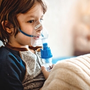
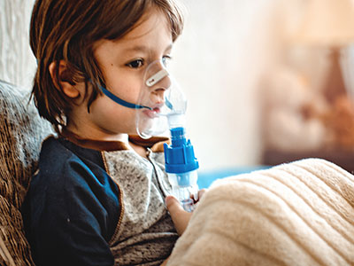
People with asthma can be indistinguishable from people who don’t have this chronic airway disease – until they have an asthma attack, also known as an exacerbation. During these events, their airways become inflamed and swollen and produce an abundance of mucus, causing dangerous narrowing of the bronchial tubes that leads to coughing, wheezing and trouble breathing. These events are a major cause of morbidity and mortality, leading to the deaths of 10 U.S. residents every day, according to the Centers for Disease Control and Prevention.

It’s long been known that colds, flu and other respiratory illnesses are major triggers for asthma exacerbations, says Children’s National in Washington, D.C., asthma expert Stephen J. Teach, M.D., MPH. Consequently, a significant body of research has focused on trying to figure out what’s happening on the cellular or molecular level as these illnesses progress to exacerbations. Targeted searches have identified several different molecular pathways that appear to be key players in this phenomenon. However, Dr. Teach says researchers have been missing a complete and unbiased snapshot of all the important pathways in illness-triggered exacerbations and how they interrelate.

To develop this big picture view, Dr. Teach and  Inner-City Asthma Consortium colleagues recruited 208 children ages 6-17 years old with severe asthma – marked by the need for daily doses of inhaled corticosteroids, two hospitalizations or systemic corticosteroid treatments over the past year, and a high concentration of asthma-associated immune cells – from nine pediatric medical centers across the country, including Children’s National. (Inhaled corticosteroids are a class of medicine that calms inflamed airways.) The researchers collected samples of nasal secretions and blood from these patients at baseline, when all of them were healthy.

Then, they waited for these children to show symptoms of respiratory illnesses. Within six days of cold symptoms, the researchers took two more samples of nasal secretions and blood. They also administered breathing tests to determine whether these respiratory illnesses led to asthma exacerbations and recorded whether these patients were treated with systemic corticosteroids to stem the associated respiratory inflammation.

The researchers examined nasal fluid samples for evidence of viral infection during illness and used analytical methods to identify the causative virus. They analyzed all the samples they collected for changes in concentrations of various immune cells. They also looked globally in these samples for changes in gene expression compared with baseline and between the two collection periods during respiratory illness.

Together, this information told the molecular story about what took place after these children got sick and after some of them developed exacerbations. Of the 208 patients recruited, 106 got respiratory illnesses during the six-month study period, leading to a total of 154 illness events. Of those, 47 caused exacerbations, and 107 didn’t.

About half the exacerbations appeared to have been triggered by a rhinovirus, a cause of common colds, the research team reports in a study published online April 8, 2019, in Nature Immunology. The other children’s cold-like symptoms could have been triggered by pollution, allergens or other irritants.

In most exacerbations, virally triggered or not, the researchers saw early activation of a network of genes that appeared to be associated with SMAD3, a signaling molecule already known to be involved in airway inflammation. At the same time, genes that control a set of immune cells known as lymphocytes were turned down. However, as the exacerbation progressed and worsened, the researchers saw gene networks turned on that related to airway narrowing, mucus hypersecretion and activation of other immune cells.

Exacerbations triggered by viruses were associated with multiple inflammatory pathways, in contrast to those in which viruses weren’t found, which were associated with molecular pathways that affected cells in the airway lining.

The researchers validated these findings in 19 patients who each got respiratory illnesses at least twice during the study period but only developed an exacerbation during one of these episodes, finding the same upregulated and downregulated molecular pathways in these patients as in the study population as a whole. They also identified a set of molecular risk factors in patients at baseline – signatures of gene activation that appeared to put patients at risk for exacerbations when they got sick. When patients were treated with systemic corticosteroids during exacerbations, these medicines appeared to restore only some of the affected molecular pathways to normal, healthy levels. Other molecular pathways remained markedly changed.

Each finding could represent a new target for drugs that could prevent or more effectively treat exacerbations, keeping more patients with asthma healthy and out of the hospital.

“Our consortium study found increased gene expression of enzymes that produce molecules that contribute to narrowed airways and dilated blood vessels,” Dr. Teach adds. “This is especially intriguing because drugs that target kallikreins or bradykinin may help treat asthma attacks that aren’t caused by viruses.”

In addition to Dr. Teach, study co-authors include Lead Author Matthew C. Altman, University of Washington; Michelle A. Gill, Baomei Shao and Rebecca S. Gruchalla, all of University of Texas Southwestern Medical Center; Elizabeth Whalen and Scott Presnell of Benaroya Research Institute; Denise C. Babineau and Brett Jepson of Rho, Inc.; Andrew H. Liu, Children’s Hospital Colorado; George T. O’Connor, Boston University School of Medicine; Jacqueline A. Pongracic, Ann Robert H. Lurie Children’s Hospital of Chicago; Carolyn M. Kercsmar and Gurjit K. Khurana Hershey, , Cincinnati Children’s Hospital; Edward M. Zoratti and Christine C. Johnson, Henry Ford Health System; Meyer Kattan, Columbia University College of Physicians and Surgeons; Leonard B. Bacharier and Avraham Beigelman, Washington University, St. Louis; Steve M. Sigelman, Peter J. Gergen, Lisa M. Wheatley and Alkis Togias, National Institute of Allergy and Infectious Diseases; and James E. Gern, William W. Busse and Senior author Daniel J. Jackson, University of Wisconsin School of Medicine and Public Health.

Funding for research described in this post was provided by the National Institute of Allergy and Infectious Diseases under award numbers 1UM1AI114271 and UM2AI117870; CTSA under award numbers UL1TR000150, UL1TR001422 and 5UL1TR001425; the National Institutes of Health under award number UL1TR000451;  CTSI under award number 1UL1TR001430; CCTSI under award numbers UL1TR001082 and 5UM1AI114271; and NCATS under award numbers UL1 TR001876 and UL1TR002345.