New method may facilitate childhood respiratory research
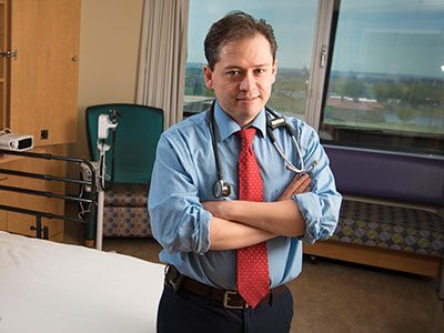
“The use of CRC is a potentially powerful translational approach to shed light on the molecular mechanisms that control airway epithelial immune responses in infants and young children. This novel approach enables us to study the origins of respiratory disease and its chronic progression through childhood and beyond,” observes Gustavo Nino, M.D., a Children’s pulmonologist and study senior author.
A new method perfected by a team at Children’s National Health System may help expand research into pulmonary conditions experienced by infants and children, an understudied but clinically important age group. The study describing the new technique was published in the December 2017 print edition of Pediatric Allergy and Immunology.
Using conditionally reprogrammed cells (CRCs), a technique that enables indefinite proliferation of cells in the lab, the team was able to produce cell cultures that have a number of advantages over standard cultures and that may make it easier and more efficient to conduct research into pediatric respiratory immune responses.
The epithelial cells that line human airways are crucial in controlling immune responses to viruses, allergens and other environmental factors. The function and dysfunction of these airway epithelial cells (AECs) play a key role in asthma, cystic fibrosis and other pulmonary conditions, many of which begin in early life.
To generate enough of these cells for research, scientists culture AECs from primary nasal and bronchial cell samples. Cells derived from adults have fueled research leading to new therapies and the discovery of key biomarkers. But little comparable research has been conducted in infants. Airway sampling in premature infants has not been reported, likely to due to airway size limitations and underlying comorbidities. Similarly, sampling in infants is limited by the need for bronchoscopy and sedation.
“A major barrier has been the lack of a good system to culture epithelial cells, since airway sampling in infants and children is a challenge,” says co-lead author, Geovanny F. Perez, M.D., co-director of Children’s Severe Bronchopulmonary Dysplasia Program. “We needed a better way to culture cells in this age group.”
While primary AECs do not survive long in the lab, that hurdle was recently overcome by a process that generates CRCs from the primary AECs of adults, making it possible to quickly generate cell cultures from specimens.
In this study, the Children’s team adapted that approach, producing CRCs from primary AECs of neonates and infants. The CRC induction successfully enabled AEC cultures from infants born prematurely and from bronchial specimens of young children.

“A major barrier has been the lack of a good system to culture epithelial cells, since airway sampling in infants and children is a challenge,” says co-lead author, Geovanny F. Perez, M.D., co-director of Children’s Severe Bronchopulmonary Dysplasia Program. “We needed a better way to culture cells in this age group.”
“We found that the CRCs have longer cell life and greater proliferation ability than standard cultures of epithelial cells. They preserved their original characteristics even after multiple experiments. And, they presented an innate immune response similar to that seen in primary human epithelial cells during viral respiratory responses in children,” says Dr. Perez.
“The use of CRC is a potentially powerful translational approach to shed light on the molecular mechanisms that control airway epithelial immune responses in infants and young children. This novel approach enables us to study the origins of respiratory disease and its chronic progression through childhood and beyond,” observes Gustavo Nino, M.D., a Children’s pulmonologist and study senior author.
The authors note that further studies are needed to define more precisely the differences and similarities in the immune responses of CRC and non-CRC derived from primary AEC. However, they conclude that CRC represents a new, effective method to study AEC innate immune responses in infants.
In addition to Drs. Perez and Nino, Children’s Center for Genetic Medicine Research co-authors include Co-Lead Author S. Wolf; Lana Mukharesh; Natalia Isaza Brando, M.D.; Diego Preciado, M.D., Ph.D.; Robert J. Freishtat, M.D., M.P.H.; Dinesh Pillai, M.D.; and M. C. Rose.
Financial support for this research was provided by the National Institute of Allergy and Infectious Diseases under grant number R21AI130502; Eunice Kennedy Shriver National Institute of Child Health and Human Development under grant number HD001399; National Heart, Lung and Blood Institute under grant number HL090020; and National Center for Advancing Translational Sciences under grant number UL1TR000075.