Tag Archive for: catheterization

E coli bacteria

Urinary bacteria in spinal cord injury cases may tip balance toward UTIs

E coli bacteria

Patients with spinal cord injuries nearly universally have bacteria present in their urine regardless of whether they have a urinary tract infection.

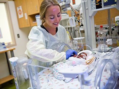
The fallout from spinal cord injury doesn’t end with loss of mobility: Patients can have a range of other issues resulting from this complex problem, including loss of bladder control that can lead to urine retention. One of the most serious implications is urinary tract infections (UTIs), the most common cause of repeat hospitalization in people with spinal cord injuries, explains Hans G. Pohl, M.D., associate chief in the division of Urology at Children’s National Health System.

Diagnosing UTIs in people with spinal cord injuries is trickier than in people who are otherwise healthy, Dr. Pohl explains. Patients with spinal cord injuries nearly universally have bacteria present in their urine regardless of whether they have a UTI. It’s unclear whether these bacteria are innocent bystanders or precursors to UTIs in patients who don’t yet show symptoms. And although antibiotics can wipe out this bacterial population, these drugs can have undesirable side effects and frequent use can promote development of antibiotic-resistant bacteria.

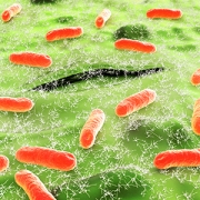
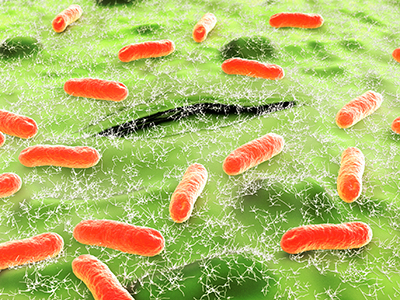
Although clinical dogma has long promoted the idea that “healthy” urine is sterile, Dr. Pohl and colleagues have shown that a variety of bacteria live in urine, even in people without symptoms. These microorganisms, like the intestinal microbiome, live in harmony with their hosts and may even help promote health. However, it’s unclear what this urinary microbiome might look like for patients with spinal cord injury before, during and after UTIs.

To start investigating this question, Dr. Pohl and co-authors recently reported a case study they published online Sept. 21, 2018, in Spinal Cord Series and Cases. The case report about a 55-year-old man who had injured the thoracic segment of his spinal cord—about the level of the bottom of his shoulder blades—in a skiing accident when he was 19 was selected as “Editor’s Choice” for the journal’s October 2018 issue.  The patient had a neurogenic bladder, which doesn’t function normally due to impaired communication with the spinal cord. To compensate for this loss of function, this patient needed to have urine removed every four to six hours by catheterization.

Over eight months Dr. Pohl, the study’s senior author, and colleagues collected 12 urine samples from this patient:

  • One was collected at a time the patient didn’t show any symptoms of a UTI
  • Nine were collected when the patient had UTI symptoms, such as bladder spasticity
  • Two samples were collected when the patient had finished antibiotic treatment for the UTI.

The researchers split each sample in half. One part was put through a standard urinalysis and culture, much like what patients with a suspected UTI would receive at the doctor’s office. The other part was analyzed using a technique that searched for genetic material to identify bacteria that might be present and to estimate their abundance.

The researchers found a variety of different bacteria present in these urine samples. Regardless of the patient’s health status and symptoms, the majority of these bacterial species are known to be pathogenic or potentially pathogenic. By contrast, this patient’s urine microbiome appeared to largely lack bacterial species known to be either neutral or with potentially probiotic properties, such as Lactobacillus.

All of the bacteria that grew in culture also were identified by their genetic material in the samples. However, genetic sequencing also identified a possible novel uropathogenic species called Burkholderia fungorum that didn’t grow in the lab in five of the samples. This bacterium is ubiquitous in the environment and has been identified in soil- and plant-based samples. It also has been discovered in the respiratory secretions of patients with cystic fibrosis, in patients with a heart condition called infectious endocarditis, in the vaginal microbiota of patients with bacterial vaginosis, and in the gut of patients with HIV who have low T-cell counts. Dr. Pohl says it’s unclear whether this species played an infectious role in this patient’s UTI or whether it’s just part of his normal urine flora.

“Consistent with our previous work, this case report demonstrates that rather than healthy urine being sterile, there is a diverse urine bacterial ecosystem during various states of health and disease,” Dr. Pohl says. “Rather than UTIs resulting from the growth or overgrowth of a single organism, it’s more likely that a change in the healthy balance of the urine ecosystem might cause these infections.”

By monitoring the relative abundance of different bacteria types present in the urine of patients with spinal cord injury and combining this information with a patient’s symptoms, Dr. Pohl says doctors may be able to make more accurate UTI diagnoses in this unique population.

In addition to Dr. Pohl, study co-authors include Marcos Pérez-Losada, Ljubica Caldovic, Ph.D., Bruce Sprague and Michael H. Hsieh, M.D., Children’s National; Emma Nally, Suzanne L. Groah and Inger Ljungberg, MedStar National Rehabilitation Hospital; and Neel J. Chandel, Montefiore Medical Center.

Baby in the NICU

Getting to the heart of cardiac output

Baby in the NICU

To keep infants in the neonatal intensive care unit (NICU) as healthy as possible, it’s important to keep close tabs on their vital signs. During their NICU stay, most babies have continuous monitoring of their blood pressure, respiratory rate and blood oxygen saturation. And although continuous monitoring of heart rate is also typically standard, other information about heart function – such as cardiac output, a measure of how well the heart is pumping blood – remains a challenge to obtain in these vulnerable babies.

Clinical markers like blood pressure, heart rate and urine output are available, but they are indirect measures of cardiac output, how much blood the heart pumps per minute. Less invasive techniques, such as Doppler ultrasound, can be imprecise. Respiratory mass spectrometry or catheterization would provide more precision by directly calculating cardiac output but carry risks or are not feasible for neonates.

Clinicians at Children’s National Health System hypothesized that COstatus monitors could offer a way to directly measure cardiac output among neonates. The COstatus monitor – a minimally invasive way to measure hemodynamics – captures cardiac output, total end diastolic volume, active circulation volume and central blood volume.

The research team tested the approach by leveraging ultrasound dilution: Injecting saline, which has an ultrasound velocity of 1533m/second, slows the ultrasound velocity of blood from its normal rate of 1580m/second and produces a dilution curve.

“It is feasible to directly measure neonatal cardiac output by ultrasound dilution via the COstatus monitor in the first two weeks of life with no adverse events,” says Khodayar Rais-Bahrami, M.D., a Children’s neonatologist and senior author for the research presented during the Pediatric Academic Societies 2018 annual meeting. “When we took consecutive measurements, we saw very little variance in the parameters.”

The COstatus monitor uses an extracorporeal loop that is connected to arterial and venous catheters. The 12 neonates included in the study already had umbilical venous catheters as well as either a peripheral arterial line or umbilical arterial catheter. The infants ranged in weight from 0.72 to 3.74 kg and were born at 24 to 41.3 gestational weeks.

The infants’ cardiac output was measured 54 times from 1 to 13 days of life. Up to two measurement sessions occurred daily for a maximum of four days. The mean cardiac output was 0.43 L/minute with a mean cardiac index of 197mL/kg/minute.

Future research will describe normal cardiac output ranges for neonates as well as how these measurements evolve during the first week of life.

In addition to Dr. Rais-Bahrami, study co-authors include Simranjeet S. Sran, M.D., and Mariam Said, M.D., a Children’s neonatologist.

Elena Grant

Interventional cardiac magnetic resonance team welcomes new specialist

elena-grant-photo

The Interventional Cardiac Magnetic Resonance (ICMR) Program at Children’s National is actively developing newer and safer ways to perform cardiac procedures on young patients, with some of the world’s leading experts in cardiac catheterization and imaging. Elena Grant, M.D., a former pediatric cardiology fellow at Children’s National, is the newest member to join the team that pioneered real-time MRI-guided radiation-free cardiac catheterization for children.

In addition to clinical work as a Children’s National Interventional Cardiologist, Dr. Grant will perform preclinical research at the National Institutes of Health to develop new procedures, techniques, and devices that can be translated to clinical practice to treat children and adults with congenital heart disease.

Dr. Grant specializes in interventional cardiology. She received her medical degree from the University of Dundee Medical School in Dundee, Scotland, followed by Foundation Training in Edinburgh, Scotland. She completed her pediatric residency at Massachusetts General Hospital, her Pediatric Cardiology fellowship at Children’s National, and she recently finished an advanced fellowship in interventional pediatric cardiology at Children’s Healthcare of Atlanta and Emory University.

Advances in interventional cardiovascular MRI

Children’s National is at the forefront of this exciting new field and is currently the only institution in the United States to perform radiation-free MRI-guided cardiac catheterization procedures in children.

ICMR is a partnership with the National Institutes of Health that brings together researchers, clinicians, engineers, and physicists to provide radiation-free, less invasive, and more precise diagnostics and treatment options for pediatric patients and adults with congenital heart disease.

The ICMR approach to heart catheterization uses real-time MRI, instead of X-ray, in pediatric research subjects undergoing medically necessary heart catheterization. This research study is intended as a step toward routine MRI-guided catheterization in children, which attempts to avoid the hazards of ionizing radiation (X-ray).

In 2015, after working with NIH to explore how interventional cardiovascular MRI could be integrated into pediatric practices, the ICMR team, including Dr. Grant, Russell Cross, M.D., Joshua Kanter, M.D., and Laura Olivieri, M.D., performed the first  radiation-free MRI-guided right heart catheterization on a 14-year-old girl at Children’s National. Since then, nearly 50 such procedures have been successfully completed, and the team is working to broaden the age range and cardiac disease complexity of patients who can undergo the procedure.

About 1 percent of newborns are born with a heart condition, and the team at Children’s performs more than 450 X-ray guided cardiac catheterizations and over 500 cardiac MRI scans per year.