Tag Archive for: brain

New study finds prenatal opioid exposure linked to smaller newborn brain volumes

brain scans of newbornA new study out of the Center for Prenatal, Neonatal & Maternal Health Research, directed by Catherine Limperopoulos, PhD, led by Yao Wu, PhD and Stephanie Merhar, MD, MS, out of Cincinnati Children’s Hospital Medical Center, sought answers to the question: “Do brain volumes differ in opioid-exposed vs. unexposed newborns?” In one of the largest studies of this kind, researchers found that prenatal exposure to opioids is associated with smaller brain volumes in newborns. These findings from the landmark Outcomes of Babies with Opioid Exposure (OBOE) study build on, reinforce smaller studies, and achieve a better understanding of the impacts of prenatal opioid exposure (POE).

Dive Deeper

The Advancing Clinical Trials in Neonatal Opioid Withdrawal (ACT-NOW)’s OBOE study is a multi-site observational study of newborns with prenatal opioid exposure and a control group of unexposed newborns from four different sites in the United States – Case Western Reserve University, Cincinnati Children’s Hospital Medical Center, University of Alabama at Birmingham, and Children’s Hospital of Philadelphia.

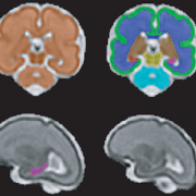
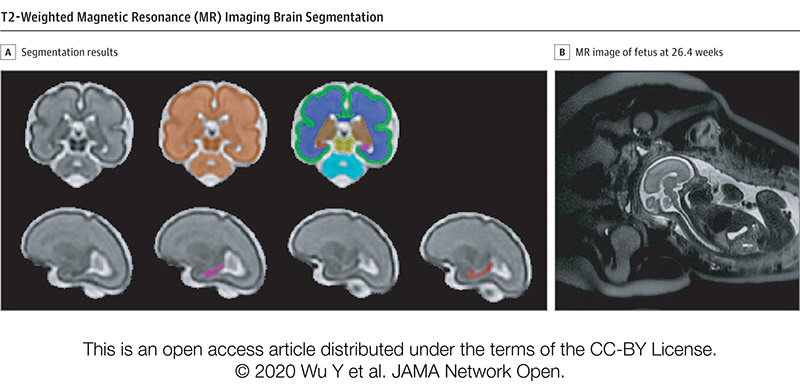
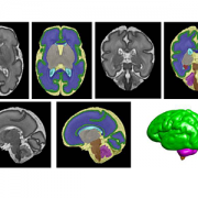
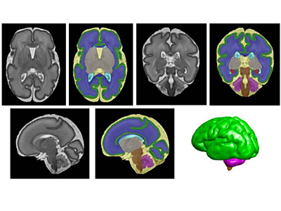
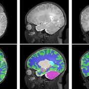
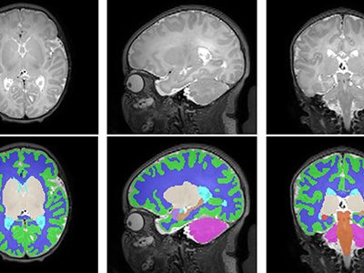
In a study involving 173 newborns who were exposed to opioids during pregnancy and 96 newborns not exposed to opioids prenatally showed smaller brain sizes in several key areas. Specifically, these exposed babies had smaller total brain volumes, as shown through MRI, as well as reduced volumes in important parts of the brain, including the cortex (outer layer of the brain), deep gray matter (areas that control movement and emotions), white matter (which helps transmit signals in the brain), cerebellum (responsible for coordination and movement), brainstem (controls basic functions like breathing), and the amygdala (involved in emotions and memory).

Further details showed that newborns exposed to medication for opioid use disorder (MOUD) during pregnancy with methadone, had smaller white matter volumes, while those exposed to MOUD with buprenorphine had smaller volumes specifically in the right amygdala. Additionally, newborns who were exposed to opioids plus additional substances such as THC and gabapentin had smaller volumes in even more brain areas compared to those who were only exposed to opioids.

What’s Next

The OBOE study sets the groundwork for further research into the long-term impact of opioid exposure during pregnancy. Additional work is necessary to expand on these findings and how they relate to functions in childhood – including exploring the way these reduced brain volumes may impact cognitive, behavioral, and motor impairments. The study raises important questions about how current guidelines for MOUD during pregnancy – specifically with methadone and buprenorphine – might evolve considering these findings. This study highlights the need for further research to assess the long-term effects of MOUD regimens on both maternal and infant outcomes.

This significant study underscores the importance of multi-disciplinary collaboration in opioid exposure research, effective regulation, and policy interventions – involving healthcare providers, researchers, policymakers, and affected families – to best mitigate the consequences and improve the health outcomes of children affected by prenatal opioid exposure.

You can read the full study, Antenatal Opioid Exposure and Global and Regional Brain Volumes in Newborns, and its companion editorial, Following the Developing Brain Affected by Opioid Exposure, in the Journal of the American Medical Association.

Additional authors from Children’s National include Kushal Kapse, BS, MS, and Josepheen De Asis-Cruz, MD, PhD. Other authors include Carla M. Bann, PhD, Jamie E. Newman, PhD4, Nicole Mack, MS, Sara B. De Mauro, MD, MSCE, Namasivayam Ambalavanan, MD, Jonathan M. Davis, MD, Scott A. Lorch, MD, MSCE5, Deanne Wilson-Costello, MD, Brenda B. Poindexter, MD and Myriam Peralta-Carcelen, MD.

AI algorithm that detects brain abnormalities could help cure epilepsy

Digital background depicting innovative technologies in (AI) artificial systems, neural interfaces and internet machine learning technologies

A new AI algorithm can detect subtle brain abnormalities that cause epileptic seizures.

An artificial intelligence (AI) algorithm that can detect subtle brain abnormalities that cause epileptic seizures has been developed by a UCL-led team of international researchers, including Children’s National Hospital.

To do this, the team quantified features from MRI scans, such as how thick or folded the brain was at nearly 300,000 locations in each case.

They then trained the AI algorithm using examples labelled by expert radiologists as either a healthy brain or one with focal cortical dysplasia (FCD) based on their patterns and features.

The results, published in Brain, showed that in the main cohort of 538 patients, the algorithm was able to detect the FCD in 67% of cases.

“We put an emphasis on creating an AI algorithm that was interpretable and could help doctors make decisions. Showing doctors how the Multicentre Epilepsy Lesion Detection project (MELD) algorithm made its predictions was an essential part of that process,” said Mathilde Ripart, research assistant at UCL and the study’s co-first author.

Around 1% of the population have epilepsy and, of these, 20-30% do not respond to medications.

“We are excited to collaborate with MELD on ways to improve the treatment of pharmacoresistant epilepsy,” said Nathan Cohen, M.D., neurologist at Children’s National Hospital and co-author of the study. “This advanced imaging platform is open source and demonstrates the benefit of team science at the broadest scale.”

In children who have had surgery to control their epilepsy, FCD is the most common cause, and in adults it is the third most common cause.

Additionally, of patients who have epilepsy that have an abnormality in the brain that cannot be found on MRI scans, FCD is the most common cause.

You can read the full UCL press release here.

Children’s National Research Institute releases annual report

Vittorio Gallo and Marc Batshaw

Children’s National Research Institute directors Vittorio Gallo, Ph.D., and Mark Batshaw, M.D.

The Children’s National Research Institute recently released its 2019-2020 academic annual report, titled 150 Years Stronger Through Discovery and Care to mark the hospital’s 150th birthday. Not only does the annual report give an overview of the institute’s research and education efforts, but it also gives a peek in to how the institute has mobilized to address the coronavirus pandemic.

“Our inaugural research program in 1947 began with a budget of less than $10,000 for the study of polio — a pressing health problem for Washington’s children at the time and a pandemic that many of us remember from our own childhoods,” says Vittorio Gallo, Ph.D., chief research officer at Children’s National Hospital and scientific director at Children’s National Research Institute. “Today, our research portfolio has grown to more than $75 million, and our 314 research faculty and their staff are dedicated to finding answers to many of the health challenges in childhood.”

Highlights from the Children’s National Research Institute annual report

  • In 2018, Children’s National began construction of its new Research & Innovation Campus (CNRIC) on 12 acres of land transferred by the U.S. Army as part of the decommissioning of the former Walter Reed Army Medical Center campus. In 2020, construction on the CNRIC will be complete, and in 2012, the Children’s National Research Institute will begin to transition to the campus.
  • In late 2019, a team of scientists led by Eric Vilain, M.D., Ph.D., director of the Center for Genetic Medicine Research, traveled to the Democratic Republic of Congo to collect samples from 60 individuals that will form the basis of a new reference genome data set. The researchers hope their project will generate better reference genome data for diverse populations, starting with those of Central African descent.
  • A gift of $5.7 million received by the Center for Translational Research’s director, Lisa Guay-Woodford, M.D., will reinforce close collaboration between research and clinical care to improve the care and treatment of children with polycystic kidney disease and other inherited renal disorders.
  • The Center for Neuroscience Research’s integration into the infrastructure of Children’s National Hospital has created a unique set of opportunities for scientists and clinicians to work together on pressing problems in children’s health.
  • Children’s National and the National Institute of Allergy and Infectious Diseases are tackling pediatric research across three main areas of mutual interest: primary immune deficiencies, food allergies and post-Lyme disease syndrome. Their shared goal is to conduct clinical and translational research that improves what we know about those conditions and how we care for children who have them.
  • An immunotherapy trial has allowed a little boy to be a kid again. In the two years since he received cellular immunotherapy, Matthew has shown no signs of a returning tumor — the longest span of time he’s been tumor-free since age 3.
  • In the past 6 years, the 104 device projects that came through the National Capital Consortium for Pediatric Device Innovation accelerator program raised $148,680,256 in follow-on funding.
  • Even though he’s watched more than 500 aspiring physicians pass through the Children’s National pediatric residency program, program director Dewesh Agrawal, M.D., still gets teary at every graduation.

Understanding and treating the novel coronavirus (COVID-19)

In a short period of time, Children’s National Research Institute has mobilized its scientists to address COVID-19, focusing on understanding the virus and advancing solutions to ameliorate the impact today and for future generations. Children’s National Research Institute Director Mark Batshaw, M.D., highlighted some of these efforts in the annual report:

  • Eric Vilain, M.D., Ph.D., director of the Center for Genetic Medicine Research, is looking at whether or not the microbiome of bacteria in the human nasal tract acts as a defensive shield against COVID-19.
  • Catherine Bollard, M.D., MBChB, director of the Center for Cancer and Immunology Research, and her team are seeing if they can “train” T cells to attack the invading coronavirus.
  • Sarah Mulkey, M.D., Ph.D., an investigator in the Center for Neuroscience Research and the Fetal Medicine Institute, is studying the effects of, and possible interventions for, coronavirus on the developing brain.

You can view the entire Children’s National Research Institute academic annual report online.

Special issue of “Neurochemical Research” honors Vittorio Gallo, Ph.D.

Vittorio Gallo

Investigators from around the world penned manuscripts that were assembled in a special issue of “Neurochemical Research” that honors Vittorio Gallo, Ph.D., for his leadership in the field of neural development and regeneration.

At a pivotal moment early in his career, Vittorio Gallo, Ph.D., was accepted to work with Professor Giulio Levi at the Institute for Cell Biology in Rome, a position that leveraged courses Gallo had taken in neurobiology and neurochemistry, and allowed him to work in the top research institute in Italy directed by the Nobel laureate, Professor Rita Levi-Montalcini.

For four years as a student and later as Levi’s collaborator, Gallo focused on amino acid neurotransmitters in the brain and mechanisms of glutamate and GABA release from nerve terminals. Those early years cemented a research focus on glutamate neurotransmission that would lead to a number of pivotal publications and research collaborations that have spanned decades.

Now, investigators from around the world who have worked most closely with Gallo penned tributes in the form of manuscripts that were assembled in a special issue of “Neurochemical Research” that honors Gallo “for his contributions to our understanding of glutamatergic and GABAergic transmission during brain development and to his leadership in the field of neural development and regeneration,” writes guest editor Arne Schousboe, of the University of Copenhagen in Denmark.

Dr. Gallo as a grad student

Vittorio Gallo, Ph.D. as a 21-year-old mustachioed graduate student.

“In spite of news headlines about competition in research and many of the negative things we hear about the research world, this shows that research is also able to create a community around us,” says Gallo, chief research officer at Children’s National Hospital and scientific director for the Children’s National Research Institute.

As just one example, he first met Schousboe 44 years ago when Gallo was a 21-year-old mustachioed graduate student.

“Research can really create a sense of community that we carry on from the time we are in training, nurture as we meet our colleagues at periodic conferences, and continue up to the present. Creating community is bi-directional: influencing people and being influenced by people. People were willing to contribute these 17 articles because they value me,” Gallo says. “This is a lot of work for the editor and the people who prepared papers for this special issue.”

In addition to Gallo publishing more than 140 peer-reviewed papers, 30 review articles and book chapters, Schousboe notes a number of Gallo’s accomplishments, including:

  • He helped to develop the cerebellar granule cell cultures as a model system to study how electrical activity and voltage-dependent calcium channels modulate granule neuron development and glutamate release.
  • He developed a biochemical/neuropharmacological assay to monitor the effects of GABA receptor modulators on the activity of GABA chloride channels in living neurons.
  • He and Maria Usowicz used patch-clamp recording and single channel analysis to demonstrate for the first time that astrocytes express glutamate-activated channels that display functional properties similar to neuronal counterparts.
  • He characterized one of the spliced isoforms of the AMPA receptor subunit gene Gria4 and demonstrated that this isoform was highly expressed in the cerebellum.
  • He and his Children’s National colleagues demonstrated that glutamate and GABA regulate oligodendrocyte progenitor cell proliferation and differentiation.
Purkinje cells

Purkinje cells are large neurons located in the cerebellum that are elaborately branched like interlocking tree limbs and represent the only source of output for the entire cerebellar cortex.

Even the image selected to grace the special issue’s cover continues the theme of continuity and leaving behind a legacy. That image of Purkinje cells was created by a young scientist who works in Gallo’s lab, Aaron Sathyanesan, Ph.D. Gallo began his career working on the cerebellum – a region of the brain important for motor control – and now studies with a team of scientists and clinician-scientists Purkinje cells’ role in locomotor adaptive behavior and how that is disrupted after neonatal brain injury.

“These cells are the main players in cerebellar circuitry,” Gallo says. “It’s a meaningful image because goes back to my roots as a graduate student and is also an image that someone produced in my lab early in his career. It’s very meaningful to me that Aaron agreed to provide this image for the cover of the special issue.”

Once overlooked cellular messengers could combat antibiotic resistance

bacterial extracellular vesicle

Children’s National Hospital researchers for the first time have isolated bacterial extracellular vesicles from the blood of healthy donors. The team theorizes that the solar eclipse lookalikes contain important signaling proteins and chromatin, DNA from the human host.

Children’s National Hospital researchers for the first time have isolated bacterial extracellular vesicles from the blood of healthy donors, a critical step to better understanding the way gut bacteria communicate with the rest of the body via the bloodstream.

For decades, researchers considered circulating bacterial extracellular vesicles as bothersome flotsam to be jettisoned as they sought to tease out how bacteria that reside in the gut whisper messages to the brain.

There is a growing appreciation that extracellular vesicles – particles that cells naturally release – actually facilitate intracellular communication.

“In the past, we thought they were garbage or noise,” says Robert J. Freishtat, M.D., MPH, associate director, Center for Genetic Medicine Research at Children’s National Research Institute. “It turns out what we throw away is not trash.”

Kylie Krohmaly, a graduate student in Dr. Freishtat’s laboratory, has isolated from blood, extracellular vesicles from Escherichia coli and Haemophilus influenzae, common bacteria that colonize the gut, and validated the results via electron microscopy.

“The images are interesting because they look like they have a bit of a halo around them or penumbra,” Krohmaly says.

The team theorizes that the solar eclipse lookalikes contain important signaling proteins and chromatin, DNA from the human host.

“It’s the first time anyone has pulled them out of blood. Detecting them is one thing. Pulling them out is a critical step to understanding the language the microbiome uses as it speaks with its human host,” Dr. Freishtat adds.

Krohmaly’s technique is so promising that the Children’s National team filed a provisional patent.

The Children’s research team has devised a way to gum up the cellular works so that bacteria no longer become antibiotic resistant. Targeted bacteria retain the ability to make antibiotic-resistance RNA, but like a relay runner dropping rather than passing a baton, the bacteria are thwarted from advancing beyond that step. And, because that gene is turned off, the bacteria are newly sensitive to antibiotics – instead of resistant bacteria multiplying like clockwork these bacteria get killed.

“Our plan is to hijack this process in order to turn off antibiotic-resistance genes in bacteria,” Dr. Freishtat says. “Ultimately, if a child who has an ear infection can no longer take amoxicillin, the antibiotic would be given in tandem with the bacteria-derived booster to turn off bacteria’s ability to become antibiotic resistant. This one-two punch could become a novel way of addressing the antibiotic resistance process.”

ISEV2020 Annual Meeting presentation
(Timing may be subject to change due to COVID-19 safety precautions)
Oral with poster session 3: Neurological & ID
Saturday May 23, 2020, 5 p.m. to 5:05 p.m. (ET)
“Detection of bacterial extracellular vesicles in blood from healthy volunteers”
Kylie Krohmaly, lead author; Claire Hoptay, co-author; Andrea Hahn, M.D., MS, infectious disease specialist and co-author; Robert J. Freishtat, M.D., MPH, associate director, Center for Genetic Medicine Research at Children’s National Research Institute and senior author.

Maternal mental health alters structure and biochemistry of developing fetal brain

Even when pregnant women have uncomplicated pregnancies and high socioeconomic status, when they experience elevated anxiety, stress or depression these prenatal stressors can alter the structure of the developing fetal brain and disrupt its biochemistry, according to Children’s National Hospital research published online Jan. 29, 2020, in JAMA Network Open.

The Children’s National research findings “have enormous scientific, clinical and public health implications,” Charles A. Nelson III, Ph.D.,  Boston Children’s Hospital, writes in a companion editorial.

“Previously we found that 65% of pregnant women who received a diagnosis of fetal congenital heart disease had elevated levels of stress. It’s concerning but not surprising that pregnant women who wonder if their baby will need open heart surgery would feel stress,” says Catherine Limperopoulos, Ph.D., director of the Center for the Developing Brain at Children’s National and the study’s senior author. “In this latest study, we ran the same panel of questionnaires and were surprised to find a high proportion of otherwise healthy pregnant women whose unborn babies are doing well also report high levels of stress.”

Anxiety and depression are the most common mental health problems during pregnancy. To learn more about the implications for the developing fetal brain, the Children’s National research team recruited 119 healthy volunteers with low-risk pregnancies from obstetric clinics in Washington, D.C., from Jan. 1, 2016, to April 17, 2019. The women’s mean age was 34.4 years old. All were high school graduates, 83% were college graduates, and 84% reported professional employment.

T2-Weighted Magnetic Resonance (MR) Imaging Brain Segmentation.

T2-Weighted Magnetic Resonance (MR) Imaging Brain Segmentation. Segmentation results of total brain (orange), cortical gray matter (green), white matter (blue), deep gray matter (brown), brainstem (yellow), cerebellum (light blue), left hippocampus (purple) and right hippocampus (red) on a 3-Dimensional reconstructed T2-weighted MR image of a fetus at 26.4 gestational weeks. The hippocampus plays a central role in memory and behavioral inhibition and contains high concentrations of corticosteroid receptors and, thus, this brain region is sensitive to stress. Credit: JAMA Network Open.

The team performed 193 fetal brain magnetic resonance imaging (MRI) sessions between 24-40 weeks gestation and measured the volume of the total fetal brain as well as the cortical gray matter, white matter, deep gray matter, cerebellum, brainstem and hippocampus volumes. On the same day as their MRI visit, the pregnant women completed validated questionnaires to measure maternal stress, anxiety and depression, answering questions such as “how do you feel right now,” “how do you generally feel” as well as the degree of stressful feelings they experienced the month prior.

Of the pregnant women in the study:

  • 27% tested positive for stress
  • 26% tested positive for anxiety
  • 11% tested positive for depression
  • Maternal anxiety and stress were associated with increased fetal cortical gyrification
  • Elevated maternal depression was associated with decreased creatine and choline levels in the fetal brain
  • Maternal stress scores decreased with increasing gestational age, while anxiety and depression did not

“We report for the first time that maternal psychological distress may be associated with increased fetal local gyrification index in the frontal and temporal lobes,” says Yao Wu, Ph.D., a research associate working with Limperopoulos at Children’s National and the study’s lead author. “We also found an association with left fetal hippocampal volume, with maternal psychological distress selectively stunting the left hippocampal volumetric growth more than the right. And elevated maternal depression was associated with decreased creatine and choline levels in the fetal brain,” Wu adds.

Late in pregnancy – at the time these women were recruited into the cohort study – the fetal brain grows exponentially and key metabolite levels also rise. Creatine facilitates recycling of adenosine triphosphate, the cell’s energy currency. Typically, levels of this metabolite rise, denoting rapid changes and higher cellular maturation; creatine also is known to support cognitive function. Choline levels also typically rise, marking cell membrane turnover as new cells are generated and support memory, mental focus and concentration.

“These women were healthy, and of high socioeconomic status and educational level, leading us to conclude that the prevalence of prenatal maternal psychological distress may be underestimated,” Limperopoulos adds. “While stress is an everyday reality for most of us, this is different because elevated stress during pregnancy can alter fetal brain programming. Our findings underscore the critical need to universally screen all pregnant women for prenatal psychological distress, even young mothers whose pregnancies wouldn’t otherwise raise red flags.”

In addition to Limperopoulos and Wu, Children’s National study co-authors include Yuan-Chiao Lu, Ph.D., research associate; Marni Jacobs, Ph.D., biostatistician; Subechhya Pradhan, Ph.D., research faculty; Kushal Kapse, MS, staff engineer; Li Zhao, Ph.D., research faculty; Nickie Niforatos-Andescavage, M.D., neonatologist; Gilbert Vezina, M.D., director of the neuroradiology program; and Adré  J. du Plessis, M.B.Ch.B., director, Fetal Medicine Institute. Research coordinators Catherine Lopez, MS, Kathryn Lee Bannantine, BSN, and Jessica Lynn Quistorff, MPH, assisted with subject recruitment.

Financial support for the research described in this post was provided by the National Institutes of Health under grant No. RO1 HL116585-01 and the Thrasher Research Fund under Early Career award No. 14764.

Journal Reference:
Yao Wu, Yuan-Chiao Lu, Marni Jacobs, Subechhya Pradhan, Kushal Kapse, Li Zhao, Nickie Niforatos-Andescavage, Gilbert Vezina, Adré J. du Plessis, Catherine Limperopoulos. “Association of prenatal maternal psychological distress with fetal brain growth, metabolism and cortical maturation,” JAMA Network Open, 3(1): e1919940, 2020

Getting to know the unstoppable Lataisia C. Jones, Ph.D.

Lataisia C. Jones on Mission Unstoppable

Children’s National Hospital neuroscientist Lataisia C. Jones, Ph.D., appears in the Jan. 18, 2020, edition of Mission Unstoppable, a Saturday morning show aired by CBS that spotlights cutting-edge women leaders in science, technology, engineering and math.

Budding neuroscientist Lataisia C. Jones, Ph.D., is unstoppable. For instance, using everyday items that families can pluck from their own kitchen cabinets, she walks kids through the steps of creating homemade lava lamps. In the process, the youngsters learn a bit of science, like the fact that oil and water do not mix provides the hypnotic magic behind their new lamps.

Jones’ infectious enthusiasm for science that Children’s National Hospital patients and families experience in person during weekly Young Scientist sessions she hosts will be shared nationwide as Jones appears in the Jan. 18, 2020, edition of “Mission Unstoppable.” The half-hour show aired by CBS on Saturday mornings is co-produced by the Geena Davis Institute on Gender in Media and spotlights cutting-edge women leaders in science, technology, engineering and math (STEM).

“I’m excited,” Jones says of the filming experience. “It’s going to be an amazing opportunity to show kids that there is a fun way of learning. This show is opening a lot of doors and a lot of eyes to the fact that science can be fun.”

Jones’ scientific inquiry focuses on the corpus callosum, a network of fibers centrally located in the middle of the brain that is responsible for transferring information from one lobe to another. Her current research leverages experimental models to better understand brain abnormalities associated with autism spectrum disorder. Or, as she tells CBS viewers, studying the brain helps the field better understand how information is processed in order for people to move, learn and think effortlessly.

Lataisia C. Jones on Mission Unstoppable

“I’m excited,” Jones says of the filming experience. “It’s going to be an amazing opportunity to show kids that there is a fun way of learning. This show is opening a lot of doors and a lot of eyes to the fact that science can be fun.”

In September 2019, Jones was selected to serve as an IF/THEN Ambassador by the American Association for the Advancement of Science, the world’s largest general scientific society, to inspire the next generation of women pursuing STEM careers. A postdoctoral fellow in the Center for Neuroscience Research lab run by Masaaki Torii, Ph.D., Jones now also serves as a role model for future scientists, connecting with middle school students in person, virtually and via the CBS network television show.

“A lot of my inspiration comes from individuals who I mentor, which also shows that I am learning as well. If I am able to teach science, translate it in different ways to different audiences, I am helping to fulfill my lifelong dream,” she adds. “I always say we all have an inner scientist.”

As the first African American to earn a Ph.D. from Florida State University’s College of Medicine, Department of Neuroscience, Jones has continued to acquire “first” experiences throughout her academic and professional career. But she’s also motivated to diversify the ranks of science to ensure she’s not the last.

“I am not the normal face you see in science,” she says. “Another reason for me to be stronger and to work harder and get more things done in science is so people who look like me know they can do the same things and know that they’re just as good.”

Stressful pregnancies can leave fingerprint on fetal brain

Catherine Limperopoulos

“We were alarmed by the high percentage of pregnant women with a diagnosis of a major fetal heart problem who tested positive for stress, anxiety and depression,” says Catherine Limperopoulos, Ph.D., director of the Center for the Developing Brain at Children’s National and the study’s corresponding author.

When a diagnosis of fetal congenital heart disease causes pregnant moms to test positive for stress, anxiety and depression, powerful imaging can detect impaired development in key fetal brain regions, according to Children’s National Hospital research published online Jan. 13, 2020, in JAMA Pediatrics.

While additional research is needed, the Children’s National study authors say their unprecedented findings underscore the need for universal screening for psychological distress as a routine part of prenatal care and taking other steps to support stressed-out pregnant women and safeguard their newborns’ developing brains.

“We were alarmed by the high percentage of pregnant women with a diagnosis of a major fetal heart problem who tested positive for stress, anxiety and depression,” says Catherine Limperopoulos, Ph.D., director of the Center for the Developing Brain at Children’s National and the study’s corresponding author. “Equally concerning is how prevalent psychological distress is among pregnant women generally. We report for the first time that this challenging prenatal environment impairs regions of the fetal brain that play a major role in learning, memory, coordination, and social and behavioral development, making it all the more important for us to identify these women early during pregnancy to intervene,” Limperopoulos adds.

Congenital heart disease (CHD), structural problems with the heart, is the most common birth defect. Still, it remains unclear how exposure to maternal stress impacts brain development in fetuses with CHD.

The multidisciplinary study team enrolled 48 women whose unborn fetuses had been diagnosed with CHD and 92 healthy women with uncomplicated pregnancies. Using validated screening tools, they found:

  • 65% of pregnant women expecting a baby with CHD tested positive for stress
  • 27% of women with uncomplicated pregnancies tested positive for stress
  • 44% of pregnant women expecting a baby with CHD tested positive for anxiety
  • 26% of women with uncomplicated pregnancies tested positive for anxiety
  • 29% of pregnant women expecting a baby with CHD tested positive for depression and
  • 9% women with uncomplicated pregnancies tested positive for depression

All told, they performed 223 fetal magnetic resonance imaging sessions for these 140 fetuses between 21 and 40 weeks of gestation. They measured brain volume in cubic centimeters for the total brain as well as volumetric measurements for key regions such as the cerebrum, cerebellum, brainstem, and left and right hippocampus.

Maternal stress and anxiety in the second trimester were associated with smaller left hippocampi and smaller cerebellums only in pregnancies affected by fetal CHD. What’s more, specific regions — the hippocampus head and body and the left cerebellar lobe – were more susceptible to stunted growth. The hippocampus is key to memory and learning, while the cerebellum controls motor coordination and plays a role in social and behavioral development.

The hippocampus is a brain structure that is known to be very sensitive to stress. The timing of the CHD diagnosis may have occurred at a particularly vulnerable time for the developing fetal cerebellum, which grows faster than any other brain structure in the second half of gestation, particularly in the third trimester.

“None of these women had been screened for prenatal depression or anxiety. None of them were taking medications. And none of them had received mental health interventions. In the group of women contending with fetal CHD, 81% had attended college and 75% had professional educations, so this does not appear to be an issue of insufficient resources,” Limperopoulos adds. “It’s critical that we routinely to do these screenings and provide pregnant women with access to interventions to lower their stress levels. Working with our community partners, Children’s National is doing just that to help reduce toxic prenatal stress for both the health of the mother and for the future newborns. We hope this becomes standard practice elsewhere.”

Adds Yao Wu, Ph.D., a research associate working with Limperopoulos at Children’s National and the study’s lead author: “Our next goal is exploring effective prenatal cognitive behavioral interventions to reduce psychological distress felt by pregnant women and improve neurodevelopment in babies with CHD.”

In addition to Limperopoulos and Wu , Children’s National study co-authors include Kushal Kapse, MS, staff engineer; Marni Jacobs, Ph.D., biostatistician; Nickie Niforatos-Andescavage, M.D., neonatologist; Mary T. Donofrio, M.D., director, Fetal Heart Program; Anita Krishnan, M.D., associate director, echocardiography; Gilbert Vezina, M.D., director, Neuroradiology Program; David Wessel, M.D., Executive Vice President and Chief Medical Officer; and Adré  J. du Plessis, M.B.Ch.B., director, Fetal Medicine Institute. Jessica Lynn Quistorff, MPH, Catherine Lopez, MS, and Kathryn Lee Bannantine, BSN, assisted with subject recruitment and study coordination.

Financial support for the research described in this post was provided by the National Institutes of Health under grant No. R01 HL116585-01 and the Thrasher Research Fund under Early Career award No. 14764.

False negatives: Delayed Zika effects in babies who appeared normal at birth

sleeping baby

Colombian infants exposed to Zika virus in the womb showed neurodevelopmental delays as toddlers, despite having “normal” brain imaging and head circumference at birth, a finding that underscores the importance of long-term neurodevelopmental follow-up for Zika-exposed infants.

Colombian infants exposed to Zika virus in the womb showed neurodevelopmental delays as toddlers, despite having “normal” brain imaging and head circumference at birth, a finding that underscores the importance of long-term neurodevelopmental follow-up for Zika-exposed infants, according to a cohort study published online Jan. 6, 2020, in JAMA Pediatrics.

“These infants had no evidence of Zika deficits or microcephaly at birth. Neurodevelopmental deficits, including declines in mobility and social cognition, emerged in their first year of life even as their head circumference remained normal,” says Sarah B. Mulkey, M.D. Ph.D., a fetal/neonatal neurologist at Children’s National Hospital and the study’s first author. “About one-third of these newborns who underwent postnatal head ultrasound had nonspecific imaging results, which we believe are the first published results finding a link between subtle brain injuries and impaired neuromotor development in Zika-exposed children.”

The multi-institutional research group led by Children’s National enrolled pregnant women in Atlántico Department, which hugs the Caribbean coast of Colombia, who had been exposed to Zika, and performed a series of fetal magnetic resonance images (MRI) and ultrasounds as their pregnancies progressed.

Even though their mothers had laboratory-confirmed Zika infections, 77 out of 82 of their offspring were born with no sign of congenital Zika syndrome, a constellation of birth defects that includes severe brain abnormalities, eye problems and congenital contractures, and 70 underwent additional testing of neurodevelopment during infancy. These apparently normal newborns were born between Aug. 1, 2016, and Nov. 30, 2017, at the height of the Zika epidemic, and had normal head circumference.

When they were 4 to 8 months or 9 to 18 months of age, the infants’ neurodevelopment was evaluated using two validated tools, the Warner Initial Developmental Evaluation of Adaptive and Functional Skills (a 50-item test of such skills as self-care, mobility, communication and social cognition) and the Alberta Infant Motor Scale (a motor examination of infants in prone, supine, sitting and standing positions). Some infants were assessed during each time point.

Women participating in the study were highly motivated, with 91% following up with appointments, even if it meant traveling hours by bus. In addition to Children’s National faculty traveling to Colombia to train staff how to administer the screening instruments, videotaped assessments, MRIs and ultrasounds were read, analyzed and scored at Children’s National. According to the study team, the U.S. scoring of Alberta Infant Motor Scale tests administered in Colombia is also unprecedented for a research study and offers the potential of remote scoring of infants’ motor skill maturity in regions of the world where pediatric specialists, like child neurologists, are lacking.

“Normally, neurodevelopment in infants and toddlers continues for years, building a sturdy neural network that they later use to carry out complex neurologic and cognitive functions as children enter school,” Dr. Mulkey adds. “Our findings underscore the recommendations by the Centers for Disease Control and Prevention (CDC) that all infants exposed to Zika in the womb undergo long-term follow-up, providing an opportunity to intervene earlier.”

An accompanying editorial by CDC staffers concurs, saying the study reported “intriguing data” that add “to the growing evidence of the need for long-term follow-up for all children with Zika virus exposure in utero to ensure they receive the recommended clinical evaluations even when no structural defects are identified at birth.”

In addition to Dr. Mulkey, study co-authors include Margarita Arroyave-Wessel, MPH, Dorothy I. Bulas, M.D., chief of Diagnostic Imaging and Radiology, JiJi Jiang, MS, Stephanie Russo, BS, Robert McCarter, ScD, research section head, design and biostatistics,  Adré J. du Plessis, M.B.Ch.B., MPH, chief of the Division of Fetal and Transitional Medicine, and co-Senior Author, Roberta L. DeBiasi, MD, MS, chief of the Division of Pediatric Infectious Diseases, all of Children’s National; Colleen Peyton, PT, DPT, of Northwestern University; Yamil Fourzali, M.D., of Sabbag Radiologos, Barranquilla, Colombia; Michael E. Msall, M.D., of University of Chicago Comer Children’s Hospital; and co-Senior Author, Carlos Cure, M.D., BIOMELab, Barranquilla, Colombia.

Funding for the research described in this post was provided by the Thrasher Research Fund, the National Institutes of Health under award Nos. UL1TR001876 and KL2TR001877, and the Leadership Education in Neurodevelopmental and Related Disorders Training Program under grant HRSA/MCHB T73 MC11047.

$5M in federal funding to help patients with urea cycle disorders

Andrea Gropman

Andrea L. Gropman, M.D.: We have collected many years of longitudinal clinical data, but with this new funding now we can answer questions about these diseases that are meaningful on a day-to-day basis for patients with urea cycle disorders.

An international research consortium co-led by Andrea L. Gropman, M.D., at Children’s National Hospital has received $5 million in federal funding as part of an overall effort to better understand rare diseases and accelerate potential treatments to patients.

Urea cycle disorder, one such rare disease, is a hiccup in a series of biochemical reactions that transform nitrogen into a non-toxic compound, urea. The six enzymes and two carrier/transport molecules that accomplish this essential task reside primarily in the liver and, to a lesser degree, in other organs.

The majority of patients have the recessive form of the disorder, meaning it has skipped a generation. These kids inherit one copy of an abnormal gene from each parent, while the parents themselves were not affected, says Dr. Gropman, chief of the Division of Neurodevelopmental Pediatrics and Neurogenetics at Children’s National. Another more common version of the disease is carried on the X chromosome and affects boys more seriously that girls, given that boys have only one X chromosome.

Regardless of the type of urea cycle disorder, when the urea cycle breaks down, nitrogen converts into toxic ammonia that builds up in the body (hyperammonemia), particularly in the brain. As a result, the person may feel lethargic; if the ammonia in the bloodstream reaches the brain in high concentrations, the person can experience seizures, behavior changes and lapse into a coma.

Improvements in clinical care and the advent of effective medicines have transformed this once deadly disease into a more manageable chronic ailment.

“It’s gratifying that patients diagnosed with urea cycle disorder now are surviving, growing up, becoming young adults and starting families themselves. Twenty to 30 years ago, this never would have seemed conceivable,” Dr. Gropman says. “We have collected many years of longitudinal clinical data, but with this new funding now we can answer questions about these diseases that are meaningful on a day-to-day basis for patients with urea cycle disorders.”

In early October 2019, the National Institutes of Health (NIH) awarded the Urea Cycle Disorders Consortium for which Dr. Gropman is co-principal investigator a five-year grant. This is the fourth time that the international Consortium of physicians, scientists, neuropsychologists, nurses, genetic counselors and researchers has received NIH funding to study this group of conditions.

Dr. Gropman says the current urea cycle research program builds on a sturdy foundation built by previous principal investigators Mendel Tuchman, M.D., and Mark Batshaw, M.D., also funded by the NIH. While previous rounds of NIH funding powered research about patients’ long-term survival prospects and cognitive dysfunction, this next phase of research will explore patients’ long-term health.

Among the topics they will study:

Long-term organ damage. Magnetic resonance elastrography (MRE) is a state-of-the-art imaging technique that combines the sharp images from MRI with a visual map that shows body tissue stiffness. The research team will use MRE to look for early changes in the liver – before patients show any symptoms – that could be associated with long-term health impacts. Their aim is spot the earliest signs of potential liver dysfunction in order to intervene before the patient develops liver fibrosis.

Academic achievement. The research team will examine gaps in academic achievement for patients who appear to be underperforming to determine what is triggering the discrepancy between their potential and actual scholastics. If they uncover issues such as learning difficulties or mental health concerns like anxiety, there are opportunities to intervene to boost academic achievement.

“And if we find many of the patients meet the criteria for depression or anxiety disorders, there are potential opportunities to intervene.  It’s tricky: We need to balance their existing medications with any new ones to ensure that we don’t increase their hyperammonemia risk,” Dr. Gropman explains.

Neurologic complications. The researchers will tap continuous, bedside electroencephalogram, which measures the brain’s electrical activity, to detect silent seizures and otherwise undetectable changes in the brain in an effort to stave off epilepsy, a brain disorder that causes seizures.

“This is really the first time we will examine babies’ brains,” she adds. “Our previous imaging studies looked at kids and adults who were 6 years and older. Now, we’re lowering that age range down to infants. By tracking such images over time, the field has described the trajectory of what normal brain development should look like. We can use that as a background and comparison point.”

In the future, newborns may be screened for urea cycle disorder shortly after birth. Because it is not possible to diagnose it in the womb in cases where there is no family history, the team aims to better counsel families contemplating pregnancy about their possible risks.

Research described in this post was underwritten by the NIH through its Rare Diseases Clinical Research Network.

Autism spectrum disorder risk linked to insufficient placental steroid

allopregnanolone molecule

A study led by Children’s National Hospital and presented during Neuroscience 2019 finds that loss of allopregnanolone, a key hormone supplied by the placenta, leads to long-term structural alterations of the cerebellum – a brain region essential for smooth motor coordination, balance and social cognition – and increases the risk of developing autism.

An experimental model study suggests that allopregnanolone, one of many hormones produced by the placenta during pregnancy, is so essential to normal fetal brain development that when provision of that hormone decreases – as occurs with premature birth – offspring are more likely to develop autism-like behaviors, a Children’s National Hospital research team reports at the Neuroscience 2019 annual meeting.

“To our knowledge, no other research team has studied how placental allopregnanolone (ALLO) contributes to brain development and long-term behaviors,” says Claire-Marie Vacher, Ph.D., lead author. “Our study finds that targeted loss of ALLO in the womb leads to long-term structural alterations of the cerebellum – a brain region that is essential for motor coordination, balance and social cognition ­– and increases the risk of developing autism,” Vacher says.

According to the Centers for Disease Control and Prevention, about 1 in 10 infants is born preterm, before 37 weeks gestation; and 1 in 59 children has autism spectrum disorder.

In addition to presenting the abstract, on Monday, Oct. 21, Anna Penn, M.D., Ph.D., the abstract’s senior author, will discuss the research with reporters during a Neuroscience 2019 news conference. This Children’s National abstract is among 14,000 abstracts submitted for the meeting, the world’s largest source of emerging news about brain science and health.

ALLO production by the placenta rises in the second trimester of pregnancy, and levels of the neurosteroid peak as fetuses approach full term.

To investigate what happens when ALLO supplies are disrupted, a research team led by Children’s National created a novel transgenic preclinical model in which they deleted a gene essential in ALLO synthesis. When production of ALLO in the placentas of these experimental models declines, offspring had permanent neurodevelopmental changes in a sex- and region-specific manner.

“From a structural perspective, the most pronounced cerebellar abnormalities appeared in the cerebellum’s white matter,” Vacher adds. “We found increased thickness of the myelin, a lipid-rich insulating layer that protects nerve fibers. From a behavioral perspective, male offspring whose ALLO supply was abruptly reduced exhibited increased repetitive behavior and sociability deficits – two hallmarks in humans of autism spectrum disorder.”

On a positive note, providing a single ALLO injection during pregnancy was enough to avert both the cerebellar abnormalities and the aberrant social behaviors.

The research team is now launching a new area of research focus they call “neuroplacentology” to better understand the role of placenta function on fetal and newborn brain development.

“Our team’s data provide exciting new evidence that underscores the importance of placental hormones on shaping and programming the developing fetal brain,” Vacher notes.

  • Neuroscience 2019 presentation
    Sunday, Oct. 20, 9:30 a.m. (CDT)
    “Preterm ASD risk linked to cerebellar white matter changes”
    Claire-Marie Vacher, lead author; Sonia Sebaoui, co-author; Helene Lacaille, co-author; Jackie Salzbank, co-author; Jiaqi O’Reilly, co-author; Diana Bakalar, co-author; Panagiotis Kratimenos, M.D., neonatologist and co-author; and Anna Penn, M.D., clinical neonatologist and developmental neuroscientist and senior author.

Preserving brain function by purposely inducing strokes

Bella when she was sick

Born to young parents, no prenatal testing had suggested any problems with Bella’s brain. But just a few hours after birth, Bella suffered her first seizure – one of many that would follow in the ensuing days. After brain imaging, her doctors in Iowa diagnosed her with hemimegalencephaly.

Strokes are neurologically devastating events, cutting off life-sustaining oxygen to regions of the brain. If these brain tissues are deprived of oxygen long enough, they die, leading to critical loss of function – and sometimes loss of life.

“As physicians, we’re taught to prevent or treat stroke. We’re never taught to inflict it,” says Taeun Chang, M.D., director of the Neonatal Neurology and Neonatal Neurocritical Care Program at Children’s National Hospital.

That’s why a treatment developed at Children’s National for a rare brain condition called hemimegalencephaly is so surprising, Dr. Chang explains. By inflicting controlled, targeted strokes, Children’s National physician-researchers have treated five newborns born with intractable seizures due to hemimegalencephaly before they’re eligible for epilepsy surgery, the standard of care. In the four surviving infants, the procedures drastically reduced or completely relieved the infants of hemimegalencephaly’s characteristic, uncontrollable seizures.

The most recent patient to receive this life-changing procedure is Bella, a 13-month-old from Iowa whose treatment at Children’s National began within her second week of life. Born to young parents, no prenatal testing had suggested any problems with Bella’s brain. But just a few hours after birth, Bella suffered her first seizure – one of many that would follow in the ensuing days. After brain imaging, her doctors in Iowa diagnosed her with hemimegalencephaly.

A congenital condition occurring in just a handful of children born worldwide each year, hemimegalencephaly is marked by one brain hemisphere growing strikingly larger and dysplastic than the other, Dr. Chang explains. This abnormal half of the brain is highly vascularized, rippled with blood vessels needed to support the seizing brain. The most conspicuous symptoms of hemimegalencephaly are the numerous seizures that it causes, sometimes several in the course of an hour, which also may prevent the normal half of the brain from developing and learning.

Prior studies suggest early surgery achieves better developmental outcomes with one study reporting as much as a drop of 10-20 IQ points with every month delay in epilepsy surgery.

The standard treatment for unilateral megalencephaly is a dramatic procedure called a hemispherectomy, in which surgeons remove and disconnect the affected half of the brain, allowing the remaining half to take over its neurological duties. However, Dr. Chang says, implementing this procedure in infants younger than 3 months of age is highly dangerous.  Excessive, potentially fatal blood loss is likely in infants younger than 3 months who have a highly vascularized brain in the setting of an immature coagulation system. That leaves their doctors with no choice but to wait until these infants are at least 3 months old, when they are more likely to survive the surgery.

However, five years ago, Dr. Chang and her colleagues came up with a different idea when a newborn continued to have several seizures per hour despite multiple IV seizure medications: Because strokes cause irreversible tissue death, it might be possible to effectively incapacitate the enlarged hemisphere from within by inflicting a stroke on purpose. At the very least, this “functional embolization” might buy time for a traditional hemispherectomy, and slow or halt ongoing brain damage until the infants are able to withstand surgery. Ideally, this procedure may be all some children need, knocking out the offending hemisphere completely so they’d never need a hemispherectomy, which has late complications, such as hydrocephalus.

A pediatrician friend of Bella’s paternal grandparents read a story on Children’s National website about Darcy, another baby who’d received functional embolization a year earlier and was doing well. She contacted Dr. Chang to see if the procedure would be appropriate for Bella.

Within days, Bella and her family headed to Washington, D.C., to prepare for functional embolization herself. Within the first weeks of life, Bella underwent three separate procedures, each three to four hours long. Under real-time fluoroscopic and angiographic guidance, interventional neuroradiologist Monica Pearl, M.D., threaded a micro-catheter up from the baby’s femoral artery through the complex network of blood vessels all the way to her brain. There, in targeted branches of her cerebral arteries, Dr. Pearl strategically placed liquid embolic agent to obstruct blood flow to the abnormal half of Bella’s brain.

Immediately after the first procedure, the team had to contend with the same consequences that come after any stroke: brain swelling that can cause bleeding and herniation, complicated further by the already enlarged hemisphere of Bella’s brain. Using neuroprotective strategies learned from treating hundreds of brain-injured newborns, the neonatal neurocritical care team and the neonatal intensive care unit (NICU) minimized the brain swelling and protected the normal half of the brain by tightly controlling the brain temperature, her sugar and electrolyte levels, her blood pressure and coagulation system.

As the brain tissue in the oversized hemisphere died, so did the seizures that had plagued Bella since birth. She has not had a seizure since she left Children’s National more than one year ago. Her adoptive parents report that Bella is hitting many of the typical developmental milestones for her age: She’s getting ready to walk, blowing kisses and saying a few words. Physical, speech and occupational therapy will keep her moving in the right direction, Dr. Chang says.

“We believe that Children’s National is the only place in the world that’s treating newborns in this way to preserve their futures,” Dr. Chang says. “We’re privileged to be able to care for Bella and other kids with this rare condition.”

Bella’s transfer and successful procedures required the support and collective efforts of many within the hospital organization including William D. Gaillard, M.D., and his surgical epilepsy team; interventional neuroradiology with Dr. Monica Pearl; Neurosurgery; Neonatology and the NICU; social work; and even approval from Robin Steinhorn, M.D., senior vice president of the Center for Hospital-Based Specialties, and David Wessel, M.D., executive vice president and Chief Medical Officer.

“While obvious credit goes to the medical team who saved Bella’s future and the neonatal intensive care nurses who provided exceptional, intensive, one-on-one care, Bella’s team of supporters extend to all levels within our hospital,” Dr. Chang adds.

Also read:

Born with hemimegalencephaly, Bella now has a bright future

bella's brain scans

PDF Version

Bella was born with a rare condition (hemimegalencephaly) in which one half of the brain developed abnormally, causing seizures. The textbook approach is to let babies grow big enough for a dramatic surgery. But Bella’s left hemisphere was triggering so many seizures each hour that waiting would mean her life would be defined by severe disability. Children’s National Hospital is believed to be the only center in the world that calms these seizures through controlled strokes.

Procedure one occurred five days after Bella came to Children’s National Hospital from Iowa, when she was 13 days old. The team first optimized control of her seizures and obtained special magnetic resonance images to plan their approach. They glued up the branches of the left posterior cerebral artery and branches of the left middle cerebral artery. Bella had a tiny bleed that was controlled immediately in the angio suite and afterwards in the Children’s National neonatal intensive care unit.

Procedure two occurred 10 days later when Bella was 23 days old. The team waited until brain swelling had subsided and brain tissue loss had occurred from the first procedure. This time, they glued up the remaining branches of the left posterior cerebral artery and some branches of the left anterior cerebral artery.

The third and final procedure was done nine days later when Bella was 29 days old.  This time the team glued and coiled, placing little wire coils where it was unsafe to use glue, getting at the remaining small and numerous branches that remained of the left anterior cerebral artery.

Also read:

Neuroimaging essential for Zika cases

zika virus

About three years ago, Zika virus emerged as a newly recognized congenital infection, and a growing body of research indicates the damage it causes differs from other infections that occur in utero.

Seventy-one of 110 Brazilian infants at the highest risk for experiencing problems due to exposure to the Zika virus in the womb experienced a wide spectrum of brain abnormalities, including calcifications and malformations in cortical development, according to a study published July 31, 2019 in JAMA Network Open.

The infants were born at the height of Brazil’s Zika epidemic, a few months after the nation declared a national public health emergency. Already, many of the infants had been classified as having the severe form of congenital Zika syndrome, and many had microcephaly, fetal brain disruption sequence, arthrogryposis and abnormal neurologic exams at birth.

These 110 infants “represented a group of ZIKV-exposed infants who would be expected to have a high burden of neuroimaging abnormalities, which is a difference from other reported cohorts,” Sarah B. Mulkey, M.D., Ph.D., writes in an invited commentary published in JAMA Network Open that accompanies the Rio de Janeiro study. “Fortunately, many ZIKV-exposed infants do not have abnormal brain findings or a clinical phenotype associated with congenital Zika syndrome,” adds Dr. Mulkey, a fetalneonatal neurologist in the Division of Fetal and Transitional Medicine at Children’s National in Washington, D.C.

Indeed, a retrospective cohort of 82 women exposed to Zika during their pregnancies led by a research team at Children’s National found only three pregnancies were complicated by severe fetal brain abnormalities. Compared with the 65% abnormal computed tomography (CT) or magnetic resonance imaging (MRI) findings in the new Brazilian study, about 1 in 10 (10%) of babies born to women living in the continental U.S. with confirmed Zika infections during pregnancy had Zika-associated birth defects, according to the Centers for Disease Control and Prevention.

“There appears to be a spectrum of brain imaging abnormalities in ZIKV-exposed infants, including mild, nonspecific changes seen at cranial US [ultrasound], such as lenticulostriate vasculopathy and germinolytic cysts, to more significant brain abnormalities, such as subcortical calcifications, ventriculomegaly and, in its most severe form, thin cortical mantle and fetal brain disruption sequence,” Dr. Mulkey writes.

About three years ago, Zika virus emerged as a newly recognized congenital infection, and a growing body of research indicates the damage it causes differs from other infections that occur in utero. Unlike congenital cytomegalovirus infection, cerebral calcifications associated with Zika are typically subcortical, Dr. Mulkey indicates. What’s more, fetal brain disruption sequence seen in Zika-exposed infants is unusual for other infections that can cause microcephaly.

“Centered on the findings of Pool, et al, and others, early neuroimaging remains one of the most valuable investigations of the Zika-exposed infant,” Dr. Mulkey writes, including infants who are not diagnosed with congenital Zika syndrome.  She recommends:

  • Cranial ultrasound as the first-line imaging option for infants, if available, combined with neurologic and ophthalmologic exams, and brainstem auditory evoked potentials
  • Zika-exposed infants with normal cranial ultrasounds do not need additional imaging unless they experience a developmental disturbance
  • Zika-exposed infants with abnormal cranial ultrasounds should undergo further neuroimaging with low-dose cranial CT or brain MRI.

Focusing on the “little brain” to rescue cognition

illustration of brain showing cerebellum

Research faculty at Children’s National in Washington, D.C., with colleagues recently published a review article in Nature Reviews Neuroscience that covers the latest research about how abnormal development of the cerebellum leads to a variety of neurodevelopmental disorders.

Cerebellum translates as “little brain” in Latin. This piece of anatomy – that appears almost separate from the rest of the brain, tucked under the two cerebral hemispheres – long has been known to play a pivotal role in voluntary motor functions, such as walking or reaching for objects, as well as involuntary ones, such as maintaining posture.

But more recently, says Aaron Sathyanesan, Ph.D., a postdoctoral research fellow at the Children’s Research Institute, the research arm of Children’s National  in Washington, D.C., researchers have discovered that the cerebellum is also critically important for a variety of non-motor functions, including cognition and emotion.

Sathyanesan, who studies this brain region in the laboratory of Vittorio Gallo, Ph.D., Chief Research Officer at Children’s National and scientific director of the Children’s Research Institute, recently published a review article with colleagues in Nature Reviews Neuroscience covering the latest research about how altered development of the cerebellum contributes to a variety of neurodevelopmental disorders.

These disorders, he explains, are marked by problems in the nervous system that arise while it’s maturing, leading to effects on emotion, learning ability, self-control, or memory, or any combination of these. They include diagnoses as diverse as intellectual disability, autism spectrum disorder (ASD), attention-deficit/hyperactivity disorder and Down syndrome.

“One reason why the cerebellum might be critically involved in each of these disorders,” Sathyanesan says, “is because its developmental trajectory takes so long.”

Unlike other brain structures, which have relatively short windows of development spanning weeks or months, the principal cells of the cerebellum – known as Purkinje cells – start to differentiate from stem cell precursors at the beginning of the seventh gestational week, with new cells continuing to appear until babies are nearly one year old.  In contrast, cells in the neocortex, a part of the brain involved in higher-order brain functions such as cognition, sensory perception and language is mostly finished forming while fetuses are still gestating in the womb.

This long window for maturation allows the cerebellum to make connections with other regions throughout the brain, such as extensive connections with the cerebral cortex, the outer layer of the cerebrum that plays a key role in perception, attention, awareness, thought, memory, language and consciousness. It also allows ample time for things to go wrong.

“Together,” Sathyanesan says, “these two characteristics are at the root of the cerebellum’s involvement in a host of neurodevelopmental disorders.”

For example, the review article notes, researchers have discovered both structural and functional abnormalities in the cerebellums of patients with ASD. Functional magnetic resonance imaging (MRI), an imaging technique that measures activity in different parts of the brain, suggests that significant differences exist between connectivity between the cerebellum and cortex in people with ASD compared with neurotypical individuals. Differences in cerebellar connectivity are also evident in resting-state functional connectivity MRI, an imaging technique that measures brain activity in subjects when they are not performing a specific task. Some of these differences appear to involve patterns of overconnectivity to different brain regions, explains Sathyanesan; other differences suggest that the cerebellums of patients with ASD don’t have enough connections to other brain regions.

These findings could clarify research from Children’s National and elsewhere that has shown that babies born prematurely often sustain cerebellar injuries due to multiple hits, including a lack of oxygen supplied by infants’ immature lungs, he adds. Besides having a sibling with ASD, premature birth is the most prevalent risk factor for an ASD diagnosis.

The review also notes that researchers have discovered structural changes in the cerebellums of patients with Down syndrome, who tend to have smaller cerebellar volumes than neurotypical individuals. Experimental models of this trisomy recapitulate this difference, along with abnormal connectivity to the cerebral cortex and other brain regions.

Although the cerebellum is a pivotal contributor toward these conditions, Sathyanesan says, learning more about this brain region helps make it an important target for treating these neurodevelopmental disorders. For example, he says, researchers are investigating whether problems with the cerebellum and abnormal connectivity could be lessened through a non-invasive form of brain stimulation called transcranial direct current stimulation or an invasive one known as deep brain stimulation. Similarly, a variety of existing pharmaceuticals or new ones in development could modify the cerebellum’s biochemistry and, consequently, its function.

“If we can rescue the cerebellum’s normal activity in these disorders, we may be able to alleviate the problems with cognition that pervade them all,” he says.

In addition to Sathyanesan and Senior Author Gallo, Children’s National study co-authors include Joseph Scafidi, D.O., neonatal neurologist; Joy Zhou and Roy V. Sillitoe, Baylor College of Medicine; and Detlef H. Heck, of University of Tennessee Health Science Center.

Financial support for research described in this post was provided by the National Institute of Neurological Disorders and Stroke under grant numbers 5R01NS099461, R01NS089664, R01NS100874, R01NS105138 and R37NS109478; the Hamill Foundation; the Baylor College of Medicine Intellectual and Developmental Disabilities Research Center under grant number U54HD083092; the University of Tennessee Health Science Center (UTHSC) Neuroscience Institute; the UTHSC Cornet Award; the National Institute of Mental Health under grant number R01MH112143; and the District of Columbia Intellectual and Developmental Disabilities Research Center under grant number U54 HD090257.

Early lipids in micropreemies’ diets can boost brain growth

preterm brain scans

Segmentation of a preterm brain T2-weighted MRI image at 30 gestational weeks [green=cortical grey matter; blue=white matter; grey=deep grey matter; cyan=lateral ventricle; purple=cerebellum; orange=brainstem; red=hippocampus; yellow=cerebrospinal fluid].

Dietary lipids, already an important source of energy for tiny preemies, also provide a much-needed brain boost by significantly increasing global brain volume as well as increasing volume in regions involved in motor activities and memory, according to research presented during the Pediatric Academic Societies 2019 Annual Meeting.

“Compared with macronutrients like carbohydrates and proteins, lipid intake during the first month of life is associated with increased overall and regional brain volume for micro-preemies,” says Catherine Limperopoulos, Ph.D., director of MRI Research of the Developing Brain at Children’s National and senior author. “Using non-invasive magnetic resonance imaging, we see increased volume in the cerebellum by 2 weeks of age. And at four weeks of life, lipids increase total brain volume and boost regional brain volume in the cerebellum, amygdala-hippocampus and brainstem.”

The cerebellum is involved in virtually all physical movement and enables coordination and balance. The amygdala processes and stores short-term memories. The hippocampus manages emotion and mood. And the brainstem acts like a router, passing messages from the brain to the rest of the body, as well as enabling essential functions like breathing, a steady heart rate and swallowing.

According to the Centers for Disease Control and Prevention, about 1 in 10 U.S. babies is born preterm, or before 37 weeks gestation. Regions of the brain that play vital roles in complex cognitive and motor activities experience exponential growth late in pregnancy, making the developing brains of preterm infants particularly vulnerable to injury and impaired growth.

Children’s research faculty examined the impact of lipid intake in the first month of life on brain volumes for very low birth weight infants, who weighed 1,500 grams or less at birth. These micro-preemies are especially vulnerable to growth failure and neurocognitive impairment after birth.

The team enrolled 68 micro-preemies who were 32 weeks gestational age and younger when they were admitted to Children’s neonatal intensive care unit during their first week of life. They measured cumulative macronutrients – carbohydrates, proteins, lipids and calories – consumed by these newborns at 2 and 4 weeks of life. Over years, Limperopoulos’ lab has amassed a large database of babies who were born full-term; this data provides unprecedented insights into normal brain development and will help to advance understanding of brain development in high-risk preterm infants.

“Even after controlling for average weight gain and other health conditions, lipid intake was positively associated with cerebellar and brainstem volumes in very low birthweight preterm infants,” adds Katherine M. Ottolini, the study’s lead author.

According to Limperopoulos, Children’s future research will examine the optimal timing and volume of lipids to boost neurodevelopment for micro-preemies.

Pediatric Academic Societies 2019 Annual Meeting presentation

  • “Early lipid intake improves brain growth in premature infants.”
    • Saturday, April 27, 2019, 1:15-2:30 p.m. (EST)

Katherine M. Ottolini, lead author; Nickie Andescavage, M.D., Attending, Neonatal-Perinatal Medicine and co-author; Kushal Kapse, research and development staff engineer and co-author; and Catherine Limperopoulos, Ph.D., director of MRI Research of the Developing Brain and senior author, all of Children’s National.

Vote for Children’s National in STAT Madness

Stat Madness 2019

Children’s National Health System has been selected to compete in STAT Madness for the second consecutive year. Our entry for the bracket-style competition is “Sensitive liquid biopsy platform to detect tumor-released mutated DNA using patient blood and CSF,” a new technique that will allow kids to get better treatment for an aggressive type of pediatric brain tumor.

In 2018, Children’s first-ever STAT Madness entry advanced through five brackets in the national competition and, in the championship round, finished second. That innovation, which enables more timely diagnoses of rare diseases and common genetic disorders, helping to improve kids’ health outcomes around the world, also was among four “Editor’s Pick” finalists, entries that spanned a diverse range of scientific disciplines.

“Children’s National researchers collaboratively work across divisions and departments to ensure that innovations discovered in our laboratories reach clinicians in order to improve patient care,” says Mark Batshaw, M.D., Children’s Executive Vice President, Chief Academic Officer and Physician-in-Chief. “It’s gratifying that Children’s multidisciplinary approach to improving the lives of children with brain tumors has been included in this year’s STAT Madness competition.”

Pediatric brain cancers are the leading cause of cancer-related death in children younger than 14. Children with tumors in their midline brain structures have the worst outcomes, and kids diagnosed with diffuse midline gliomas, including diffuse intrinsic pontine glioma, have a median survival of just 12 months.

“We heard from our clinician colleagues that many kids were coming in and their magnetic resonance imaging (MRI) suggested a particular type of tumor. But it was always problematic to identify the tumor’s molecular subtype,” says Javad Nazarian, Ph.D., MSC, a principal investigator in Children’s Center for Genetic Medicine Research. “Our colleagues wanted a more accurate measure than MRI to find the molecular subtype. That raised the question of whether we could actually look at their blood to determine the tumor subtype.”

Children’s liquid biopsy, which remains at the research phase, starts with a simple blood draw using the same type of needle as is used when people donate blood. When patients with brain tumors provide blood for other laboratory testing, a portion of it is used for the DNA detective work. Just as a criminal leaves behind fingerprints, tumors shed telltale clues in the blood. The Children’s team searches for the histone 3.3K27M (H3K27M), a mutation associated with worse clinical outcomes.

“With liquid biopsy, we were able to detect a few copies of tumor DNA that were hiding behind a million copies of healthy DNA,” Nazarian says. “The blood draw and liquid biopsy complement the MRI. The MRI gives the brain tumor’s ZIP code. Liquid biopsy gives you the demographics within that ZIP code.”

Working with collaborators around the nation, Children’s National continues to refine the technology to improve its accuracy. The multi-institutional team published findings online Oct. 15, 2018, in Clinical Cancer Research.

Even though this research technique is in its infancy, the rapid, cheap and sensitive technology already is being used by people around the globe.

“People around the world are sending blood to us, looking for this particular mutation, H3K27M, ” says Lindsay B. Kilburn, M.D., a Children’s neurooncologist, principal investigator at Children’s National for the Pacific Pediatric Neuro-Oncology Consortium, and study co-author. “In many countries or centers, children do not have access to teams experienced in taking a biopsy of tumors in the brainstem, they can perform a simple blood draw and have that blood processed and analyzed by us. In only a few days, we can provide important molecular information on the tumor subtype previously only available to patients that had undergone a tumor biopsy.”

“With that DNA finding, physicians can make more educated therapeutic decisions, including prescribing medications that could not have been given previously,” Nazarian adds.

The STAT Madness round of 64 brackets opened March 4, 2019, and the championship round voting concludes April 5 at 5 p.m. (EST).

In addition to Nazarian and Dr. Kilburn, study co-authors include Eshini Panditharatna, Madhuri Kambhampati, Heather Gordish-Dressman, Ph.D., Suresh N. Magge, M.D., John S. Myseros, M.D., Eugene I. Hwang, M.D. and Roger J. Packer, M.D., all of Children’s National; Mariam S. Aboian, Nalin Gupta, Soonmee Cha, Michael Prados and Co-Senior Author Sabine Mueller, all of University of California, San Francisco; Cassie Kline, UCSF Benioff Children’s Hospital; John R. Crawford, UC San Diego; Katherine E. Warren, National Cancer Institute; Winnie S. Liang and Michael E. Berens, Translational Genomics Research Institute; and Adam C. Resnick, Children’s Hospital of Philadelphia.

Financial support for the research described in the report was provided by the V Foundation for Cancer Research, Goldwin Foundation, Pediatric Brain Tumor Foundation, Smashing Walnuts Foundation, The Gabriella Miller Kids First Data Resource Center, Zickler Family Foundation, Clinical and Translational Science Institute at Children’s National under award 5UL1TR001876-03, Piedmont Community Foundation, Musella Foundation for Brain Tumor Research, Matthew Larson Foundation, The Lilabean Foundation for Pediatric Brain Cancer Research, The Childhood Brain Tumor Foundation, the National Institutes of Health and American Society of Neuroradiology.

Eradicating epilepsy with Visualase

Chima Oluigbo examines a patient

Chima Oluigbo, M.D., and his team are using Visualase to identify and eliminate seizure foci and provide patients with a minimally invasive procedure for treating epilepsy.

About one in 26 people will be diagnosed with epilepsy in their lifetime. That adds up to about 3.4 million people in the U.S., or about 1 percent of the population nationwide. This condition can have huge consequences on quality of life, affecting whether children will learn well in school, eventually drive a car, hold down a job or even survive into adulthood.

For most of those that develop epilepsy, medications can keep seizures in check. However, for about a third of patients, this strategy doesn’t work, says Chima Oluigbo, M.D., an attending neurosurgeon at Children’s National Health System. That’s when he and his team offer a surgical fix.

Epilepsy surgery has come a long way, Dr. Oluigbo explains. When he first began practicing in the early 2000s, most surgeries were open, he says – they involved making a long incision in the scalp that can span half a foot or more. After drilling out a window of skull that can be as long as five inches, surgeons had to dig through healthy brain to find the abnormal tissue and remove it.

Each part of this “maximally invasive” procedure can be traumatic on a patient, Dr. Oluigbo says. That leads to significant pain after the procedure, extended hospital stays of at least a week followed by a long recovery. There are also significant risks for neurological complications including stroke, weakness, paralysis, speech problems and more.

However, open surgery isn’t the only option for epilepsy surgery anymore. Several new minimally invasive alternatives are now available to patients and the most promising, Dr. Oluigbo says, is called Visualase. He and his team are the only surgeons in the region who perform this procedure.

In Visualase surgeries, Dr. Oluigbo and his colleagues start by making a tiny incision, about 5 millimeters, on the scalp. Through this opening, they bore an even tinier hole into the skull and thread a needle inside that’s about 1.6 millimeters wide. “The brain barely notices that it’s there,” he says.

The tip of this wire holds a laser. Once this tip is placed directly at the seizure foci – the cluster of nerve cells responsible for generating a seizure – the patient is placed in an intraoperative magnetic resonance imaging (MRI) device. There, after checking the tip’s precise placement, the surgeons turn the laser on. Heat from the laser eradicates the foci, which the surgeons can see in real time using MRI thermography technology. The margins of the destroyed tissue are well-defined, largely sparing healthy tissue.

After the wire is removed, the incision is closed with a single stitch, and patients go home the next day. The majority of patients are seizure free, with rates as high as 90 percent for some types of epilepsy, Dr. Oluigbo says. Although seizure-free rates are also high for open procedures, he adds, Visualase spares them many of open surgeries’ painful and difficult consequences.

“Having done both open surgeries and Visualase,” Dr. Oluigbo says, “I can tell you the difference is night and day.”

Although open procedures will still be necessary for some patients with particularly large foci that are close to the surface, Dr. Oluigbo says that Visualase is ideal for treating medication-resistant cases in which the foci are buried deep within the brain. A typical example is a condition called hypothalamic hamartoma, in which tumors on the hypothalamus lead to gelastic seizures, an unusual seizure type characterized by uncontrollable laughing. He also uses Visualase for another condition called tuberous sclerosis, in which waxy growths called tubers develop in the brain, and for cancerous and benign brain tumors.

It’s gratifying to be able to help these children become seizure-free for the rest of their lives, says Dr. Oluigbo – even more so with the numerous updates he receives from families telling him how much this procedure has improved their children’s lifestyle.

“Visualase has completely changed the way that we approach these patients,” Dr. Oluigbo says. “It’s extraordinary to see the effects that this one procedure can have on the quality of life for patients here at Children’s National.”

Tracking oxygen saturation with vital signs to identify vulnerable preemies


Khodayar-Rais-Bahrami

PDF Version

What’s known

Critically ill infants in neonatal intensive care units (NICU) require constant monitoring of their vital signs. Invasive methods, such as using umbilical arterial catheters to check blood pressure, are the gold standard but pose significant health risks. Low-risk noninvasive monitoring, such as continuous cardiorespiratory monitors, can measure heart rate, respiratory rate and blood oxygenation. A noninvasive technique called near-infrared spectroscopy (NIRS) can gauge how well tissues, including the brain, are oxygenated. While NIRS long has been used to monitor oxygenation in conditions in which blood flow is altered, such as bleeding in the brain, how NIRS values relate to other vital sign measures in NICU babies was unknown.

What’s new

A research team led by Khodayar Rais-Bahrami, M.D., a neonatologist at Children’s National Health System, investigated this question in 27 babies admitted to Children’s NICU. The researchers separated these subjects into two groups: Low birth weight (LBW, less than 1.5 kg or 3.3 pounds) and moderate birth weight (MBW, more than 1.5 kg). Then, they looked for correlations between information extracted from NIRS, such as tissue oxygenation (specific tissue oxygen saturation, StO2) and the balance between oxygen supply and consumption (fractional tissue oxygen extraction, FTOE), and various vital signs. They found that StO2 increased with blood pressure for LBW babies but decreased with blood pressure for MBW babies. Brain and body FTOE in LBW babies decreased with blood pressure. In babies with abnormal brain scans, brain StO2 increased with blood pressure and brain FTOE decreased with blood pressure. Together, the researchers suggest, these measures could give a more complete picture of critically ill babies’ health.

Questions for future research

Q: Can NIRS data be used as a surrogate for other forms of monitoring?

Q: How could NIRS data help health care professionals intervene to improve the health of critically ill infants in the NICU?

Source: Significant correlation between regional tissue oxygen saturation and vital signs of critically ill infants.” B. Massa-Buck, V. Amendola, R. McCloskey and K. Rais-Bahrami. Published by Frontiers in Pediatrics Dec. 21, 2017.

Children’s National Fetal Medicine Institute hosts 2nd annual International Symposium on the Fetal Brain

The Children’s National Health System Fetal Medicine Institute hosted the 2nd annual International Symposium on the Fetal Brain in August 2017 in Washington, D.C.

Speakers at this year’s symposium focused on the following four areas:

  • Brain Development in an Unsupportive In Utero Environment – Diagnosis and Consequences
  • Supporting Brain Development in the Ex Utero Fetus: How Far Are We From Optimal?
  • Genomic and Epigenomic Mechanisms Underlying Differences in Brain Development
  • The Emergence of Consciousness and Pain Sensation

Adré J. du Plessis, M.B.Ch.B., M.P.H., Director of the Fetal Medicine Institute and Division Chief of Fetal and Transitional Medicine hosted the conference. In his opening remarks Dr. du Plessis noted “Our goal has been to gather together a diverse group from across the spectrum of disciplines focused on the well-being of the fetal brain and to engage all disciplines together.”

Diana-Bianchi-at-ISFB

Diana Bianchi, MD gives her keynote presentation on non-invasive fetal testing at the second annual International Symposium on the Fetal Brain.

Invited, internationally renowned speakers presented on diverse topics, including Diana Bianchi, Director of the Eunice Kennedy Shriver National Institute of Child Health and Human Development who spoke on the “Non-Invasive Fetal Testing Beyond Karyotype: What’s in it for the Fetal Brain?”

A new component to the symposium was the clinically-focused breakfast breakout sessions, created based on feedback from attendees of the 2016 Symposium. Sessions covered varied topics such as “Fetal Ultrasound: the Cornerstone of Fetal Neurodiagnosis,” “The Essentials of Neurogenetic Testing,” “Developing a Transitional Fetal-Neonatal Program” and “Using MRI to Advance Fetal Neurodiagnosis.”

The conference started with an exciting discussion by Alistair Gunn, M.B.Ch.B., Head of the Department of Physiology at the University of Auckland. His presentation “Fetal Heart Rate: What It Does and Does Not Tell Us” explored the considerable body of evidence that essentially all decelerations are mediated by chemoreflex responses to repeated hypoxia and that the parasympathetic autonomic nervous system is the critical regulator of both fetal heart rate and heart rate variation in labor.

Following a voting process from the symposium’s external speakers, the inaugural Andrea Poretti Abstract Award was presented to Katherine Ottolini for her poster titled: Breastmilk Feeds Improve Brain Microstructural Development in Very Premature Infants.

For more information about the sessions and speakers at the 2017 Symposium, please visit our website.