Tag Archive for: brain volume

Brain food for preemies

Preemie Baby

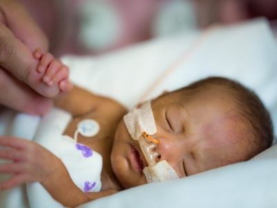
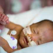
Babies born prematurely – before 37 weeks of pregnancy – often have a lot of catching up to do. Not just in size. Preterm infants typically lag behind their term peers in a variety of areas as they grow up, including motor development, behavior and school performance.

New research suggests one way to combat this problem. The study, led by Children’s researchers and presented during the Pediatric Academic Societies 2018 annual meeting, suggests that the volume of carbohydrates, proteins, lipids and calories consumed by very vulnerable premature infants significantly contributes to increased brain volume and white matter development, even though additional research is needed to determine specific nutritional approaches that best support these infants’ developing brains.

During the final weeks of pregnancy, the fetal brain undergoes an unprecedented growth spurt, dramatically increasing in volume as well as structural complexity as the fetus approaches full term.

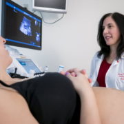
One in 10 infants born in the U.S. in 2016 was born before 37 weeks of gestation, according to the Centers for Disease Control and Prevention. Within this group, very low birthweight preemies are at significant risk for growth failure and neurocognitive impairment. Nutritional support in the neonatal intensive care unit (NICU) helps to encourage optimal brain development among preterm infants. However, their brain growth rates still lag behind those seen in full-term newborns.

“Few studies have investigated the impact of early macronutrient and caloric intake on microstructural brain development in vulnerable preterm infants,” says Katherine Ottolini, lead author of the Children’s-led study. “Advanced quantitative magnetic resonance imaging (MRI) techniques may help to fill that data gap in order to better direct targeted interventions to newborns who are most in need.”

The research team at Children’s National Health System enrolled 69 infants who were born younger than 32 gestational weeks and weighed less than 1,500 grams. The infants’ mean birth weight was 970 grams and their mean gestational age at birth was 27.6 weeks.

The newborns underwent MRI at their term-equivalent age, 40 weeks gestation. Parametric maps were generated for fractional anisotropy in regions of the cerebrum and cerebellum for diffusion tensor imaging analyses, which measures brain connectivity and white matter tract integrity. The research team also tracked nutritional data: Grams per kilogram of carbohydrates, proteins, lipids and overall caloric intake.

“We found a significantly negative association between fractional anisotropy and cumulative macronutrient/caloric intake,” says Catherine Limperopoulos, Ph.D., director of Children’s Developing Brain Research Laboratory and senior author of the research. “Curiously, we also find significantly negative association between macronutrient/caloric intake and regional brain volume in the cortical and deep gray matter, cerebellum and brainstem.”

Because the nutritional support does contribute to cerebral volumes and white matter microstructural development in very vulnerable newborns, Limperopoulos says the significant negative associations seen in this study may reflect the longer period of time these infants relied on nutritional support in the NICU.

In addition to Ottolini and Limperopoulos, study co-authors include Nickie Andescavage, M.D., Attending, Children’s Neonatal-Perinatal Medicine; and Kushal Kapse.

The brain’s fluid-filled spaces during growth

Catherine Limperopoulous

Catherine Limperopoulous, Ph.D., and her colleagues used volumetric MRIs to assess how the ventricles, cerebrospinal fluid and the rest of the fetal brain normally change over time.

The human brain is not one solid mass. Buried within its gray and white matter are a series of four interconnected chambers, called ventricles, which produce cerebrospinal fluid. These ventricles are readily apparent on the fetal ultrasounds that have become the standard of prenatal care in the United States and most developed countries around the world. Abnormalities in the ventricles’ size or shape – or both – can give doctors an early warning that fetal brain development might be going awry.

But what is abnormal? It is not always clear, says Catherine Limperopoulos, Ph.D., director of the Developing Brain Research Laboratory at Children’s National Health System. Limperopoulos explains that despite having many variations in fetal ventricles, some infants have completely normal neurodevelopmental outcomes later. On the other hand, some extremely subtle variations in shape and size can signal problems.

On top of these complications are the tools clinicians typically use to assess the ventricles. Limperopoulos explains that most early indications of ventricle abnormalities come from ultrasounds, but the finer resolution of magnetic resonance imaging (MRI) can provide a more accurate assessment of fetal brain development. Still, both standard MRI and ultrasound provide only two-dimensional pictures, making it difficult to quantify slight differences in the volume of structures.

To help solve these problems, Limperopoulos and her colleagues recently published a paper in Developmental Neuroscience that takes a different tack. The team performed volumetric MRIs – a technique that provides a precise three-dimensional measure of structural volumes – on the brains of healthy fetuses to assess how the ventricles, cerebrospinal fluid and the rest of the brain normally change over time. Limperopoulos’ team recently performed a similar study to assess normal volumetric development in the brain’s solid tissues.

Previous studies published on comparable topics typically used information gathered from subjects who initially had clinical concerns but eventually were dismissed from these studies for not having worrisome diagnoses in the end. This might not truly reflect a typical population of pregnant women, Limperopoulos says.

Working with 166 pregnant women with healthy pregnancies spanning from 18 to 40 weeks gestation, the researchers performed volumetric MRIs on their singleton fetuses that covered every week of this second half of pregnancy. This technique allowed them to precisely calculate the volumes of structures within the fetal brain and get an idea of how these volumes changed over time within the group.

Their results show that over the second and third trimester:

  • The lateral ventricles, the largest ventricles found in the cerebrum with one for each brain hemisphere, grew about two-fold;
  • The third ventricle, found in the forebrain, grew about 23-fold;
  • The fourth ventricle, found in the hindbrain, grew about eight-fold;
  • And the extra-axial cerebrospinal fluid, found under the lining of the brain, increased about 11-fold.

The total brain volume increased 64-fold over this time, with the parenchyma – the solid brain tissue that encompasses gray and white matter – growing significantly faster than the cerebrospinal fluid-filled spaces.

Limperopoulos points out that the ability to measure the growth of the brain’s fluid-filled spaces relative to the surrounding brain tissue can provide critical information to clinicians caring for developing fetuses. In most cases, knowing what is normal allows doctors to reassure pregnant women that their fetus’ growth is on track. Abnormalities in these ratios can provide some of the first signals to alert doctors to blockages in cerebrospinal fluid flow, abnormal development, or the loss of brain tissue to damage or disease. Although the neurodevelopmental outcomes from each of these conditions can vary significantly, traditional ultrasounds or MRIs might not be able to distinguish these possibilities from each other. Being able to differentiate why cerebrospinal fluid spaces have abnormal shapes or sizes might allow doctors to better counsel parents, predict neurological outcomes, or potentially intervene before or after birth to mitigate brain damage.

“By developing a better understanding of what’s normal,” Limperopoulos says, “we can eventually identify reliable biomarkers of risk and guide interventions to minimize risks for vulnerable fetuses.”

Fetal medicine update: fetal brain development, zika virus

May 2, 2016 -Impaired global and tissue-specific brain development in the growth-restricted fetus.
A team of researchers applied an advanced imaging technique, three-dimensional MRI, to study brain development in high-risk pregnancies and are the first to report regional, tissue-specific volume delays for the developing fetal brain in FGR-affected pregnancies. The team compared overall fetal brain volume as well as regional brain volumes for a control group of healthy young pregnant women with a group of young women whose pregnancies were complicated by FGR. While fetuses in both groups grew exponentially as pregnancies progressed, the researchers began to see dramatic differences when they compared the volumes of specific regions of the brain, including the cerebellum, which coordinates balance and smooth movement; the deep gray matter, which also is involved in complex functions, such as memory and emotion; and the white matter, which is made up of millions of nerve fibers that connect to neurons in different regions.

March 30, 2016Congenital Zika viral infection linked to significant fetal brain abnormalities, despite ‘normal’ ultrasounds.
Infectious Zika virus was isolated from the brain of a 21-week-old fetus after causing extensive damage to brain tissue – despite ultrasounds that showed no sign of microcephaly at weeks 13, 16, and 17, according to a report published online in The New England Journal of Medicine. “While this is a single case, it poses troubling questions that could inform future research,” says the study’s co-senior author, Adre du Plessis, M.B.Ch.B., Director of the Fetal Medicine Institute and Chief of the Fetal and Transitional Medicine Division at Children’s National Health System. “Evidence is mounting that the Zika virus can persist in pregnant women’s bloodstreams weeks after their initial infection, arguing for changes to how these pregnancies are monitored,” Dr. du Plessis said. Six of the named authors are affiliated with Children’s National, where the pregnant woman sought more thorough assessment after testing positive for the Zika virus herself following international travel.