Tag Archive for: birth defect

DNA Molecule

Decoding cellular signals linked to hypospadias

DNA Molecule

“By advancing our understanding of the genetic causes and the anatomic differences among patients, the real goal of this research is to generate knowledge that will allow us to take better care of children with hypospadias,” Daniel Casella, M.D. says.

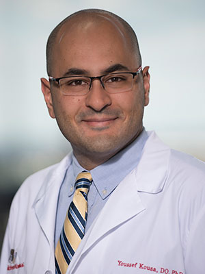
Daniel Casella, M.D., a urologist at Children’s National, was honored with an AUA Mid-Atlantic Section William D. Steers, M.D. Award, which provides two years of dedicated research funding that he will use to better understand the genetic causes for hypospadias.

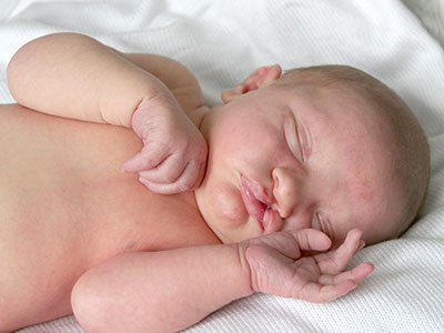
With over 7,000 new cases a year in the U.S., hypospadias is a common birth defect that occurs when the urethra, the tube that transports urine out of the body, does not form completely in males.

Dr. Casella has identified a unique subset of cells in the developing urethra that have stopped dividing but remain metabolically active and are thought to represent a novel signaling center. He likens them to doing the work of a construction foreman. “If you’re constructing a building, you need to make sure that everyone follows the blueprints.  We believe that these developmentally senescent cells are sending important signals that define how the urethra is formed,” he says.

His project also will help to standardize the characterization of hypospadias. Hypospadias is classically associated with a downward bend to the penis, a urethra that does not extend to the head of the penis and incomplete formation of the foreskin. Still, there is significant variability among patients’ anatomy and to date, no standardized method for documenting hypospadias anatomy.

“Some surgeons take measurements in the operating room, but without a standardized classification system, there is no definitive way to compare measurements among providers or standardize diagnoses from measurements that every surgeon makes,” he adds. “What one surgeon may call ‘distal’ may be called ‘midshaft’ by another.” (With distal hypospadias, the urethra opening is near the penis head; with midshaft hypospadias, the urethra opening occurs along the penis shaft.)

“By advancing our understanding of the genetic causes and the anatomic differences among patients, the real goal of this research is to generate knowledge that will allow us to take better care of children with hypospadias,” he says.

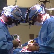
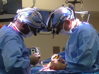
Parents worry about lingering social stigma, since some boys with hypospadias are unable to urinate while standing, and in older children the condition can be associated with difficulties having sex. Surgical correction of hypospadias traditionally is performed when children are between 6 months to 1 year old.

When reviewing treatment options with family, “discussing the surgery and postoperative care is straight forward. The hard part of our discussion is not having good answers to questions about long-term outcomes,” he says.

Dr. Casella’s study hopes to build the framework to enable that basic research to be done.

“Say we wanted to do a study to see how patients are doing 15-20 years after their surgery.  If we go to their charts now, often we can’t accurately describe their anatomy prior to surgery.  By establishing uniform measurement baselines, we can accurately track long-term outcomes since we’ll know what condition that child started with and where they ended up,” he says.

Dr. Casella’s research project will be conducted at Children’s National under the mentorship of Eric Vilain, M.D., Ph.D., an international expert in sex and genitalia development; Dolores J. Lamb, Ph.D., HCLD, an established leader in urology based at Weill Cornell Medicine; and Marius George Linguraru, DPhil, MA, MSc, an expert in image processing and artificial intelligence.

Baby with Cleft Palate

Understanding genetic synergy in cleft palate

Baby with Cleft Palate

Like mechanics fixing a faulty engine, Youssef A. Kousa, M.S., D.O., Ph.D., says researchers will not be able to remedy problems related to IRF6, a gene implicated in cleft palate, until they better understand how the gene works.

Like all of the individual elements of fetal development, palate growth is a marvel of nature. In part of this process, ledges of tissue on the sides of the face grow downwards on each side of the tongue, then upward, fusing at the midline at the top of the mouth. The vast majority of the time, this process goes correctly. However, some part of it goes awry for the 2,650 babies born in the United States each year with cleft palates and the thousands more born worldwide with the defect.

For nearly two decades, researchers have known that a gene known as IRF6 is involved in palate formation. Studies have shown that this gene contributes about 12 percent to 18 percent of the risk of cleft palate, more than any other gene identified thus far. IRF6 is active in epithelial tissues – those that line cavities and surfaces throughout the body – including the periderm, a tissue that lines the mouth cavity and plays an important role during development.

According to Youssef A. Kousa, M.S., D.O., Ph.D., a child neurology fellow at Children’s National Health System, the periderm acts like a nonstick layer, preventing the tongue or other structures from adhering to the growing palate and preventing it from sealing at the midline. While researchers have long suspected that IRF6 plays a strong role in promoting this nonstick quality, exactly how it exerts its influence has not been clear.

“Gaining a better understanding of this gene might help us to eventually address deficits or perturbations in the system that creates the palate,” Dr. Kousa says. “Like a mechanic fixing a faulty engine, we will not be able to remedy problems related to this gene until we know how the gene works.”

Youssef Kousa

“Gaining a better understanding of this gene might help us to eventually address deficits or perturbations in the system that creates the palate,” Dr. Kousa says. “Like a mechanic fixing a faulty engine, we will not be able to remedy problems related to this gene until we know how the gene works.”

In a study published July 19, 2017 by the Journal of Dental Research, Dr. Kousa and colleagues seek to decipher one piece of this puzzle by investigating how this key gene might interact with others that are active during fetal development. The researchers were particularly interested in genes that work together in a cascade of activity known as the tyrosine kinase receptor signaling pathway.

Because this pathway includes a large group of genes, Dr. Kousa and colleagues reasoned that they could answer whether IRF6 interacts with this pathway by looking at whether the gene interacts with the last member of the cascade, a gene called SPRY4. To do this, the researchers worked with experimental models that had mutations in IRF6, SPRY4 or both. If these two genes interact, the scientists hypothesized, carrying mutations in both genes at the same time should result in a dramatically different outcome compared with animals that carried mutations in just one gene.

Using selective breeding techniques, the researchers created animals that had mutations in either of these genes or in both. Their results suggest that IRF6 and SPRY4 indeed do interact: Significantly more of the oral surface was adhered to the tongue during fetal development in experimental models that had mutations in both genes compared with those that had just one single gene mutated. Examining the gene activity in the periderm cells of these affected animals, the researchers found that doubly mutated experimental models also had decreased activity in a third gene known as GRHL3, which also has been linked with cleft lip and palate.

Dr. Kousa says the research team plans to continue exploring this interaction to better understand the flow of events that lead from perturbations in these genes to formation of cleft palate. Some of the questions they would like to answer include exactly which gene or genes in the tyrosine kinase receptor signaling pathway specifically interact with IRF6 – since SPRY4 represents just the end of that pathway, others genes earlier in the pathway are probably the real culprits responsible for driving problems in palate formation. They also will need to verify if these interactions take place in humans in the same way they occur in preclinical models.

Eventually, Dr. Kousa adds, the findings could aid in personalized prenatal counseling, diagnosis and screening related to cleft palate, as well as preventing this condition during pregnancy. Someday, doctors might be able to advise couples who carry mutations in these genes about whether they are more likely to have a baby with a cleft palate or determine which select group of pregnancies need closer monitoring. Additionally, because research suggests that GRHL3 might interact with nutrients, including inositol, it might be possible to prevent some cases of cleft palate by taking additional supplements during pregnancy.

“The more we know about how these genes behave,” Dr. Kousa says, “the more we can potentially avoid fetal palate development going down the wrong path.”

two doctors perform surgery

Working miracles to control seizures and preserve brain power in newborns

Oluigbo and Myseros neurosurgery

In the spring of 2017, a multidisciplinary team applied an innovative approach to help preserve function in the working right hemisphere of a baby who experienced her first seizure hours after birth.

When orderly early fetal brain development is disturbed in one half of the brain, infants can be born with hemimegalencephaly—a rare occurrence—that results in one of the brain’s two hemispheres being oversized, heavy and malformed. This brain malformation arises early in the fetal period of life, is not inherited and is associated with seizures early in life.

Children with hemimegalencephaly can develop horrible seizures within the first hours or days of life. According to published research, every month these infants experience uncontrolled seizures correlates to a steep decline in IQ.

Because these types of seizures do not respond to multiple anti-seizure medications—medicines which may also cause worrisome side effects of their own in neonates—care teams attempt to schedule surgery as soon as feasible to remove or disconnect the hemisphere triggering the damaging seizures. “The ‘bad’ brain does not sustain any function and it interferes with the ‘good’ brain doing what it needs to do,” says William D. Gaillard, M.D., chief of Children’s division of Epilepsy and Neurophysiology and chief of Neurology.

Hemispherectomy is intricate surgery on an organ that is softer than normal and crisscrossed with a tangle of blood vessels that supply the damaged hemisphere with blood. Because of the risks of life-threatening blood loss in very young infants, the dramatic surgery is usually not performed until babies are at least 3 months old and weigh at least 10 pounds.

The challenge: The vulnerable babies who most need relief, infants who have been seizing since early life, are too young for the operation.

Neurosurgeons have clamped the carotid artery that supplies blood to the brain to minimize blood loss when the hemisphere is surgically removed. Dr. Gaillard says knowledge of that approach led the team to think: What if we use embolization—blocking blood supply to targeted locations in the brain—to achieve the same effect?  The plan effectively destroys the malformed brain from within, neutralizing its ability to cause the seizures.

“It was eye-opening for us to think about actually inflicting brain injury as a way of treating something in the brain that was causing seizures. That is really novel in itself: We’re thinking out of the box in applying existing techniques in a different age group. The conventional thinking with newborns is to let them be; their seizures don’t look that bad,” says Taeun Chang, M.D., director of Children’s Neonatal Neurology and Neonatal Neurocritical Care Program.

“We have evidence to suggest this is a safe and effective way of avoiding recurrent seizures and minimizing the need to give these infants potentially toxic medications so early in life. Ultimately, this helps a select group of babies who need the surgery to get to the point of being old enough to have it—all the while, sparing the healthy part of their brain,” Dr. Gaillard adds.

Darcy hemimegalencephaly

Once the embolization ended Darcy’s most severe seizures, the little girl could make eye contact, started smiling, and then graduated from smiling to full laughs. In weekly physical therapy, the infant works on tummy time, head control and ensuring her eyes track.

In the spring of 2017, the multidisciplinary team applied the innovative approach to help preserve function in the working right hemisphere of a baby named Darcy Murphy. Darcy experienced her first seizure hours after she was born, and when she arrived at Children’s National had been in and out of two different emergency rooms in another state for the first few weeks of her life.

The team explained to the Murphy family that Darcy was on multiple medications, but her seizures continued unabated. The options included inducing a coma, sending Darcy home despite ongoing seizures or minimally invasive embolization.

“We would not have even posed this if we were not confident in our ability to do the procedure and deal with potential complications,” Dr. Chang says.

“Oh my gosh, as a parent you know what you’re doing is permanent,” says Rachel Murphy, 29, Darcy’s mom said of the decisions that she and husband Ryan, 33, faced for the youngest of their three children. “What if it’s not the right decision? What if in a week they come out with a new procedure you could have done? We were horrified all the time. The nice part with this procedure is the reward is apparent very quickly, and it just gets better. You don’t have to wait two years to know you made the right decision. You can see half a brain is better than the whole thing for this specific child.”

Once the embolization ended Darcy’s most severe seizures, the little girl could initiate and maintain eye contact with family members, started smiling and then graduated from smiling to full laughs. In weekly physical therapy, the infant works on tummy time, head control and ensuring her eyes track.

Children’s multidisciplinary care team includes experts in newborn intensive care (neonatologists) to aggressively manage seizures in the traditional fashion as they occur and to monitor vital signs; a neonatal neurologist/neurointensivist at the bedside and in the Angio suite monitoring Darcy’s brain activity; a neonatal epileptologist; a surgical epilepsy team; an interventional neuroradiologist; neurosurgeons to perform the delicate functional hemispherectomy to remove any residual brain tissue from the bad hemisphere; and physical therapists working to help Darcy achieve maximum function after surgery.

“We were just like one unit in the sense of being able to provide coherent, comprehensive care. It’s about blood pressure management, breathing, electrolytes, making sure everything is right for going to the operating room,” Dr. Chang explains. “Darcy’s case highlights the ways in which Children’s National is different and offers personalized care that is superior to other centers.”

The team, which recently published a case report of two previous serial embolizations followed by hemispherectomy, plans follow-up papers describing EEG manifestations during an acute stroke in a newborn, advice to the field on best practices for the embolization and using cooling to control the planned brain injury during embolization hemispherectomy.

Revised Nov. 7, 2017

Related resources

Chima Oluigbo

A novel way to treat intractable epilepsy caused by hemimegalencephaly

Chima Oluigbo

A multidisciplinary team led by Chima Oluigbo, M.D., F.R.C.S.C., pioneered a novel technique to preserve newborns’ healthy brain tissue, buying time until the infants became old enough to undergo a hemispherectomy.

PDF Version

What’s known

Hemimegalencephaly is an extremely rare birth defect in which one side of the brain grows larger than the other. This anomaly typically leads to severe, recurrent seizures that can be difficult to control solely with medications. While the seizures themselves are detrimental to the developing brain, the amount of medications used to reduce seizure frequency often come with significant side effects and have the potential to hamper brain growth. Hemispherectomy, a radical surgery in which one half of the brain is removed, is often the most successful way to treat severe and intractable epilepsy. However, this surgery can be challenging to perform successfully in very young babies.

What’s new

In this case report, the Children’s National Health System Epilepsy Team led by Chima Oluigbo, M.D., F.R.C.S.C., a pediatric neurosurgeon; Tammy N. Tsuchida, M.D., PhD., a pediatric surgical epileptologist; Monica Pearl, M.D., a pediatric interventional neuroradiologist; Taeun Chang, M.D., a neonatal neurointensivist; and the neonatal intensive care team explored the possibility of using minimally invasive surgery to cut off the blood supply to the brain hemisphere responsible for generating seizures in newborns with hemimegalencephaly. This procedure, they reasoned, could buy time for babies to mature and become more resilient to withstand the future hemispherectomy while also lessening the damage caused by uncontrolled, recurrent seizures. The case report focused on the first two patients with hemimegalencephaly who had sequential procedures to gradually restrict blood flow to the affected brain hemisphere within their first few weeks of life, followed by hemispherectomies at a few months of age. This novel approach significantly lessened their seizures until hemispherectomy, allowing these children to continue to grow and develop seizure-free.

Questions for future research

Q: Which patients are best suited for this surgical procedure?
Q: How can surgeons reduce the risk of excessive blood loss during hemispherectomy caused by the growth of additional blood vessels after flow through the brain’s major vessels has been blocked?
Q: What are the long-term outcomes for infants who undergo these procedures?

Source: “ ‘Endovascular embolic hemispherectomy’: A strategy for the initial management of catastrophic holohemispheric epilepsy in the neonate.” Oluigbo, C., M.S. Pearl, T.N. Tsuchida, T. Chang, C.-Y. Ho and W. D. Gaillard. Published by Child’s Nervous System October 29, 2016.