pregnancy

New Children’s National and Inova collaboration

pregnancy

A new research collaboration will streamline completion of retrospective and prospective research studies, shedding light on myriad conditions that complicate pregnancies.

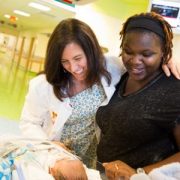
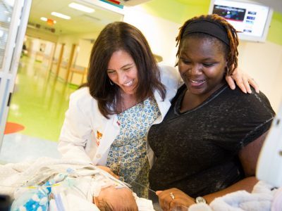
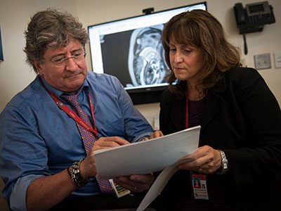
A new three-year, multi-million dollar research and education collaboration in maternal, fetal and neonatal medicine aims to improve the health of pregnant women and their children. The partnership between Children’s National Health System and Inova will yield a major, nationally competitive research and academic program in these areas that will leverage the strengths of both health care facilities and enhance the quality of care available for these vulnerable populations.

The collaboration will streamline completion of retrospective and prospective research studies, shedding light on a number of conditions that complicate pregnancies. It is one of several alliances between the two institutions aimed at improving the health and well-being of children in Northern Virginia and throughout the region.

“The Washington/Northern Virginia region has long had the capability to support a major, nationally competitive research and academic program in maternal and fetal medicine,” says Adre du Plessis, M.B.Ch.B., Director of the Fetal Medicine Institute at Children’s National and a co-Principal Investigator for this partnership. “The Children’s National/Inova maternal-fetal-neonatal research education program will fill this critical void.

“This new partnership will help to establish a closer joint education program between the two centers, working with the OB/Gyn residents at Inova and ensuring their involvement in Children’s National educational programs and weekly fetal case review meetings,” Dr. du Plessis adds.

Larry Maxwell, M.D., Chairman of Obstetrics and Gynecology at Inova Fairfax Medical Campus and a co-Principal Investigator for the collaboration, further emphasizes that “Inova’s experience in caring for women and children — combined with genomics- and proteomics-based research — will synergize with Children National’s leadership in neonatal pediatrics, placental biology and fetal magnetic resonance imaging (MRI) to create an unprecedented research consortium. This will set the stage for developing clinically actionable interventions for mothers and babies in metropolitan District of Columbia.”

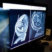
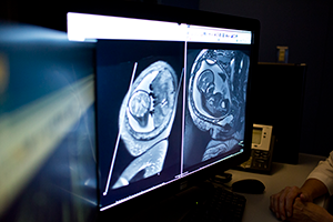
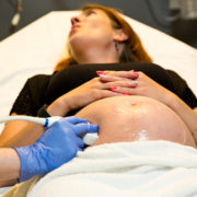
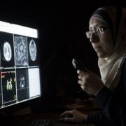
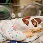
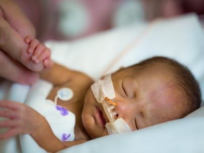
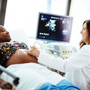
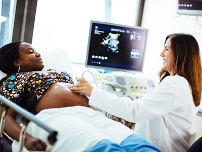
Children’s National, ranked No. 3 nationally in neonatology, has expertise in pediatric neurology, fetal and neonatal neurology, fetal and pediatric cardiology, infectious diseases, genetics, neurodevelopment and dozens of additional pediatric medical subspecialties. Its clinicians are national leaders in next-generation imaging techniques, such as MRI. Eighteen specialties and 50 consultants evaluate more than 700 cases per year through its Fetal Medicine Institute. In mid-2016, Children’s National created a Congenital Zika Virus Program to serve as a dedicated resource for referring clinicians and pregnant women. The hospital performs deliveries in very high-risk, complex situations, but does not offer a routine labor and delivery program.

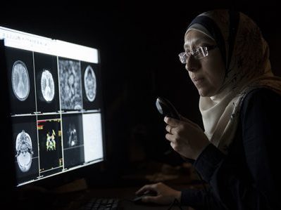
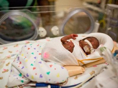
Inova Fairfax Medical Campus is home to both Inova Women’s Hospital and Inova Children’s Hospital. Inova Women’s Hospital is the region’s most comprehensive and highest-volume women’s hospital — delivering more than 10,000 babies in 2016. Inova Children’s Hospital serves as Northern Virginia’s children’s hospital —providing expert care in pediatric and fetal cardiology, cardiac surgery, genetics, complex general surgery, neurology, neurosurgery and other medical and surgical specialties. Its 108-bed Level IV neonatal intensive care unit is one of the largest and most comprehensive in the nation. Inova’s Translational Medicine Institute includes a genomics lab, as well as a research Institute focused on studies designed to build genetic models that help answer questions about individual disease. Each of these specialties is integrated into the Inova Fetal Care Center — which serves as a connection point between Inova Women’s and Children’s Hospitals. The Inova Fetal Care Center provides complex care coordination for women expecting infants with congenital anomalies or with other fetal concerns. Because Inova Women’s Hospital and Inova Children’s Hospital are co-located, women are able to deliver their babies in the same building where their children will receive care.

The research collaboration will support research assistants; tissue technicians; a placental biologist; as well as support for biomedical engineering, fetal-neonatal imaging, telemedicine, regulatory affairs and database management. The joint research projects that will take place under its auspices include:

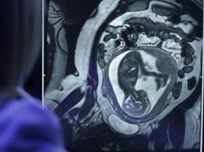
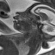
  • Fetal growth restriction (FGR), which occurs when the failing placenta cannot support the developing fetus adequately. FGR is a major cause of stillbirth and death, and newborns who survive face numerous risks for multiple types of ailments throughout their lives. A planned study will use quantitative MRI to identify signs of abnormal brain development in pregnancies complicated by FGR.
  • Placental abnormalities, including placenta accreta. A planned study will combine quantitative MRI studies on the placenta during the third trimester and other points in time with formal histopathology to identify MRI signals of placenta health and disease.
  • Microcephaly, a condition that is characterized by babies having a much smaller head size than expected due to such factors as interrupted brain development or brain damage during pregnancy. While the global Zika virus epidemic has heightened awareness of severe microcephaly cases, dozens of pregnancies in the region in recent years have been complicated by the birth defect for reasons other than Zika infection. A planned study will examine the interplay between MRI within the womb and head circumference and weight at birth to examine whether brain volume at birth correlates with the baby’s developmental outcomes.

Using fMRI for assessment prior to neurosurgery

For more than 20 years, Children’s National has explored the use of non-invasive fMRI as an alternative to more invasive testing to assess children’s language and memory.

A new Practice Guideline Summary published in Neurology, the journal of the American Academy of Neurology, contains the first complete, objective assessment of available data on the efficacy of functional magnetic resonance imaging (fMRI) to assess baseline language and memory, brain hemisphere dominance and to predict postsurgical impacts prior to surgery in patients with epilepsy.

According to contributing author William D. Gaillard, M.D., chief of Child Neurology, Epilepsy and Neurophysiology, and director of the Comprehensive Pediatric Epilepsy Program at Children’s National Health System, the report outlines several cases in which fMRI presents an effective alternative to the current standard of care, intracarotid amobarbital procedure (IAP). In IAP, medication is injected through the carotid artery to isolate one hemisphere of the brain at a time, followed by the patient performing memory or language tasks. The approach requires catheterization via a major artery. While minimally invasive, the procedure still carries the standard risks of vascular catheter procedures and requires recovery time.

“This publication took six years to complete,” Dr. Gaillard notes, “but we are happy to finally have the practice parameters that will make the case for the use of fMRI in an evidence-based way.”

Though the Practice Guidelines focus on adults, the evidence assessment included all available pediatric data as well, says Dr. Gaillard. A great deal of that data were contributed by Children’s National faculty, who lead the nation in clinical applications of fMRI. More than 20 years ago, Dr. Gaillard and his team began studying fMRI as a viable alternative to IAP to collect accurate language assessments in children, particularly those with epilepsy. Today, Children’s National is at the forefront of clinical application of fMRI, having performed about 1,000 pediatric assessments in the last two decades — more than nearly every other institution.

An 11-member panel of international experts conducted the analyses for the Practice Guidelines. Overall, the report indicates:

  • fMRI is a viable option for measuring lateralized language functions in place of IAP in medial temporal lobe epilepsy, temporal epilepsy in general or extratemporal epilepsy.
  • Evidence was insufficient to recommend fMRI over IAP for patients with temporal neocortical epilepsy or temporal tumors.
  • Pre-surgical fMRI can serve as an adequate alternative to IAP memory testing for predicting verbal memory outcome.

In closing, the authors also explicitly recommend that clinicians carefully advise every patient of the risks and benefits of both fMRI and IAP before recommending either approach.

Related resources: Use of fMRI in the presurgical evaluation of patients with epilepsy

Setting a baseline for healthy brain development

Catherine Limperopoulos, Ph.D., and colleagues performed the largest magnetic resonance imaging study of normal fetal brains in the second and third trimesters of pregnancy.

Starting as a speck barely visible to the naked eye and ending the in utero phase of its journey at an average weight of 7.5 pounds, the growth of the human fetus is one of the most amazing events in biology. Of all the organs, the fetal brain undergoes one of the most rapid growth trajectories, expanding over 40 weeks from zero to 100 billion neurons — about as many brain cells as there are stars in the Milky Way Galaxy.

This exponential growth is part of what gives humans our unique abilities to use language or have abstract thoughts, among many other cognitive skills. It also leaves the brain extremely vulnerable should disruptions occur during fetal development. Any veering off the developmental plan can lead to a cascade of results that have long-lasting repercussions. For example, studies have shown that placental insufficiency, or the inability of the placenta to supply the fetus with oxygen and nutrients in utero, is associated with attention deficit hyperactivity disorder, autism, and schizophrenia.

Recent research has identified differences in the brains of people with these disorders compared with those without. Despite the almost certain start of these conditions within the womb, they have remained impossible to diagnose until children begin to show clinical symptoms. If only researchers could spot the beginnings of these problems early in development, says Children’s National Health System researcher Catherine Limperopoulos, Ph.D., they might someday be able to develop interventions that could turn the fetal brain back toward a healthy developmental trajectory.

“Conventional tools like ultrasound and magnetic resonance imaging (MRI) can identify structural brain abnormalities connected to these problems, but by the time these differences become apparent, the damage already has been done,” Limperopoulos says. “Our goal is to be able to pick up very early deviations from normal in the high-risk pregnancy before an injury to the fetus might become permanent.”

Before scientists can recognize abnormal, she adds, they first need to understand what normal looks like.

In a new study published in Cerebral Cortex, Limperopoulos and colleagues begin to tackle this question through the largest MRI study of normal fetal brains in the second and third trimesters of pregnancy. While other studies have attempted to track normal fetal brain growth, that research has not involved nearly as many subjects and typically relied on data collected when fetuses were referred for MRIs for a suspected problem. When the suspected abnormality was ruled out by the scan, these “quasi-controls” were considered “normal” — even though they may be at risk for problems later in life, Limperopoulos explains.

By contrast, the study she led recruited 166 healthy pregnant women from nearby low-risk obstetrics practices. Each woman had an unremarkable singleton pregnancy and ended up having a normal full-term delivery, with no evidence of problems affecting either the mother or fetus over the course of 40 weeks.

At least one time between 18 and 39 gestational weeks, the fetuses carried by these women underwent an MRI scan of their brains. The research team developed complex algorithms to account for movement (since neither the mothers nor their fetuses were sedated during scans) and to convert the two-dimensional images into three dimensions. They used the information from these scans to measure the increasing volumes of the cerebellum, an area of the brain connected to motor control and known to mediate cognitive skills; as well as regions of the cerebrum, the bulk of the brain, that is pivotal for movement, sensory processing, olfaction, language, and learning and memory.

Their results in uncomplicated, full-term pregnancies show that over 21 weeks in the second half of pregnancy, the cerebellum undergoes an astounding 34-fold increase in size. In the cerebrum, the fetal white matter, which connects various brain regions, grows 22-fold. The cortical gray matter, key to many of cerebrum’s functions, grows 21-fold. And the deep subcortical structures (thalamus and basal ganglia), important for relaying sensory information and coordination of movement and behavior, grow 10-fold. Additional examination showed that the left hemisphere has a larger volume than the right hemisphere early in development, but sizes of the left and right brain halves were equal by birth.

By developing similar datasets on high-risk pregnancies or births—for example, those in which fetuses are diagnosed with a problem in utero, mothers experience a significant health problem during pregnancy, babies are born prematurely, or fetuses have a sibling diagnosed with a health problem with genetic risk, such as autism—Limperopoulos says that researchers might be able to spot differences during gestation and post-natal development that lead to conditions such as schizophrenia, attention deficit hyperactivity disorder and autism spectrum disorder.

Eventually, researchers may be able to develop fixes so that babies grow up without life-long developmental issues.

“Understanding ‘normal’ is really opening up opportunities for us to begin to precisely pinpoint when things start to veer off track,” Limperopolous says. “Once we do that, opportunities that have been inaccessible will start to present themselves.”

Altered blood flow may contribute to preemie brain injuries

A Children’s National research team for the first time mapped abnormalities in blood flow that may contribute to brain injury suffered by preterm infants.

Advanced noninvasive imaging permitted Children’s National Health System researchers to measure the lasting impact of abnormalities in blood flow on the immature brains of premature babies. Blood flow to the brain, or perfusion, has been studied previously to understand its role in other health conditions, but this is the first time a research team has mapped how these changes may contribute to brain injury suffered by babies born before 32 weeks’ gestation.

Preterm birth is a major risk factor for brain injury. The prospective study examined infants weighing less than 1,500 grams who were born prior to 32 gestational weeks.

Of 78 infants studied, 47 had structural brain injuries categorized as either mild or moderate to severe, and 31 had no brain injury. While global cerebral blood flow decreased with advancing postnatal age, the blood flow decreased more significantly among preterm infants with brain injury, says Eman S. Mahdi, M.D., M.B.Ch.B. Dr. Mahdi is a pediatric radiology fellow at Children’s National and lead author of the abstract.

“In addition to differences in global brain blood flow, we saw a marked decrease in regional blood flow to the thalamus and the pons, regions known to be metabolically active during this time,” Dr. Mahdi says. The thalamus helps to process information from the senses and relays it elsewhere within the brain. Located at the base of the brain, the pons is part of the central nervous system and also is a critical relay of information between the cerebrum and cerebellum. “These regional variations in blood flow reflect vulnerability of the cerebral-cerebellar circuitry,” she adds.

The Radiological Society of North America (RSNA) recognized Dr. Mahdi with its Trainee Research Prize. She presented the work, “Cerebral Perfusion Is Perturbed by Preterm Birth and Brain Injury,” during the RSNA Scientific Assembly and Annual Meeting, held from Nov. 27 to Dec. 2.

The findings point to the need for additional research to explore how cerebral blood flow trends evolve as preemies grow older and whether abnormal blood flow is linked to differences in health outcomes. In addition, the technique used by the research team, arterial spin labeling perfusion imaging – a type of magnetic resonance imaging – represents a useful and non-invasive technology for identifying early cerebral perfusion abnormalities in preterm infants, says Catherine Limperopoulos, Ph.D., director of the Developing Brain Research Laboratory at Children’s National and abstract senior author.

Premature birth may alter critical cerebellar development linked to learning and language

 Diffusion tensor imaging teases out subtle injury to cerebral and cerebellar white matter that is not evident with conventional MRI, allowing researchers to quantify brain tissue microstructure and classify white matter integrity.

Diffusion tensor imaging teases out subtle injury to cerebral and cerebellar white matter that is not evident with conventional MRI, allowing researchers to quantify brain tissue microstructure and classify white matter integrity.

Premature birth can interrupt a key period of brain development that occurs in the third trimester, which has the potential to impact a child’s long-term learning, language, and social skills. A recent case-control study published in The Journal of Pediatrics applied diffusion tensor magnetic resonance imaging (DTI) to zoom in on the microstructures comprising the critical cerebellar neural networks related to learning and language, and found significant differences between preterm and full-term newborns.

“The third trimester, during which many premature births occur, is typically when the developing cerebellum undergoes its most dramatic period of growth. Normally, the cerebellar white matter tracts that connect to the deep nuclei are rich in pathways where nerve fibers cross. Those connections permit information to flow from one part of the brain to another. It is possible that premature birth leads to aberrant development of these critical neural networks,” says Catherine Limperopoulos, Ph.D., director of the Developing Brain Research Laboratory at Children’s National Health System and senior study author.

One in 10 American babies is born prematurely. The brain injury that infants born prematurely experience is associated with a range of neurodevelopmental disabilities, including some whose influence isn’t apparent until years later, when the children begin school. Nearly half of extremely preterm infants go on to experience long-term learning, social, and behavioral impairments.

While conventional magnetic resonance imaging (MRI) can detect many brain abnormalities in newborns, a newer technique called DTI can tease out even subtle injury to cerebral and cerebellar white matter that is not evident with conventional MRI. White matter contains axons, which are nerve fibers that transmit messages. With DTI, researchers can quantify brain tissue microstructure and describe the integrity of white matter.

The research team compared imaging from 73 premature infants born before 32 weeks gestation who weighed less than 1,500 grams with 73 healthy newborns born to mothers who delivered at full term after 37 weeks. After the newborns had been fed, swaddled, and fitted with double ear protection, the imaging was performed as they slept. Nurses monitored their heart rates and oxygen saturation. Their brain abnormalities were scored as normal, mild, moderate, or severe.

All of the full-term newborns had normal brain MRIs as did 44 (60.3 percent) of the preemies.

The preemies had significantly higher fractional anisotropy in the cerebellum, the part of the brain that processes incoming information from elsewhere in the brain, permitting coordinated movement as well as modulating learning, language, and social skills. Alterations in cerebellar microarchitecture was associated with markers for illness severe enough to require surgery – such as correcting abnormal blood flow caused by the failure of the ductus arteriosus to close after birth and to remedy a bowel disease known as necrotizing enterocolitis. The risk factors also are associated with compromised cardiorespiratory function and low Apgar score at five minutes, Limperopoulos and co-authors write. The Apgar score is a quick way to gauge, one minute after birth, how well the newborn withstood the rigors of childbirth. It is repeated at five minutes to describe how the newborn is faring outside of the womb.

“In previous studies, we and others have associated cerebellar structural injury in preterm infants with long-term motor, cognitive, and socio-affective impairments. This is one of the first studies to provide a detailed report about these unexpected alterations in cerebellar microstructural organization,” she adds. “We postulate that the combination of premature birth and early exposure of the immature developing cerebellum to the extrauterine environment results in disturbed micro-organization.”

Additional research is warranted in larger groups of patients as well as long-term follow up of this cohort of newborns to determine whether this microstructural disorganization predicts long-term social, behavioral, and learning impairments.

“A large number of these prematurely born newborns had MRI readings in the normal range. Yet, we know that these children are uniquely at risk for developing neurodevelopmental disabilities later in life. With additional study, we hope to identify interventions that could lower those risks,” Limperopoulos says.

Related resources: The Journal of Pediatrics editorial

Fetal Cardiac Health

Managing transposition of the great arteries in the womb

Fetal Cardiac Health

Monitoring and managing fetuses’ heart health in the womb can greatly improve their chances of living long and productive lives

Over the 22 years that Mary T. Donofrio, M.D., has been practicing fetal cardiology, the field has changed radically. The goal once had been simply to offer parents an accurate diagnosis and prepare them for sometimes devastating outcomes. Now, Dr. Donofrio, who directs the Fetal Heart Program and Critical Care Delivery Program at Children’s National Health System, says specialists can follow fetuses throughout the pregnancy and manage many conditions in the womb, greatly improving their chances of living long and productive lives.

Case in point: Transposition of the great arteries, a congenital defect characterized by reversal of the heart’s two main arteries—the aorta, which distributes oxygenated blood throughout the body, and the pulmonary artery, which carries deoxygenated blood from the heart to the lungs. The single abnormality means that the oxygenated “red” blood flows back to the lungs while deoxygenated “blue” blood flows out to the body.

After birth, when the cord is clamped and the connection to the placenta severed, the baby’s cardiovascular system must adjust. If the fetal connections between the two sides of the heart no longer remain, the brain and other organs in infants with this defect are severely deprived of oxygen. The condition may be fatal if something is not done immediately to reopen the fetal connections to stabilize the circulation before surgery can be done. But if the fetal cardiologist can keep tabs on what’s happening to the heart over time and prepare a specialty team of cardiologists to treat the problem immediately after birth, chances of survival are significantly improved.

More than a decade ago, as a young attending physician, Dr. Donofrio witnessed a case that has stuck with her to this day. The baby’s diagnosis of transposition of the great arteries was not made until shortly before birth. In addition, the two fetal blood flow connections that allow blood to circulate had closed, causing severe heart failure. Although the care team performed an emergency delivery and immediate cardiac procedure, including initiation of a heart-lung machine in the delivery room to try to stabilize the circulation, the baby ultimately died due to complications from a very low oxygen level. “I always wonder what happened,” Dr. Donofrio says. “Was the baby’s heart always that bad and nobody noticed it, or did it change over time?”

In a paper published recently in the Journal of Neonatal-Perinatal Medicine, she and colleagues illustrate the dramatic transformation in care that has taken place in the 14 years since this unforgettable case. The new publication describes the case of a different fetus diagnosed at 22 weeks gestation with transposition of the great arteries in 2015 at Children’s National. Unlike many congenital heart disorders, the heart’s four chambers appear misleadingly normal at the typical mid-pregnancy ultrasound. Despite the challenging diagnosis for many obstetricians, this fetus’ heart condition was recognized early by looking at the arteries leaving the heart in addition to the chambers.

While such a defect is fatal if left untreated, Dr. Donofrio explains there are two pathways that can allow the blood to get to where it needs to go such that the circulation is stabilized and the damage mitigated. One is the fetal blood vessel known as the ductus arteriosus that typically stays open for a day or two after birth. The second is an opening between the heart’s two upper chambers, known as the foramen ovale, which usually closes upon delivery. By keeping those two pathways open, blood can cross from one side of the heart to the other, buying time in the delivery room so that babies can be stabilized before they receive surgery to permanently move the arteries back to their normal position.

In the 2015 case, Dr. Donofrio and colleagues had the chance to monitor the fetus and the fetal heart at follow-up appointments every four weeks after diagnosis. What they saw completely changed the course of their treatment plan and likely saved the baby’s life. With each ultrasound, they saw that the ductus arteriosus and the foramen ovale—the critical connections needed for survival—were gradually closing.

Dr. Donofrio noted at the fetal evaluation at 38 weeks that the structures had closed, and the heart was showing signs that it was not functioning well.  She and her team realized that the only way to save this baby was to deliver earlier than planned and to have cardiac specialists standing by ready to perform a life-saving procedure to open the connections right after the baby was separated from the placenta. The baby was delivered by Cesarean section in the cardiac operating room at Children’s. The cardiac intervention team immediately created a hole where the foramen ovale should have been by using a balloon to open the tissue that had closed. The care team also administered a prostaglandin infusion, a drug that can keep the ductus arteriosis open. This time, however, the medicine did not work. The baby was stabilized with several cardiac medications and, with little time to spare, the cardiac surgeons operated on the one-day-old baby to switch his great arteries back to the normal position, saving his life.

The baby is now 1-year-old, Dr. Donofrio says, and is healthy—a scenario that likely wouldn’t have happened had the fetal team not made the diagnosis and continually monitored the condition in the womb.

“I remember back to that first case when we were really scrambling to do everything we could at the last minute because we didn’t have the information we needed until the very end,” Dr. Donofrio says. “Now, we can spot problems early and do something about it. For me, that’s amazing. We’re making a difference, and that’s a really great thing.”

Every day fetuses remain in utero critical to preserving normal brain development

preemieimage

If it does not jeopardize the health of the pregnant mother or her fetus, pregnancies should be carried as close to full term as possible to avoid vulnerable preemies experiencing a delay in brain development, study results published October 28 in Pediatrics indicate.

Some 15 million infants around the world – and 1 in 10 American babies – are born prematurely. While researchers have known that preemies’ brain growth is disturbed when compared with infants born at full term, it remained unclear when preemies’ brain development begins to veer off course and how that impairment evolves over time, says Catherine Limperopoulos, Ph.D., Director of the Developing Brain Research Laboratory at Children’s National Health System and senior study author.

A look at the research

In order to shine a spotlight on this critical phase of fetal brain development, Limperopoulos and colleagues studied 75 preterm infants born prior to the 32th gestational week who weighed less than 1,500 grams who had no evidence of structural brain injury. These preemies were matched with 130 fetuses between 27 to 39 weeks gestational age.

The healthy fetal counterparts are part of a growing database that the Children’s National Developing Brain Research Laboratory has assembled. The research lab uses three-dimensional magnetic resonance imaging to carefully record week-by-week development of the normal in utero fetal brains as well as week-by-week characterizations of specific regions of the fetal brain.

The availability of time-lapsed images of normally developing brains offers a chance to reframe research questions in order to identify approaches to prevent injuries to the fetal brain, Limperopoulos says.

“Up until now, we have been focused on examining what is it about being born too early? What is it about those first few hours of life that leaves preemies more vulnerable to brain injury?” she says. “What is really unique about these study results is for the very first time we have an opportunity to better understand the ways in which we care for preemies throughout their hospitalization that optimize brain development and place more emphasis those activities.”

When the research team compared third-trimester brain volumes, preemies showed lower volumes in the cerebrum, cerebellum, brainstem, and intracranial cavity. The cerebrum is the largest part of the brain and controls speech, thoughts, emotions, learning, as well as muscle function. The cerebellum plays a role in learning and social-behavioral functions as well as complex motor functions; it also controls the balance needed to stand up and to walk. The brainstem is like a router, ferrying information between the brain, the cerebellum, and the spinal cord.

“What this study shows us is that every day and every week of in utero development is critical. If at all possible, we need to keep fetuses in utero to protect them from the hazards that can occur in the extra uterine environment,” she says.

New program provides science-driven answers about zika virus’s impact on pregnancies

Drs. DeBiasi and du Plessis

Each week, as temperatures rise, the likelihood increases that the United States will experience domestic Zika virus transmission. Indeed, such domestic Zika transmission already is occurring in Puerto Rico and the U.S. Virgin Islands. The Children’s National Health System Fetal Medicine Institute and Division of Pediatric Infectious Disease announced the formation of a Congenital Zika Virus Program to serve as a dedicated resource for referring clinicians and for pregnant women to receive counseling and science-driven answers about the impact of the Zika virus on their pregnancies.

Over years, Children’s National has invested in equipment and highly trained personnel, building expertise in infectious diseases, pediatric neurology, pediatric cardiology, genetics, neurodevelopment, and other specialties. Children’s clinicians are recognized as national leaders in next-generation imaging techniques, such as fetal MRI, and a variety of divisions work together to offer multidisciplinary support and coordinated care to infants born with special needs. As the nation prepares for the Zika virus, Children’s National is facilitating the multi-step process of blood testing, helping to ensure timely and precise information. Children’s National specialists are able to guide Zika-affected pregnancies through the fetal period and can oversee the care of Zika-affected infants after delivery. Care and clinical support is provided by a multidisciplinary team of pediatric neurologists, physical therapists, infectious disease experts, and neurodevelopmental physicians.