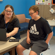
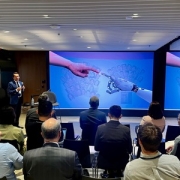
Cell & Gene Therapy in the DMV: Experts collaborate for cures
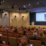
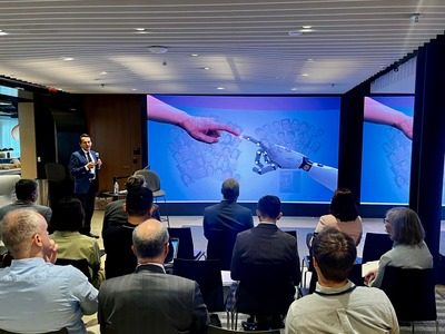
Leaders in medicine, academia, industry and state and local government came together for the first annual Cell and Gene Therapy in the DMV Symposium, hosted at the Children’s National Research & Innovation Campus. The mission: Connect the local scientific community – bursting with expertise and collaboration potential – to develop these cutting-edge therapies for cancers, sickle cell disease and immune-mediated disorders.
The daylong event drew over 100 experts from a range of organizations in the D.C, Maryland and Virginia region, sometimes called the DMV: Children’s National Hospital, the Food and Drug Administration, the National Institute of Standards and Technology, the National Institutes of Health, the General Accounting Office, Virginia Tech, MaxCyte, AstraZeneca, Kite Pharma, Montgomery College, the Maryland State Department of Commerce and more. Together, they unraveled a host of topics including the regulatory environment, workforce development and training, research standards and the promise of these therapies.
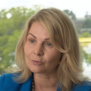
“Our Cell & Gene Therapy Symposium brings together our current collaborators and future partners in the D.C., Maryland and Virginia space, which is an incredibly rich area. We see tremendous opportunity and breakthroughs in our future,” said Catherine Bollard, M.D., M.B.Ch.B., interim chief academic officer and chief of Pediatrics at Children’s National Hospital. “Many different diseases rely on the immune system to either be ramped up or to be controlled, and we can seize on these biological processes. Cell and gene therapies are at the heart of where medicine is going.”
The big picture
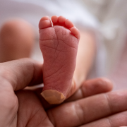
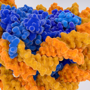
For decades, oncologists largely have turned to the same menu of treatments to fight cancer, including surgery, chemotherapy and radiation. Cell and gene therapies offer the promise of training the immune system to fight diseases with fewer side effects and potentially higher success rates. Early work has shown progress in liquid cancers, like leukemia, raising the possibility that the therapies could be used on solid tumors and other disorders, such as lupus and sickle cell disease. However, many disciplines must come together to yield discoveries.
“Nobel Prize-winning work doesn’t necessarily translate into available therapies for patients. It takes a whole community like this to make it happen,” said Cenk Sumen, chief scientific officer at MaxCyte Inc., an international cell engineering company based in Rockville, Md. “It has been exciting to see this diverse group of stakeholders come together, which is probably unmatched anywhere on the planet.”
Why we’re excited
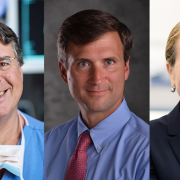
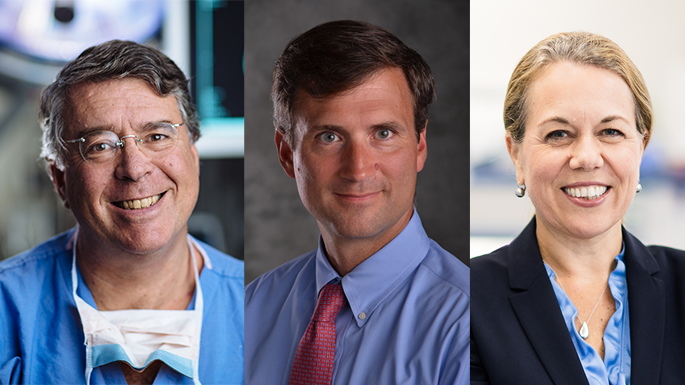
Symposium host Patrick Hanley, Ph.D., chief and director of the Cellular Therapy Program at Children’s National, said the goal was to cement the region as the No. 1 location for this highly technical research and development. He believes Children’s National can offer essential elements to this success, given its clinical and research expertise, workforce training opportunities and geographic proximity to the scientific leadership of the federal government. “What makes us unique is our proximity to all the players who can help create new treatment options for patients. We truly are the biomedical capital of the world,” he said.
Michael Friedlander, vice president for health sciences at Virginia Tech, notes that the earliest stages of invention will emanate from academic labs including those at Virginia Tech and Children’s National. “You have basic scientists who are doing fundamental research on properties and procedures that will lead to the new therapies of tomorrow,” he said. “We are putting in place the fundamental pieces to advance children’s health in all dimensions.”
What’s ahead
One challenge is developing a workforce to help prepare cell therapies for patients, following precise standards to ensure the therapy works as designed. Children’s National does this training, as do others in the region. Lori Kelman, Ph.D., M.B.A., biotechnology coordinator and professor at Montgomery College, said that the area is full of people who want to help people and who like science.
“The thing that people might not know is that you don’t need a Ph.D. to work in cell and gene therapy,” she said. “There are opportunities at all levels, including the entry level, which is where a great career often starts.”
 The hospital will advocate for the unique needs of children as part of nationwide network working to accelerate transformative health solutions.
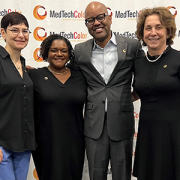
 Children’s National Hospital was awarded nearly $7.5 million in a five-year grant to continue its leadership of an FDA-funded pediatric device consortium. Building upon a decade of previous consortium leadership, the new consortium is Alliance for Pediatric Device Innovation (APDI) and features a new and expanded roster of partners that reflects its added focus on providing pediatric innovators with expert support on evidence generation, including the use of real-world evidence (RWE), for pediatric device development.


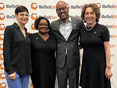
 Children’s National Hospital named
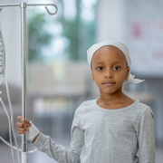
 Children’s National Hospital is joining a team of global health researchers to use large language models (LLMs) like ChatGPT to help Kenyan youth learn about their health and adopt lifestyles that may prevent cancer, diabetes and other non-communicable diseases.