Anna Penn

Protecting the fetal brain from harm

Anna Penn

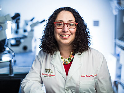
Ongoing placental dysfunction and allopregnanolone loss, not the increase that was expected due to stress, may alter cortical development in complicated pregnancies and put babies at risk, says Anna Penn, M.D., Ph.D.

Researchers long have known that allopregnanolone (ALLO), a derivative of the hormone progesterone, is produced in adults’ brains during times of acute stress and modulates how easily the brain’s neurons fire. ALLO also is produced in the placenta during fetal development, one of more than 200 different hormones that each uniquely contribute to fostering a smooth pregnancy and maintaining a fetus’ overall health. Although ALLO is thought to protect the developing brain in pregnancies complicated by conditions that might harm it, such as high blood pressure, how its levels evolve during pregnancy and in newborns shortly after birth has remained unknown.

Now, a new study presented during the Pediatric Academic Societies (PAS) 2018 annual meeting suggests that the placenta ramps up ALLO production over the second trimester, peaking just as fetuses approach full term.

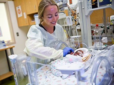
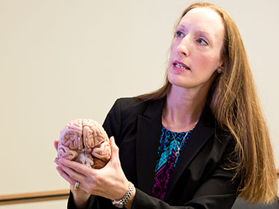
To investigate this phenomenon, Anna Penn, M.D., Ph.D., a neonatologist/neuroscientist at Children’s National Health System, and colleagues created a designer experimental model to study how premature loss of ALLO alters orderly brain development. Knowing more about the interplay between ALLO and normal development of the cortex, the outer layer of the cerebrum, is a first step that could lead to strategies to rescue this vital brain region.

“The cortex is basically the brain’s command-and-control center for higher functions. In our experimental model, it develops from the middle of gestation through to the end of gestation. If ALLO levels are disrupted just as these cells are being born, neurons migrating to the cortex are altered and the developing neural network is compromised,” says Dr. Penn, senior author of the research presented at PAS 2018. “We’re concerned this same phenomenon occurs in human infants whose preterm birth disrupts their supply of this essential hormone.”

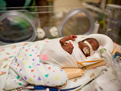
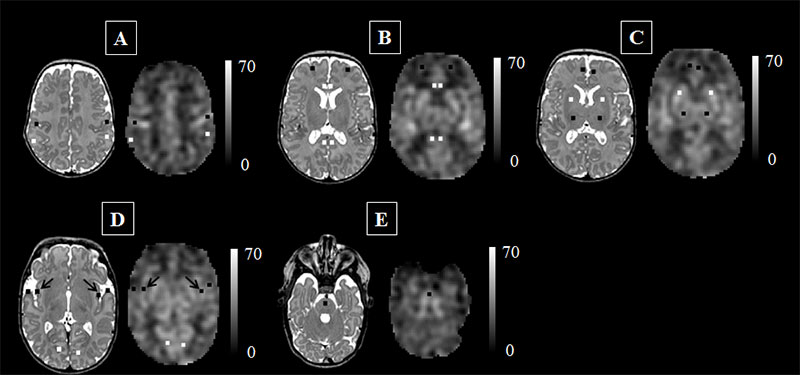
To better understand the human placental hormone pattern, the research team analyzed cord blood or serum samples collected within the first 36 hours of life for 61 preterm newborns born between 24 to 36 gestational weeks. They compared those preemie samples with samples drawn from 61 newborns carried to term who were matched by race, gender, size for gestational age, delivery method and maternal demographics.

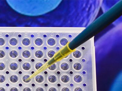
They used liquid-chromatography-tandem mass spectrometry, a technique that can precisely analyze trace levels of compounds, to compare levels of 27 different steroids, including ALLO and its precursors as well as better-known adrenal gland hormones, such as cortisol and 17-Hydroxyprogesterone.

“Pregnancies complicated by hypertension tended to correlate with lower ALLO levels, though this finding did not reach statistical significance. This suggests that ongoing placental dysfunction and ALLO loss, not the increase that we expected to be caused by stress, may alter cortical development in these pregnancies and put babies at risk,” Dr. Penn adds. “In addition, having the largest neonatal sample set to date in which multiple steroid hormones have been measured can provide insight into the shifting hormone patterns that occur around 36 weeks gestation, just prior to term. Hopefully, restoring the normal hormonal milieu for preemies or other at-risk newborns will improve neurological outcomes in the future.”

In addition to Dr. Penn, study co-authors include Caitlin Drumm, MedStar Georgetown University Hospital; Sameer Desale, MedStar Health Research Institute; and Kathi Huddleston, Benjamin Solomon and John Niederhuber, Inova Translational Medicine Institute.

Baby in the NICU

Getting to the heart of cardiac output

Baby in the NICU

To keep infants in the neonatal intensive care unit (NICU) as healthy as possible, it’s important to keep close tabs on their vital signs. During their NICU stay, most babies have continuous monitoring of their blood pressure, respiratory rate and blood oxygen saturation. And although continuous monitoring of heart rate is also typically standard, other information about heart function – such as cardiac output, a measure of how well the heart is pumping blood – remains a challenge to obtain in these vulnerable babies.

Clinical markers like blood pressure, heart rate and urine output are available, but they are indirect measures of cardiac output, how much blood the heart pumps per minute. Less invasive techniques, such as Doppler ultrasound, can be imprecise. Respiratory mass spectrometry or catheterization would provide more precision by directly calculating cardiac output but carry risks or are not feasible for neonates.

Clinicians at Children’s National Health System hypothesized that COstatus monitors could offer a way to directly measure cardiac output among neonates. The COstatus monitor – a minimally invasive way to measure hemodynamics – captures cardiac output, total end diastolic volume, active circulation volume and central blood volume.

The research team tested the approach by leveraging ultrasound dilution: Injecting saline, which has an ultrasound velocity of 1533m/second, slows the ultrasound velocity of blood from its normal rate of 1580m/second and produces a dilution curve.

“It is feasible to directly measure neonatal cardiac output by ultrasound dilution via the COstatus monitor in the first two weeks of life with no adverse events,” says Khodayar Rais-Bahrami, M.D., a Children’s neonatologist and senior author for the research presented during the Pediatric Academic Societies 2018 annual meeting. “When we took consecutive measurements, we saw very little variance in the parameters.”

The COstatus monitor uses an extracorporeal loop that is connected to arterial and venous catheters. The 12 neonates included in the study already had umbilical venous catheters as well as either a peripheral arterial line or umbilical arterial catheter. The infants ranged in weight from 0.72 to 3.74 kg and were born at 24 to 41.3 gestational weeks.

The infants’ cardiac output was measured 54 times from 1 to 13 days of life. Up to two measurement sessions occurred daily for a maximum of four days. The mean cardiac output was 0.43 L/minute with a mean cardiac index of 197mL/kg/minute.

Future research will describe normal cardiac output ranges for neonates as well as how these measurements evolve during the first week of life.

In addition to Dr. Rais-Bahrami, study co-authors include Simranjeet S. Sran, M.D., and Mariam Said, M.D., a Children’s neonatologist.

distressed woman holding baby

When depression lingers after the NICU

distressed woman holding baby

Roughly half a million babies end up in the neonatal intensive care unit (NICU) each year in the U.S., often sending their parents on a wild emotional rollercoaster. Like other new parents, many parents feel symptoms of depression when their child leaves the NICU. For the majority, these depressive symptoms lift over time. But for others, depression can persist, affecting their well-being and relationships, including those with their new babies.

Thus far, it’s been unclear which parents are at a higher risk for this lasting depression. However, a new study led by Children’s researchers and presented at the Pediatric Academic Societies 2018 annual meeting suggests that parents whose depression lingers six months after their child’s NICU discharge tend to share certain demographic characteristics: They’re younger, have less education and care for more than one child.

“Using a validated screening tool, we found that 40 percent of parents in our analyses were positive for depression at the time their newborn was discharged from the NICU,” says Karen Fratantoni, M.D., M.P.H., a Children’s pediatrician and the lead study author. “It’s reassuring that, for many parents, these depressive symptoms ease over time. However for a select group of parents, depression symptoms persisted six months after discharge. Our findings help to ensure that we target mental health screening and services to these more vulnerable parents,” Dr. Fratantoni adds.

The study is an offshoot from “Giving Parents Support (GPS) after NICU discharge,” a large, randomized clinical trial exploring whether providing peer-to-peer parental support after NICU discharge improves babies’ overall health as well as their parents’ mental health.

Mothers of preterm and full-term infants who are hospitalized in NICUs are at risk for peripartum mood disorders, including postpartum depression. The Children’s research team sought to determine how many parents of NICU graduates experience depression and which characteristics are shared by parents with elevated depression scores.

They included 125 parents who had enrolled in the GPS clinical trial in their exploratory analyses and assessed depressive symptoms using a 10-item, validated screening tool, the Center for Epidemiological Studies Depression Scale (CES-D). Eighty-four percent of the parents were women. Nearly 61 percent of their infants were male and were born at a median gestational age of 37.7 weeks and mean birth weight of 2,565 grams. The median length of time these newborns remained in the NICU was 18 days.

When the newborns were discharged, 50 parents (40 percent) had elevated CES-D scores. By six months after discharge, that number dropped to 17 parents (14 percent).Their mean age ranged from 26.5 to 30.6 years old.

“Parents of NICU graduates who are young, have less education and are caring for other children are at higher risk for persistent symptoms of depression,” says Dr. Fratantoni. “We know that peripartum mood disorders can persist for one year or more after childbirth so these findings will help us to better match mental health care services to parents who are most in need.”

An American College of Obstetricians and Gynecologists’ committee opinion issued May 2018 calls for all women to have contact with a maternal care provider within the first three weeks postpartum and to undergo a comprehensive postpartum visit no later than 12 weeks after birth that includes screening for postpartum depression and anxiety using a validated instrument.

Study co-authors include Lisa Tuchman, M.D., MPH, chief, Children’s Adolescent and Young Adult Medicine Division; Randi Streisand, Ph.D., Children’s interim chief of Psychology and Behavioral Health; Nicole S. Herrera; Katherine Kritikos and Lamia Soghier, M.D., Children’s neonatologist.

Preemie Baby

Brain food for preemies

Preemie Baby

Babies born prematurely – before 37 weeks of pregnancy – often have a lot of catching up to do. Not just in size. Preterm infants typically lag behind their term peers in a variety of areas as they grow up, including motor development, behavior and school performance.

New research suggests one way to combat this problem. The study, led by Children’s researchers and presented during the Pediatric Academic Societies 2018 annual meeting, suggests that the volume of carbohydrates, proteins, lipids and calories consumed by very vulnerable premature infants significantly contributes to increased brain volume and white matter development, even though additional research is needed to determine specific nutritional approaches that best support these infants’ developing brains.

During the final weeks of pregnancy, the fetal brain undergoes an unprecedented growth spurt, dramatically increasing in volume as well as structural complexity as the fetus approaches full term.

One in 10 infants born in the U.S. in 2016 was born before 37 weeks of gestation, according to the Centers for Disease Control and Prevention. Within this group, very low birthweight preemies are at significant risk for growth failure and neurocognitive impairment. Nutritional support in the neonatal intensive care unit (NICU) helps to encourage optimal brain development among preterm infants. However, their brain growth rates still lag behind those seen in full-term newborns.

“Few studies have investigated the impact of early macronutrient and caloric intake on microstructural brain development in vulnerable preterm infants,” says Katherine Ottolini, lead author of the Children’s-led study. “Advanced quantitative magnetic resonance imaging (MRI) techniques may help to fill that data gap in order to better direct targeted interventions to newborns who are most in need.”

The research team at Children’s National Health System enrolled 69 infants who were born younger than 32 gestational weeks and weighed less than 1,500 grams. The infants’ mean birth weight was 970 grams and their mean gestational age at birth was 27.6 weeks.

The newborns underwent MRI at their term-equivalent age, 40 weeks gestation. Parametric maps were generated for fractional anisotropy in regions of the cerebrum and cerebellum for diffusion tensor imaging analyses, which measures brain connectivity and white matter tract integrity. The research team also tracked nutritional data: Grams per kilogram of carbohydrates, proteins, lipids and overall caloric intake.

“We found a significantly negative association between fractional anisotropy and cumulative macronutrient/caloric intake,” says Catherine Limperopoulos, Ph.D., director of Children’s Developing Brain Research Laboratory and senior author of the research. “Curiously, we also find significantly negative association between macronutrient/caloric intake and regional brain volume in the cortical and deep gray matter, cerebellum and brainstem.”

Because the nutritional support does contribute to cerebral volumes and white matter microstructural development in very vulnerable newborns, Limperopoulos says the significant negative associations seen in this study may reflect the longer period of time these infants relied on nutritional support in the NICU.

In addition to Ottolini and Limperopoulos, study co-authors include Nickie Andescavage, M.D., Attending, Children’s Neonatal-Perinatal Medicine; and Kushal Kapse.

newborn in incubator

How EPO saves babies’ brains

newborn in incubator

Researchers have discovered that treating premature infants with erythropoietin can help protect and repair their vulnerable brains.

The drug erythropoietin (EPO) has a long history. First used more than three decades ago to treat anemia, it’s now a mainstay for treating several types of this blood-depleting disorder, including anemia caused by chronic kidney disease, myelodysplasia and cancer chemotherapy.

More recently, researchers discovered a new use for this old drug: Treating premature infants to protect and repair their vulnerable brains. However, how EPO accomplishes this feat has remained unknown. New genetic analyses presented at the Pediatric Academic Societies 2018 annual meeting that was conducted by a multi-institutional team that includes researchers from Children’s National show that this drug may work its neuroprotective magic by modifying genes essential for regulating growth and development of nervous tissue as well as genes that respond to inflammation and hypoxia.

“During the last trimester of pregnancy, the fetal brain undergoes tremendous growth. When infants are born weeks before their due dates, these newborns’ developing brains are vulnerable to many potential insults as they are supported in the neonatal intensive care unit during this critical time,” says An Massaro, M.D., an attending neonatologist at Children’s National Health System and lead author of the research. “EPO, a cytokine that protects and repairs neurons, is a very promising therapeutic approach to support the developing brains of extremely low gestational age neonates.”

The research team investigated whether micro-preemies treated with EPO had distinct DNA methylation profiles and related changes in expression of genes that regulate how the body responds to such environmental stressors as inflammation, hypoxia and oxidative stress.  They also investigated changes in genes involved in glial differentiation and myelination, production of an insulating layer essential for a properly functioning nervous system. The genetic analyses are an offshoot of a large, randomized clinical trial of EPO to treat preterm infants born between 24 and 27 gestational weeks.

The DNA of 18 newborns enrolled in the clinical trial was isolated from specimens drawn within 24 hours of birth and at day 14 of life. Eleven newborns were treated with EPO; a seven-infant control group received placebo.

DNA methylation and whole transcriptome analyses identified 240 candidate differentially methylated regions and more than 50 associated genes that were expressed differentially in infants treated with EPO compared with the control group. Gene ontology testing further narrowed the list to five candidate genes that are essential for normal neurodevelopment and for repairing brain injury:

“These findings suggest that EPO’s neuroprotective effect may be mediated by epigenetic regulation of genes involved in the development of the nervous system and that play pivotal roles in how the body responds to inflammation and hypoxia,” Dr. Massaro says.

In addition to Dr. Massaro, study co-authors include Theo K. Bammler, James W. MacDonald, biostatistician, Bryan Comstock, senior research scientist, and Sandra “Sunny” Juul, M.D., Ph.D., study principal investigator, all of University of Washington.

Research and Education Week awardees embody the diverse power of innovation

cnmc-research-education-week

“Diversity powers innovation” was brought to life at Children’s National April 16 to 20, 2018, during the eighth annual Research and Education Week. Children’s faculty were honored as President’s Award winners and for exhibiting outstanding mentorship, while more than 360 scientific poster presentations were displayed throughout the Main Atrium.

Two clinical researchers received Mentorship Awards for excellence in fostering the development of junior faculty. Lauren Kenworthy, Ph.D received the award for Translational Science and Murray M. Pollack, M.D., M.B.A., was recognized in the Clinical Science category as part of Children’s National Health System’s Research and Education Week 2018.

Dr. Kenworthy has devoted her career to improving the lives of people on the autism spectrum and was cited by former mentees as an inspirational and tireless counselor. Her mentorship led to promising new lines of research investigating methods for engaging culturally diverse families in autism studies, as well as the impact of dual language exposure on cognition in autism.

Meanwhile, Dr. Pollack was honored for his enduring focus on motivating early-career professionals to investigate outcomes in pediatric critical care, emergency medicine and neonatology. Dr. Pollack is one of the founders of the Collaborative Pediatric Critical Care Research Network. He developed PRISM 1 and 2, which has revolutionized pediatric intensive care by providing a methodology to predict mortality and outcome using standardly collected clinical data. Mentees credit Dr. Pollack with helping them develop critical thinking skills and encouraging them to address creativity and focus in their research agenda.

In addition to the Mentorship and President’s Awards, 34 other Children’s National faculty, residents, interns and research staff were among the winners of Poster Presentation awards. The event is a celebration of the commitment to improving pediatric health in the form of education, research, scholarship and innovation that occurs every day at Children’s National.

Children’s Research Institute (CRI) served as host for the week’s events to showcase the breadth of research and education programs occurring within the entire health system, along with the rich demographic and cultural origins of the teams that make up Children’s National. The lineup of events included scientific poster presentations, as well as a full slate of guest lectures, educational workshops and panel discussions.

“It’s critical that we provide pathways for young people of all backgrounds to pursue careers in science and medicine,” says Vittorio Gallo, Ph.D., Children’s chief research officer and CRI’s scientific director. “In an accelerated global research and health care environment, internationalization of innovation requires an understanding of cultural diversity and inclusion of different mindsets and broader spectrums of perspectives and expertise from a wide range of networks,” Gallo adds.

“Here at Children’s National we want our current and future clinician-researchers to reflect the patients we serve, which is why our emphasis this year was on harnessing diversity and inclusion as tools to power innovation,” says Mark L. Batshaw, M.D., physician-in-chief and chief academic officer of Children’s National.

“Research and Education Week 2018 presented a perfect opportunity to celebrate the work of our diverse research, education and care teams, who have come together to find innovative solutions by working with local, national and international partners. This event highlights the ingenuity and inspiration that our researchers contribute to our mission of healing children,” Dr. Batshaw concludes.

Awards for the best posters were distributed according to the following categories:

  • Basic and translational science
  • Quality and performance improvement
  • Clinical research
  • Community-based research and
  • Education, training and program development.

Each winner illustrated promising advances in the development of new therapies, diagnostics and medical devices.

Diversity powers innovation: Denice Cora-Bramble, M.D., MBA
Diversity powers innovation: Vittorio Gallo, Ph.D.
Diversity powers innovation: Mark L. Batshaw, M.D.

Robin Steinhorn

Children’s National senior vice president elected to American Pediatric Society leadership

Robin Steinhorn

Robin Steinhorn, M.D., Senior Vice President of Center for Hospital-Based Specialties at Children’s National Health System, was elected by her peers to become vice president and president-elect of the American Pediatric Society (APS) beginning May 2018 at the annual Pediatric Societies Meeting in Toronto, Canada. Dr. Steinhorn will serve in this role for one year and will then become the Society’s president in May 2019 for a one-year term.

Dr. Steinhorn is a globally recognized physician-leader, researcher and clinician in the fields of neonatal perinatal medicine and fetal pulmonary development. She was elected to the APS Council in 2015 and currently holds a seat on the American Board of Pediatrics’ Board of Directors.

“Dr. Steinhorn has devoted her professional career to advancing the field of pediatrics through exemplary leadership in related societies, as well as editorial oversight of cutting-edge research,” says David Wessel, M.D., executive vice president and chief medical officer of Hospital and Specialty Services at Children’s National. “This elevated role with the APS will enable her to further share her expertise to benefit children on a national and international level.”

Dr. Steinhorn serves as associate editor of the Journal of Pediatrics and is also a contributing editor for NEJM Journal Watch’s Pediatric and Adolescent Medicine.  Additionally, she sits on the editorial boards of Pediatric Critical Care Medicine and Pulmonary Circulation. Dr. Steinhorn is an elected fellow of the American Heart Association and a member of both the Perinatal Research Society and the American Thoracic Society.

Founded in 1888, the American Pediatric Society is the oldest and most prestigious academic pediatric organization in North America. Members are elected to APS based on their accomplishments as academic leaders in pediatrics and goal to shape the future of academic pediatrics. Mark L. Batshaw, M.D., physician-in-chief and chief academic officer of Children’s National preceded Dr. Steinhorn as APS President from 2016-2017.

“This is a tremendous honor, and it is a special privilege to follow Dr. Batshaw’s sound leadership. I look forward to leveraging the collective leadership and research accomplishments by our members to improve the health of infants and children throughout the U.S.,” said Dr. Steinhorn.

Dr. Steinhorn joined Children’s National in 2015 after a successful tenure as professor and chair of the department of pediatrics at the University of California, Davis (UCD) School of Medicine and as physician-in-chief, UCD Children’s Hospital. Previously, she was vice chair of the department of pediatrics and chief of the division of neonatology at Northwestern University and the Ann & Robert H. Lurie Children’s Hospital of Chicago.

Dr. Steinhorn’s clinical and academic interests have focused primarily on fetal and neonatal pulmonary vascular development. Her translational work has spanned from in vitro studies, to experimental models and clinical trials. In addition to her own translational research program, she has participated in numerous multicenter trials that have helped define the clinical treatment of pulmonary hypertension during the neonatal period. Her clinical research work also has addressed other topics, such as harmonization of electronic health records for clinical research and telemedicine support of neonatal care in small rural hospitals.

Additionally, Dr. Steinhorn is particularly passionate about mentoring faculty and supporting the growth and career development of young neonatologists and scientists, with several having developed their own research laboratories and assumed division leadership positions. She was selected as a “Top Doctor” by Northern Virginia  Magazine in 2018.

Sudeepta Basu

GABA concentration in pre-term brain increases with gestational age

Sudeepta Basu

“A more complete understanding of the diagnostic and prognostic importance of GABA and glutamate in the preterm brain will help us to direct treatment strategies for the most vulnerable preterm infants at risk of brain injury,” says Sudeepta K. Basu, M.D.

The major neurotransmitters gamma-aminobutyric acid (GABA) and glutamate are pivotal to fetal and newborn brain development and influence evolution of brain injury and repair following preterm birth. Magnetic resonance spectroscopy (MRS) enables in vivo measurement of brain metabolites. However, GABA and glutamate are found in the developing brain in low concentrations, and their weak signal can be swamped by the stronger signal of more dominant metabolites.

A Children’s research team reports findings from a pilot study utilizing an innovative technique of MRS to reliably measure in vivo GABA in the developing preterm brain. The groundbreaking research done by the team that includes Principal Investigator Sudeepta K. Basu, M.D., neonatology attending at Children’s National Health System, is very unique and original since there are no existing data of in vivo GABA concentrations in the developing cerebellum. Under the mentorship of Catherine Limperopoulos, Ph.D., director of Children’s Developing Brain Research Laboratory, the team of multi-disciplinary specialists is pursuing cutting-edge technologies in advanced MRI neuroimaging to explore brain development and injury in preterm infants.

The research, presented at the Eastern Society for Pediatric Research (ESPR) annual meeting by Dr. Basu, was honored with the “2018 Meritorious Poster Award.” The research titled “Distinct temporal trends of GABA and glutamate in the cerebellum and frontal cortex of preterm infants” reports, for the first time, positive temporal trends in the specific regions of the developing brain intricately involved in cognitive and motor functions. This work lays the foundation for developing novel ways to diagnose, monitor and investigative brain protective therapies for vulnerable prematurely born infants.

The Children’s team performed non-sedated MRS in 44 preterm infants whose mean gestational age at birth was 26.5 weeks, placing voxels at the middle of the cerebellum and the right frontal cortex. GABA and GIx (glutamate combined with glutamine) were positively correlated with post-menstrual age in the frontal cortex, but not the cerebellum.  At the ESPR meeting, the team also presented for the first time that caffeine, a neuroprotective agent in preemies, leads to increased in vivo GABA concentration in the developing frontal cortex.

“Open questions include whether these findings reflect varying paces of maturation and vulnerability to injury among specific regions of the brain. Also, the relationship between clinical factors and medication exposure and changes in the concentration of these neurotransmitters may guide brain protective therapies in future,” Dr. Basu says. “A more complete understanding of the diagnostic and prognostic importance of GABA and glutamate in the preterm brain will help us to direct treatment strategies for the most vulnerable preterm infants at risk of brain injury.”

Children’s senior fellows from Division of Neonatology made four platform presentations during the ESPR conference:

  • “Caffeine increases GABA/Cr ratio in frontal cortex of preterm infants on spectroscopy.” Aditi Gupta; Sudeepta K. Basu, M.D.; Mariam Said, M.D.; Subechhya Pradhan, Linda White; Kushal Kapse; Jonathan Murnick, M.D., Ph.D.; Taeun Chang, M.D.; and Catherine Limperopoulos, Ph.D.
  • “Impact of early nutrition on microstructural brain development in VLBW Infants.” Katherine M. Ottolini, Nickie Andescavage, M.D.; Kushal Kapse; and Catherine Limperopoulos, Ph.D.
  • “Direct measurement of neonatal cardiac output utilizing the CO status monitor.” Simranjeet S. Sran, Mariam Said, M.D.; and Khodayar Rais-Bahrami, M.D.
  • “Cerebro-cerebellar diaschisis in preterm infants following unilateral cerebral parenchymal injury.” Huma Mirza, Yao Wu, Kushal Kapse, Jonathan Murnick, M.D., Ph.D.; Taeun Chang, M.D.; and Catherine Limperopoulos, Ph.D.
Sean Donahue

Pediatric ophthalmology celebrates 75th anniversary in Washington, D.C.

Sean Donahue

Angeline M. Parks Visiting Professor Sean P. Donahue, M.D., Ph.D., (front left) enjoys a light moment during the celebration of the 75th anniversary while Anthony Sandler, M.D., Children’s surgeon in chief, senior vice president of the Joseph E. Robert Jr. Center for Surgical Care and director of the Sheikh Zayed Institute, speaks to the group.

After 75 years dedicated to the eyes of children, the world’s pediatric ophthalmologists gathered in Washington, D.C., the specialty’s birthplace, to share the latest research and innovation in the field. The group gathered for a joint meeting of the International Strabismological Association (ISA) and the American Association for Pediatric Ophthalmology and Strabismus (AAPOS), which was held March 18-22, 2018.

“This year marks the 75th anniversary of our specialty, which was founded right here, at Children’s National, in Washington, D.C., when Dr. Frank Costenbader restricted his practice exclusively to children and began to train residents in the nuance of treating children’s eyes,” says Mohamad S. Jaafar, M.D., chief of the Division of Ophthalmology at Children’s National Health Center. “It is a tremendous honor to welcome my colleagues back to the birthplace of pediatric ophthalmology on this grand occasion.”

In advance of the larger meeting, Children’s Division of Ophthalmology welcomed some of the international attendees to Children’s National for a special gathering on Saturday, March 17, 2018.

The event at Children’s featured a special lecture by this year’s Angeline M. Parks Visiting Professor, Sean P. Donahue, M.D., Ph.D. Dr. Donahue is the Sam and Darthea Coleman Chair in Pediatric Ophthalmology and Chief of Pediatric Ophthalmology at the Children’s Hospital at Vanderbilt. This Annual Visiting Professorship was established by the members of the Costenbader Society (The Children’s National Pediatric Ophthalmology Alumni Society) in memory of Angeline M. Parks, the wife of pediatric ophthalmologist Marshall M. Parks, M.D., to carry on her legacy of establishing a warm and supportive environment between physician and spouse, which benefits the physicians and their young patients.

Three former division chiefs of Ophthalmology at Children’s National, Drs. Costenbader, Parks and Friendly, have national lectureships established in their names to reflect their contributions to the field. Dr. Frank Costenbader, the society’s namesake, established the sub-specialty of pediatric ophthalmology. Dr. Parks founded the Children’s Eye Foundation and the AAPOS, and David S. Friendly, M.D., codified pediatric ophthalmology fellowship training across the United States.

Honor Awards for Children’s pediatric ophthalmologists at ISA-AAPOS

During the ISA-AAPOS meeting, two current Children’s National pediatric ophthalmologists were recognized with Honor Awards for their long-term dedication to pediatric ophthalmology, their patients, and their engagement in the AAPOS to advance the field.

William Madigan, M.D., vice chief of Ophthalmology at Children’s, a professor of surgery at the Uniformed Services University of the Health Sciences, and a clinical professor of Ophthalmology and Pediatrics at the George Washington University School of Medicine and Health Sciences. He was recognized by AAPOS for his long-time service, including:

  • Chair of the organization’s audit committee and the Costenbader Lecture selection committee.
  • Membership on the fellowship directors’ committee that developed nationwide requirements for pediatric ophthalmology fellowships and established the certification process to insure high quality and uniform education in the specialty.
  • Invited lectures in Shanghai, China; Geneva, Switzerland; and Sao Paolo, Brazil, among others.
  • Many posters and presentations about clinical and research topics of importance for members of the AAPOS and other distinguished professional societies.

Marijean Miller, M.D., director of Neonatal Ophthalmology, division research director at Children’s National and clinical professor of Ophthalmology and Pediatrics at the George Washington University School of Medicine and Health Sciences, was recognized by AAPOS for her cumulative contributions to the society, including:

  • Multiple memberships on vital committees, including AAPOS’s training and accreditation committee and audit committee.
  • Presentation of original research via posters and oral presentations on topics including best practices in neonatal clinical care, innovative tools and applications and advocacy for patients and their families.

“We are so grateful to have a team that continues the tradition of excellence in pediatric ophthalmology here at Children’s National,” Dr. Jaafar says. “Drs. Madigan and Miller exemplify the dedication of our division to caring for the children we serve, and to advancing our field. Congratulations to both!”

chromosome

X-linked genes help explain why boys of all ages face higher respiratory risk

chromosome

A multi-institution research team that includes Children’s National Health System attempted to characterize gender-based epigenomic signatures in the human airway early in children’s lives with a special attention to defining DNA methylation patterns of the X chromosome.

Human airways already demonstrate gender-based differences in DNA methylation signatures at birth, providing an early hint of which infants may be predisposed to develop respiratory disorders like asthma later in life, a research team reports in a paper published online April 3, 2018, in Scientific Reports.

It’s clear that boys and young men are more likely to develop neonatal respiratory distress syndrome, bronchopulmonary dysplasia, viral bronchiolitis, pneumonia, croup and childhood asthma. Unlike boys, girls have an additional copy of the X chromosome, which is enriched with immune-related genes, some of which play key roles in the development of respiratory conditions. Methylation prevents excessive gene activity in X-linked genes, however much remains unknown about how this process influences infants’ risk of developing airway diseases.

A multi-institution research team that includes Children’s National Health System attempted to characterize gender-based epigenomic signatures in the human airway early in children’s lives with a special attention to defining DNA methylation patterns of the X chromosome.

“It’s clear as we round in the neonatal intensive care unit that baby boys remain hospitalized longer than girls and that respiratory ailments are quite common. Our work provides new insights about gender differences in airway disease risk that are pre-determined by genetics,” says Gustavo Nino, M.D., a Children’s pulmonologist and the study’s senior author.

“Characterizing early airway methylation signatures holds the promise of clarifying the nature of gender-based disparities in respiratory disorders and could usher in more personalized diagnostic and therapeutic approaches.”

The research team enrolled 12 newborns and infants in the study and obtained their nasal wash samples. Six of the infants were born preterm, and six were born full term. The researchers developed a robust gender classification algorithm to generate DNA methylation signals. The machine learning algorithm identified X-linked genes with significant differences in methylation patterns in boys, compared with girls.

As a comparison group, they retrieved pediatric nasal airway epithelial cultures from a study that looked at genomic DNA methylation patterns and gene expression in 36 children with persistent atopic asthma compared with 36 heathy children.

The team went on to classify X-linked genes that had significant gender-based X methylation and those genes whose X methylation was variable.

“These results confirm that the X chromosome contains crucial information about gender-related genetic differences in different airway tissues,” Dr. Nino says. “More detailed knowledge of the genetic basis for gender differences in the respiratory system may help to predict, prevent and treat respiratory disorders that can affect patients over their entire lifetimes.”

In addition to Dr. Nino, study co-authors include Lead Author Cesar L. Nino, bioinformatics scientist, Pontificia Universidad Javeriana; Geovanny F. Perez, M.D., co-director of Children’s Severe Bronchopulmonary Dysplasia Program; Natalia Isaza Brando, M.D., Children’s neonatology attending; Maria J. Gutierrez, Johns Hopkins University School of Medicine; and Jose L. Gomez, Yale University School of Medicine.

Financial support for this research was provided by the National Institutes of Health under award numbers
AI130502-01A1, HL090020, HL125474-03, HD001399, UL1TR000075 and KL2TR000076.

Sarah Mulkey

MRI finds novel brain defects in Zika-exposed newborns

Sarah Mulkey

“Imaging is constantly helping us make new discoveries with this virus, and in these two cases we found things that had not been previously described,” says Sarah Mulkey, M.D., Ph.D.

Magnetic resonance imaging (MRI) has identified two brain abnormalities never before reported in newborns with prenatal exposure to the Zika virus. Children’s National Health System researchers reported these findings from a study of more than 70 fetuses or newborns with Zika exposure in utero. The study was published in the January 2018 edition of Pediatric Neurology.

The two novel defects – cranial nerve enhancement and cerebral infarction – may join the growing list of neurological findings associated with congenital Zika infection.

“Imaging is constantly helping us make new discoveries with this virus, and in these two cases we found things that had not been previously described,” says Sarah Mulkey, M.D., Ph.D., the study’s lead author and a fetal-neonatal neurologist at Children’s National. Dr. Mulkey works in Children’s Congenital Zika Virus Program, one of the nation’s first comprehensive, dedicated Zika programs.

The research team recommends that postnatal brain MRI be considered in addition to ultrasound for newborns exposed to Zika in utero. “Brain MRI can be performed in the newborn often without sedation and provides an opportunity to look for brain abnormalities we might not catch otherwise – or might not detect until much later,” says Dr. Mulkey.

Birth defects are seen in 6 to 11 percent of pregnancies affected by Zika, and some of the neurological complications in infants are not apparent until well after birth.

Of the two infants in which the new abnormalities were observed, both had normal head size at birth. Neither had smaller-than-normal head size (microcephaly), one of the more severe effects associated with congenital Zika syndrome.

One infant had a normal neurological evaluation at 2 days of age. However, a brain MRI conducted the following day, using gadolinium contrast due to concern of infection, showed enhancement of multiple cranial nerves. “Nerve root enhancement is very rare in a newborn and had not been described with Zika before,” Dr. Mulkey says. “Yet, there was no neurological deficit that we could identify by physical exam.”

The research team acknowledges that the clinical significance of this finding is not yet known.

In the second patient, brain MRI conducted without contrast at 16 days of age revealed a small area consistent with chronic infarction (ischemic stroke) that likely occurred during the third trimester.

“We followed the mother throughout her pregnancy, and both MRI and ultrasound imaging were normal at 28 weeks gestation,” Dr. Mulkey says. “A postnatal ultrasound was also normal, but the postnatal MRI showed a stroke that had occurred at least one month prior to the MRI and after the last fetal study.”

She adds: “This is the first published report of fetal stroke associated with Zika infection, and it may add to our knowledge of what can occur with congenital Zika infection.”

Unlike most congenital infections, Zika virus does not appear to cause viral-induced placental inflammation, which can lead to fetal stroke. So, the authors say they cannot be sure that congenital Zika contributed to the infarct in this case. However, they write, “Given the relatively low incidence of perinatal ischemic infarct and the lack of other maternal- or birth-related risk factors for this patient, Zika infection is considered a possible etiology.”

In both patients, neonatal brain MRI identified subclinical findings that had not previously been described as part of congenital Zika syndrome. As the body of evidence about the Zika virus has grown, the spectrum of associated brain abnormalities has expanded to include considerably more findings than isolated microcephaly.

Data gathered in 2017 from the Centers for Disease Control and Prevention’s Zika pregnancy and infant registry indicates that 25 percent of eligible U.S. infants receive recommended postnatal imaging. Dr. Mulkey said this represents many possible missed opportunities for earlier identification of brain abnormalities.

“Brain MRI should be considered in all newborns exposed to Zika virus in utero, even in the presence of normal birth head circumference, normal cranial ultrasound and normal fetal imaging,” she says. “In both of these patients, the changes we observed were not evident on cranial ultrasound or on fetal MRI and fetal ultrasound.”

In addition to Dr. Mulkey, Children’s co-authors include L. Gilbert Vezina, M.D., Neuroradiology Program director; Dorothy I. Bulas, M.D., chief of Diagnostic Imaging and Radiology; Zarir Khademian, M.D., radiologist; Anna Blask, M.D., radiologist; Youssef A. Kousa, M.S., D.O., Ph.D., child neurology fellow; Lindsay Pesacreta, FNP; Adré  J. du Plessis, M.B.Ch.B., M.P.H., Fetal Medicine Institute director; and Roberta L. DeBiasi, M.D., M.S., senior author and Pediatric Infectious Disease division chief; and Caitlin Cristante, B.S.

Financial support for this research was provided by the Thrasher Research Fund.

banner year

2017: A banner year for innovation at Children’s National

banner year

In 2017, clinicians and research faculty working at Children’s National Health System published more than 850 research articles about a wide array of topics. A multidisciplinary Children’s Research Institute review group selected the top 10 articles for the calendar year considering, among other factors, work published in high-impact academic journals.

“This year’s honorees showcase how our multidisciplinary institutes serve as vehicles to bring together Children’s specialists in cross-cutting research and clinical collaborations,” says Mark L. Batshaw, M.D., Physician-in-Chief and Chief Academic Officer at Children’s National. “We’re honored that the National Institutes of Health and other funders have provided millions in awards that help to ensure that these important research projects continue.”

The published papers explain research that includes using imaging to describe the topography of the developing brains of infants with congenital heart disease, how high levels of iron may contribute to neural tube defects and using an incisionless surgery method to successfully treat osteoid osteoma. The top 10 Children’s papers:

Read the complete list.

Dr. Batshaw’s announcement comes on the eve of Research and Education Week 2018 at Children’s National, a weeklong event that begins April 16, 2018. This year’s theme, “Diversity powers innovation,” underscores the cross-cutting nature of Children’s research that aims to transform pediatric care.

baby in arms

Breast-feeding, anesthesia and analgesics: What’s safe?

baby in arms

Breast-feeding is safe even just after moms have woken from anesthesia or while they take most pain medications, says Sarah Reece-Stremtan, M.D., lead author of an expanded protocol about the topic.

Moms can safely continue breast-feeding even just after waking from anesthesia and while taking most pain medications, according to a newly expanded clinical guidance, “Clinical Protocol No. 15: Analgesia and Anesthesia for the Breastfeeding Mother,” from the Academy of Breastfeeding Medicine (ABM).

In general, mothers who are beyond the postpartum stage do not need to avoid breast-feeding or to pump and discard breast milk while taking analgesics or receiving local or general anesthesia. The protocol was published in the journal Breastfeeding Medicine.

Sarah Reece-Stremtan, M.D., an anesthesiologist and acute pain medicine specialist at Children’s National Health System, co-chairs ABM’s protocol committee and is the lead author of the expanded protocol. A specialist in the intersection of anesthesia, pain medicine and breast-feeding medicine, Dr. Reece-Stremtan led the drafting of the recommendations.

“The key recommendation in this protocol is after waking up from anesthesia, most moms can breast-feed right away,” says Dr. Reece-Stremtan. “The standard thinking has been ‘pump and dump’ – discarding the breast milk for 24 hours after anesthesia. As an outdated practice, it is not evidence-based and is potentially harmful for babies. The evidence shows that this breast milk is safe.”

The authors’ main note of caution relates to opioids: “The most concerning class of medications used for anesthesia and analgesia in breast-feeding mothers is opioids, as these medications transfer into breast milk,” they write. “Judicious use of opioids for short periods is likely to be safe for most breast-feeding mothers and infants.”

The protocol recommendations cover pain medications, brief procedures, regional and general anesthesia and perioperative considerations. They provide more granular detail about specific anesthesia and analgesic agents.

For each recommendation, the protocol notes the strength or weakness of the evidence base. The authors note there is little rigorous information in the scientific literature about anesthesia or procedural sedation in breast-feeding mothers.

“For obvious reasons, it is unethical to conduct randomized, controlled clinical trials for this area, so we rely on expert opinion and on observational studies that do exist,” says Dr. Reece-Stremtan.

The protocol is intended to be relevant to a broad range of medical fields, from anesthesiology to general pediatrics, and to help any physician who may care for a new mother.

For instance, it includes a perioperative plan with suggestions that surgeons or physicians can share with their patients to make things easier for a breast-feeding mom who needs local or general anesthesia – and safer for their babies. “It’s important to acknowledge that medication isn’t the only or even the most important thing,” says Dr. Reece-Stremtan. Tips to aid breast-feeding can ease the minds of mothers and their physicians alike.

Dr. Reece-Stremtan has long been interested in breast-feeding and has seen a need for more education about where her areas of expertise, pediatric anesthesia and pain medicine, intersect. Few physicians specialize in this area, so she often gives talks to other clinicians on the topic.

“I know that most anesthesiologists do not encounter this scenario often, so many have questions about the impact of anesthesia agents on breast-feeding,” says Dr. Reece-Stremtan. “Likewise, general pediatricians, neonatal specialists and other health professionals who care for moms and newborns may have limited knowledge about the safety of pain medicine or anesthesia for breast-feeding infants.”

In developing this new set of recommendations, ABM’s protocol committee aimed to provide practical clinical guidance for two scenarios: Postpartum, and moms and babies who are past that stage. The committee divided a previous ABM protocol into these two areas and expanded them to offer clinicians more complete guidance that is clinically relevant yet concise. Dr. Reece-Stremtan attributes this expansion to a growing appreciation of the importance of breast-feeding to both individual and public health. She is helping to finalize ABM’s new birth-postpartum protocol on anesthesia and analgesics, which will be published in early 2018.

To build on these protocols, Dr. Reece-Stremtan is helping the Academy develop a set of free patient education materials that will inform mothers about the use of pain medications or the need for anesthesia while breast-feeding, so they can feel at ease that they are doing the best thing for their baby’s health.

newborn in incubator

Tracking oxygen saturation with vital signs to identify vulnerable preemies


Khodayar-Rais-Bahrami

PDF Version

What’s known

Critically ill infants in neonatal intensive care units (NICU) require constant monitoring of their vital signs. Invasive methods, such as using umbilical arterial catheters to check blood pressure, are the gold standard but pose significant health risks. Low-risk noninvasive monitoring, such as continuous cardiorespiratory monitors, can measure heart rate, respiratory rate and blood oxygenation. A noninvasive technique called near-infrared spectroscopy (NIRS) can gauge how well tissues, including the brain, are oxygenated. While NIRS long has been used to monitor oxygenation in conditions in which blood flow is altered, such as bleeding in the brain, how NIRS values relate to other vital sign measures in NICU babies was unknown.

What’s new

A research team led by Khodayar Rais-Bahrami, M.D., a neonatologist at Children’s National Health System, investigated this question in 27 babies admitted to Children’s NICU. The researchers separated these subjects into two groups: Low birth weight (LBW, less than 1.5 kg or 3.3 pounds) and moderate birth weight (MBW, more than 1.5 kg). Then, they looked for correlations between information extracted from NIRS, such as tissue oxygenation (specific tissue oxygen saturation, StO2) and the balance between oxygen supply and consumption (fractional tissue oxygen extraction, FTOE), and various vital signs. They found that StO2 increased with blood pressure for LBW babies but decreased with blood pressure for MBW babies. Brain and body FTOE in LBW babies decreased with blood pressure. In babies with abnormal brain scans, brain StO2 increased with blood pressure and brain FTOE decreased with blood pressure. Together, the researchers suggest, these measures could give a more complete picture of critically ill babies’ health.

Questions for future research

Q: Can NIRS data be used as a surrogate for other forms of monitoring?

Q: How could NIRS data help health care professionals intervene to improve the health of critically ill infants in the NICU?

Source: Significant correlation between regional tissue oxygen saturation and vital signs of critically ill infants.” B. Massa-Buck, V. Amendola, R. McCloskey and K. Rais-Bahrami. Published by Frontiers in Pediatrics Dec. 21, 2017.

Pregnant-Mom

MRI opens new understanding of fetal growth restriction

Pregnant-Mom

Quantitative MRI can identify placental dysfunction complicated by fetal growth restriction earlier, creating the possibility for earlier intervention to minimize harm to the developing fetus.

A team of researchers has found that quantitative magnetic resonance imaging (MRI) can identify pregnancies where placental dysfunction results in fetal growth restriction (FGR), creating the possibility for earlier FGR detection and intervention to augment placental function and thus minimize harm to the fetal brain.

The study, published online in the Journal of Perinatology, reports for the first time that in vivo placental volume is tied to global and regional fetal brain volumes.

Placental insufficiency is a known risk factor for impaired fetal growth and neurodevelopment. It may cause the fetus to receive inadequate oxygen and nutrients, making it difficult to grow and thrive. The earlier placental insufficiency occurs in a pregnancy, the more serious it can be. But detecting a failing placenta before the fetus is harmed has been difficult.

One additional challenge is that a fetus may be small because the placenta is not providing adequate nourishment. Or the fetus simply may be genetically predisposed to be smaller. Being able to tell the difference early can have a lifelong impact on a baby. Infants affected by FGR can experience behavioral problems, learning difficulties, memory and attention deficits, and psychiatric issues as the child grows into adolescence and adulthood.

“Our study proved that MRI can more accurately determine which pregnancies are at greater risk for impaired fetal health or compromised placenta function,” says Nickie Andescavage, M.D., the study’s lead author and a specialist in neonatology and neonatal neurology and neonatal critical care at Children’s National Health System. “The earlier we can identify these pregnancies, the more thoughtful we can be in managing care.”

Dr. Andescavage’s research focus has been how fetal growth affects labor, delivery and postnatal complications.

Nickie-Andescavage-Niforatos

“Our study proved that MRI can more accurately determine which pregnancies are at greater risk for impaired fetal health or compromised placenta function,” says Nickie Andescavage, M.D., the study’s lead author.

“We don’t have a good understanding of why FGR happens, but we do know it’s hard to identify during pregnancy because often there are no signs,” says Dr. Andescavage. “Even when detected, it’s hard to follow. But if we’re aware of it, we can better address important questions, like when to deliver an at-risk fetus.”

In the study, the team measured placental and fetal brain growth in healthy, uncomplicated pregnancies and in pregnancies complicated by FGR. A total of 114 women participated, undergoing ultrasound, Doppler ultrasound and MRI imaging to measure placental volume and fetal brain volume.

An ultrasound test is often what detects FGR, but the measurements generated by ultrasound can be non-specific. In addition, reproducibility issues with 3D sonography limit its use as a standalone tool for placental assessment. Once FGR is detected via ultrasound, this study showed that complementary MRI provides more accurate structural measures of the fetal brain, as well as more detail and insight into placental growth and function.

“Our team has studied FGR for a few years, using imaging to see that’s happening with the fetus in real time,” says Dr. Andescavage. “The relationship of placental volume and fetal brain development had not been previously studied in utero.”

In pregnancies complicated by FGR, MRI showed markedly decreased placental and brain volumes. The team observed significantly smaller placental, total brain, cerebral and cerebellar volumes in these cases than in the healthy controls. The relationship between increasing placental volume and increasing total brain volume was similar in FGR and in normal pregnancies. However, the study authors write “the overall volumes were smaller and thus shifted downward in pregnancies with FGR.”

In addition, FGR-complicated pregnancies that also showed abnormalities in Doppler ultrasound imaging had even smaller placental, cerebral and cerebellar volumes than pregnancies complicated by FGR that did not have aberrations in Doppler imaging.

Since this study showed that quantitative fetal MRI can accurately detect decreased placental and brain volumes when FGR is present, Dr. Andescavage believes this imaging technique may give doctors important new insights into the timing and possibly the mechanisms of brain injury in FGR.  “Different pathways can lead to FGR. With this assessment strategy, we could potentially elucidate those,” she adds.

Using quantitative MRI to identify early deviations from normal growth may create opportunities for future interventions to protect the developing fetal brain. New treatments on the horizon promise to address placental health. MRI could be used to investigate these potential therapies in utero. When those therapies become available, it could allow doctors to monitor treatment effects in utero.

Study co-authors include Adré J. du Plessis, M.B.Ch.B., M.P.H., Director of Children’s Fetal Medicine Institute; Marina Metzler; Dorothy Bulas, M.D., FACR, FAIUM, FSRU, Chief of Children’s Division of Diagnostic Imaging and Radiology; L. Gilbert Vezina, M.D., Director of Children’s Neuroradiology Program; Marni Jacobs; Catherine Limperopoulos, Ph.D., Director of Children’s Developing Brain Research Laboratory and study senior author; Sabah N. Iqbal, MedStar Washington Hospital Center; and Ahmet Alexander Baschat, Johns Hopkins Center for Fetal Therapy.

Research reported in this post was supported by the Canadian Institutes of Health Research, MOP-81116; the National Institutes of Health under award numbers UL1TR000075 and KL2TR000076; and the Clinical and Translational Science Institute at Children’s National.

Neonatal baby

Multidisciplinary experts help CDC’s Zika research

“We are very excited about this next phase in our Zika research,” says Roberta L. DeBiasi, M.D., M.S. “It is a natural extension of our earlier participation as subject matter experts assisting as the CDC developed and published guidelines to inform the care of Zika-exposed and Zika-infected infants across the nation and U.S. territories.”

The Centers for Disease Control and Prevention (CDC) is funding three multidisciplinary experts from the Congenital Zika Virus Program at Children’s National Health System to collaborate on two of the CDC’s longitudinal Zika research projects in Colombia, South America.

“Zika en embarazadas y niños en Colombia” (ZEN) is a research study jointly designed by Colombia’s Instituto Nacional de Salud (INS) and the CDC to evaluate the association between Zika virus infection and adverse maternal, fetal and infant health outcomes. The study is following a large cohort of Colombian women from the first trimester of pregnancy, their male partners and their infants.

Under the six-month contract, Roberta L. DeBiasi, M.D., M.S., Sarah B. Mulkey, M.D., Ph.D., and Cara Biddle, M.D., M.P.H., will serve as consultants for the ZEN study providing expertise in pediatric infectious diseases, neurology, neurodevelopment and coordination of the complex care needs of Zika-affected infants.

The federal funding will underwrite the consultants’ work effort, as well as travel to the CDC’s headquarters in Atlanta and to research sites in Colombia. To that end, Drs. DeBiasi, Mulkey and Biddle participated in a December 2017 kickoff meeting, joining ZEN team leaders based in the U.S. at the CDC, as well as the INS in Colombia, with whom they will conduct research and collaborate academically.

Cara-Biddle-and-Sarah-Mulkey

Cara Biddle, M.D., M.P.H., and Sarah B. Mulkey, M.D., Ph.D., also will serve as consultants for the ZEN study.

“We are very excited about this next phase in our Zika research,” says Dr. DeBiasi, chief of the Division of Pediatric Infectious Diseases and co-director of the Children’s Zika program. “It is a natural extension of our earlier participation as subject matter experts assisting as the CDC developed and published guidelines to inform the care of Zika-exposed and Zika-infected infants across the nation and U.S. territories.”

Children’s National is leading its own longitudinal studies in Colombia that explore such questions as whether Zika-exposed infants whose neuroimaging appears normal when they are born experience any longer-term neurological issues and the role of genetics in neurologic injury following congenital Zika virus exposure and infection.

Carlos Ferreira Lopez

Researchers discover new gene variant for inherited amino acid-elevating disease

Carlos Ferreira Lopez

PDF Version

What’s known

Hypermethioninemia is a rare condition that causes elevated levels of methionine, an essential amino acid in humans. This condition stems from genetic variations inherited from one or both parents. Some forms of hypermethioninemia are recessive, meaning that two copies of defective genes are necessary to cause this disease. Other forms are dominant, meaning that only one copy can cause hypermethioninemia. Recessive forms of the disease tend to have more serious consequences, causing elevated methionine levels throughout life and leading to changes in the brain’s white matter visible on magnetic resonance imaging that can cause neurological problems. The dominant forms are generally thought to be largely benign and require minimal follow-up.

What’s new

A research team led by Carlos Ferreira Lopez, M.D., a medical geneticist at Children’s National Health System, discovered a new gene variant that had not been associated with hypermethioinemia previously when an infant who had tested positive for elevated methionine on newborn blood-spot screening came in for a follow-up evaluation. While the majority of dominant hypermethioninemia are caused by a genetic mutation known as MAT1A p.Arg264His, the child didn’t have this or any of the common recessive hypermethioninemia mutations. Genetic testing showed that she carried a different mutation to the MAT1A gene known as p.Ala259Val, of which she carried only a single copy. The child fit the typical profile of having the dominant form of the disease, with methionine levels gradually declining over time. Testing of her mother showed that she carried the same gene variant, with few consequences other than a hepatitis-like illness as a child. Because liver disease can accompany dominant hypermethioninemia, the infant’s doctors will continue periodic follow-up to ensure she remains healthy.

Questions for future research

Q: Besides the potential for harmful liver effects, does dominant hypermethioninemia have other negative consequences?

Q: How common is this gene variant, and are certain people at more risk for carrying it?

Source: Confirmation that MAT1A p.Ala259Val mutation causes autosomal dominant hypermethioninemia. Muriello, M.J., S. Viall, T. Bottiglieri, K. Cusmano-Ozog and C. R. Ferreira. Published by Molecular Genetics and Metabolism Reports December 2017.

Volumetric imaging of upper airways

Preemies’ narrowed upper airways may explain higher OSA risk

Volumetric imaging of upper airways

The airway structures of interest to the Children’s National research team included the nasopharynx (labeled red), oropharynx (labeled purple), hypopharynx (labeled green), adenoids (labeled yellow) and tonsils (labeled blue). The team displayed the volumetric imaging in three perpendicular planes and a three-dimensional model.
Credit: A. Smitthimedhin, et al, Clinical Imaging.

Infants born preterm have significantly lower nasopharyngeal and oropharyngeal volumes, compared with newborn peers carried to full term, and those lower airway volumes are independent of the infants’ gender, ethnicity or weight, according to a study published online Dec. 16, 2017 in Clinical Imaging.

According to the Centers for Disease Control and Prevention, 1 in 10 babies born in the United States is preterm, or born prior to the 37th gestational week. Premature birth leaves these children more susceptible to disordered breathing while sleeping, including obstructive sleep apnea (OSA), an ailment characterized by increased upper-airway resistance that narrows airways.

“In addition to finding some airway volumes were smaller in preterm infants, our results indicated both sets of newborns had similar hypopharyngeal volumes. This suggests that risk factors that lead to OSA are confined to the uppermost airway and do not appear to be explained by enlarged adenoids and tonsils,” says Anilawan Smitthimedhin, a Children’s National Health System radiology research fellow at the time the study was performed and lead author of the paper.

In order to diagnose OSA, clinicians now use bronchoscopy, but the method has limitations, including the need to insert a lighted instrument into the airway, which can affect pressure and resistance within the airway.

The Children’s National research team theorized that magnetic resonance imaging (MRI) could offer a non-invasive way to evaluate the upper airway, determine its anatomy and dynamic function, while shielding infants from radiation exposure that can accompany other imaging techniques.

They enrolled 96 infants who had undergone brain MRIs as part of an unrelated study about neonatal brain development. The newborns had a range of medical conditions, including suspected hypoxic ischemic encephalopathy, cardiac disease and seizures/movement disorders.

Forty-nine of the infants were born preterm; at the time of the MRI, their corrected mean gestational age was 38.4 weeks. Forty-seven of the newborns were born full term; they received MRIs at 1.7 weeks of age. The airway structures of interest included the nasopharynx (the upper part of the pharynx), oropharynx (located at the back of the mouth behind the oral cavity), hypopharynx (the entrance into the esophagus), adenoids and tonsils. The team displayed the volumetric imaging in three perpendicular planes and a three-dimensional model.

“Nasopharyngeal volume of full-term infants was 495.6 mm, compared with 221.1 mm in preterm infants. Oropharyngeal volume of full-term infants was 313.6 mm, compared with 179.3 mm in preterm infants,” Smitthimedhin says.

Aided by volumetric 3D data that more accurately measures airway and lymphoid tissue, the team proposes to study a larger group of infants to determine whether narrowing of the uppermost airways predisposes very young children to experiencing OSA later in life.

“Ultimately, our goal is to incorporate dedicated, dynamic MR imaging of the airway while children sleep, which would provide real-time, detailed information about the changes associated with sleep. This innovation holds the promise of leading to more accurate, non-invasive diagnosis of OSA in infants,” says Dorothy Bulas, M.D., chief of Diagnostic Imaging and Radiology at Children’s National.

Children’s National study co-authors include Radiologist Matthew Whitehead, M.D.; University of Maryland student Mahya Bigdeli; Pulmonologist Gustavo Nino Barrera, M.D.; Pulmonologist Geovanny Perez, M.D,; and Hansel Otero, who was at Children’s National when the research work was performed but now works at Children’s Hospital of Philadelphia.

the cerebral blood flow (CBF) maps, corresponding anatomical image aligned to the CBF map, and the regions of interest examined

Tracking preemies’ blood flow to monitor brain maturation

Blood is the conduit through which our cells receive much of what they need to grow and thrive. The nutrients and oxygen that cells require are transported by this liquid messenger. Getting adequate blood flow is especially important during the rapid growth of gestation and early childhood – particularly for the brain, the weight of which roughly triples during the last 13 weeks of a typical pregnancy. Any disruption to blood flow during this time could dramatically affect the development of this critical organ.

Now, a new study by Children’s National Health System researchers finds that blood flow to key regions of very premature infants’ brains is altered, providing an early warning sign of disturbed brain maturation well before such injury is visible on conventional imaging. The prospective, observational study was published online Dec. 4, 2017 by The Journal of Pediatrics.

“During the third trimester of pregnancy, the fetal brain undergoes an unprecedented growth spurt. To power that growth, cerebral blood flow increases and delivers the extra oxygen and nutrients needed to nurture normal brain development,” says Catherine Limperopoulos, Ph.D., director of the Developing Brain Research Laboratory at Children’s National and senior author of the study. “In full-term pregnancies, these critical brain structures mature inside the protective womb where the fetus can hear the mother and her heartbeat, which stimulates additional brain maturation. For infants born preterm, however, this essential maturation process happens in settings often stripped of such stimuli.”

The challenge: How to capture what goes right or wrong in the developing brains of these very fragile newborns? The researchers relied on arterial spin labeling (ASL) magnetic resonance (MR) imaging, a noninvasive technique that labels the water portion of blood to map how blood flows through infants’ brains in order to describe which regions do or do not receive adequate blood supply. The imaging work can be done without a contrast agent since water from arterial blood itself illuminates the path traveled by cerebral blood.

“In our study, very preterm infants had greater absolute cortical cerebral blood flow compared with full-term infants. Within regions, however, the insula (a region critical to experiencing emotion), anterior cingulate cortex (a region involved in cognitive processes) and auditory cortex (a region involved in processing sound) for preterm infants received a significantly decreased volume of blood, compared with full-term infants. For preterm infants, parenchymal brain injury and the need for cardiac vasopressor support both were correlated with decreased regional CBF,” Limperopoulos adds.

The team studied 98 preterm infants who were born June 2012 to December 2015, were younger than 32 gestational weeks at birth and who weighed less than 1,500 grams. They matched those preemies by gestational age with 104 infants who had been carried to term. The brain MRIs were performed as the infants slept.

Blood flows where it is needed most with areas of the brain that are used more heavily commandeering more oxygen and nutrients. Thus, during brain development, CBF is a good indicator of functional brain maturation since brain areas that are the most metabolically active need more blood.

the cerebral blood flow (CBF) maps, corresponding anatomical image aligned to the CBF map, and the regions of interest examined

This figure represents the cerebral blood flow (CBF) maps, corresponding anatomical image aligned to the CBF map, and the regions of interest examined. The scale indicates the quantitative value of the CBF map and is expressed in mL/100g/min. The data are from a preterm infant scanned at term age without evidence of brain injury. The insula (see black arrows in panel ‘D’) may be particularly vulnerable to the added stresses of the preterm infant’s life outside the womb.
Credit: M. Bouyssi-Kobar, et al., The Journal of Pediatrics.

“The ongoing maturation of the newborn’s brain can be seen in the distribution pattern of cerebral blood flow, with the greatest volume of blood traveling to the brainstem and deep grey matter,” says Marine Bouyssi-Kobar, M.S., the study’s lead author. “Because of the sharp resolution provided by ASL-MR images, our study finds that in addition to the brainstem and deep grey matter, the insula and the areas of the brain responsible for sensory and motor functions are also among the most oxygenated regions. This underscores the critical importance of these brain regions in early brain development. In preterm infants, the insula may be particularly vulnerable to the added stresses of life outside the womb.”

Of note, compromised regional brain structures in adults are implicated in multiple neurodevelopmental disorders. “Altered development of the insula and anterior cingulate cortex in newborns may represent early warning signs of preterm infants at greater risk for long-term neurodevelopmental impairments,” Limperopoulos says.

Research reported in this post was supported by the Canadian Institutes of Health Research, MOP-81116; the SickKids Foundation, XG 06-069; and the National Institutes of Health under award number R01 HL116585-01.

Human Rhinovirus

When a common cold may trigger early supportive care

Human Rhinovirus

A new study led by Children’s National Health System shows that in infants who were born severely premature, human rhinovirus infections appear to trigger airway hyper-reactivity, which leads to wheezing, hyperinflation and more severe respiratory disease.

Human rhinovirus (HRV), the culprit behind most colds, is the leading cause of hospitalization for premature babies. However, in very preterm children, exactly how HRV causes severe respiratory disease – and which patients may need more intensive observation and treatment – is less well understood.

A new study led by Children’s National Health System research-clinicians showed in children who were born severely premature, HRV infections seem to trigger an airway hyper-reactivity (AHR) type of disease, which leads to wheezing and air-trapping (hyperinflation) and more severe respiratory disease. This, in turn, increases the risk for hospitalization.

The study, published online Oct. 21, 2017 in Pediatrics and Neonatology, found that other signs of respiratory distress, such as low arterial blood oxygen or rapid shallow breathing, were no more common in severely premature children (less than 32 weeks of gestational age) than in kids born preterm or full-term. The findings have implications for administering supportive care sooner or more intensively for severely premature children than for other infants.

“When it comes to how they respond to such infections, severely premature children are quite different,” says Geovanny Perez, M.D., a specialist in pulmonary medicine at Children’s National and lead study author. “We’ve known they are more susceptible to human rhinovirus infection and have more severe disease. However, our study findings suggest that severely premature kids have an ‘asthma’ type of clinical picture and perhaps should be treated differently.”

The study team sought to identify clinical phenotypes of HRV infections in young children hospitalized for such infections. The team theorized that severely premature babies would respond differently to these infections and that their response might resemble symptoms experienced by patients with asthma.

“For a number of years, our team has studied responses to viruses and prematurity, especially HRV and asthma,” Dr. Perez says. “We know that premature babies have an immune response to HRV from the epithelial cells, similar to that seen in older patients with asthma. But we wanted to address a gap in the research to better understand which children may need closer monitoring and more supportive care during their first HRV infection.”

Geovanny Perez

“When it comes to how they respond to such infections, severely premature children are quite different,” says Geovanny Perez, M.D. “We’ve known they are more susceptible to human rhinovirus infection and have more severe disease. However, our study findings suggest that severely premature kids have an ‘asthma’ type of clinical picture and perhaps should be treated differently.”

In a retrospective cross-sectional analysis, the study looked at 205 children aged 3 years or younger who were hospitalized at Children’s National in 2014 with confirmed HRV infections. Of these, 71 percent were born full-term (more than 37 gestational weeks), 10 percent were preterm (32 to 37 gestational weeks) and 19 percent were severely premature (less than 32 gestational weeks).

Dr. Perez and his team developed a special respiratory distress scoring system based on physical findings in the children’s electronic medical records to assess the degree of lower-airway obstruction or AHR (as occurs in asthma) and of parenchymal lung disease. The physical findings included:

  • Wheezing;
  • Subcostal retraction (a sign of air-trapping/hyperinflation of the lungs), as can occur in pneumonia;
  • Reduced oxygen levels (hypoxemia); and
  • Increased respiratory rate (tachypnea).

The research team assigned each case an overall score. The severely premature children had worse overall scores – and significantly worse scores for AHR and hyperinflated lungs relative to children born late preterm or full-term.

“What surprised us, though, in this study was that the phenotypical characterization using individual parameters for parenchymal lung disease, such as hypoxemia or tachypnea, were not different in severe preterm children and preterm or full term,” says Dr. Perez. “On the other hand, our study found that severely preterm children had a lower airway obstruction phenotype associated with retractions and wheezing. Moreover there was a ‘dose effect’ of prematurity: Children who were born more premature had a higher risk of wheezing and retractions.”

Among the implications of this study, Dr. Perez sees the potential to use phenotypical (clinical markers, such as retractions and wheezing) and biological biomarkers to better personalize patients’ treatments. Dr. Perez and his team have identified biological biomarkers in nasal secretions of children with rhinovirus infection that they plan to combine with clinical biomarkers to identify which patients with viral infections will benefit from early supportive care, chronic treatments or long-term monitoring.

Dr. Perez says further research in this area should pursue a number of paths, including:

  • A longitudinal study to elucidate which children will benefit from asthma-like treatment, such as bronchodilators or corticosteroids;
  • A study of biomarkers, including microRNAs and other inflammatory molecules; or
  • Alternatively, a longitudinal study exploring the mechanism by which wheezing develops, perhaps looking at first and subsequent rhinovirus infections in babies born at different gestational ages.