Tag Archive for: Sheikh Zayed Institute

doctors doing heart surgery

Novel dye may improve outcomes for liver surgery

Researchers at Children’s National Hospital and the National Cancer Institute (NCI) have developed a novel, near‐infrared dye that can help surgeons identify structures and detect leakage during liver surgery, offering a promising tool that may someday improve outcomes for patients undergoing gastroenterology procedures.

The problem has vexed the medical community for some time: Despite advances in bile leak detection, only a third of bile duct injuries are found at the time of surgery, extending hospital stays and increasing the risk of liver failure, sepsis and even death.

Why we’re excited

The new dye – known as Bile Label Dye 760 (BL-760) – provided several promising advantages over existing surgical tools during non-clinical testing. When administered into the liver, BL‐760 was excreted and visible in bile ducts within minutes, without significant or prolonged impact on organ tissue. Its fluorescence against the surgical field also provided a superior view of leaks, offering an opportunity to treat the patient while still in the operating room. Details were published recently in Lasers in Surgery and Medicine.

“BL-760 is a promising option for monitoring the health of the liver during surgery, and we are excited to continue our testing and hopefully see first-in-human trials in the future,” said Richard Cha, Ph.D., principal investigator at the Sheikh Zayed Institute of Pediatric Surgical Innovation, part of the NIH-funded team that developed the dye.

doctors doing heart surgery

The new dye – known as Bile Label Dye 760 (BL-760) – provided several promising advantages over existing surgical tools during non-clinical testing.

The big picture

The dye could significantly advance hepatobiliary and pancreatic (HPB) procedures in years to come. More than 40,000 new cases of liver cancer are diagnosed each year, causing more than 30,000 deaths in the U.S. alone. Gallbladder disease is also one of the most common conditions in the U.S., with more than 20 million people affected annually. In pediatrics, gall bladder removal, or cholecystectomy, is on the rise.

Procedures to treat these diseases have many challenges. During minimally invasive surgery, including laparoscopic cholecystectomy or robot-assisted hepatectomy, surgeons can struggle to precisely identify the bile ducts because of a narrow field of view or because they are embedded in fat or other tissues. Existing FDA-approved contrast agents that can enhance the biliary anatomy such as indocyanine green (ICG) aren’t well tailored for HPB surgeries because of the timing of their administration and their inferior ability to highlight biliary structures. In addition, while pre-operative imaging has improved outcomes, it cannot be used to predict leaks from the surgery itself.

What’s ahead

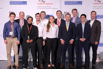
BL-760 was created at Children’s National and NCI by a team of experts in surgery and engineering, led by Anthony Sandler, M.D., senior vice president and surgeon-in-chief. They hope to continue their testing on the dye in the months ahead. The team was encouraged when Michele Saruwatari, M.D., a Joseph E. Robert Fellow in the Sheik Zayed Institute, recently won first place in the resident and fellow abstract presentation competition at the annual meeting of the Society of American Gastrointestinal and Endoscopic Surgeons.

“Having this tool in the operating room will change outcomes for our pediatric patients,” Sandler said.  “This dye has the potential to become an essential step in liver cancer surgery, cholecystectomy and treating other pediatric diseases like biliary atresia. I look forward to the day when we can get it in the hands of surgical teams.”

NCC-PDI Finalists

Pediatric medical device competition names finalists

Five finalists have been named in the prestigious annual “Make Your Medical Device Pitch for Kids!” competition presented by the National Capital Consortium for Pediatric Device Innovation (NCC-PDI). Representing innovations in pediatric technologies that aim to address unmet medical needs for children, these five finalists now have access to a pediatric accelerator program led by MedTech Innovator and will compete for a share of $150,000 in grant funding from the U.S. Food and Drug Administration (FDA) in the final virtual pitch event in October 2022. The pediatric pitch event is part of the 10th Annual Symposium on Pediatric Device Innovation, co-located with the MedTech Conference, powered by AdvaMed.

“Addressing unmet needs across pediatric populations is critical to advancing children’s health and we are delighted to once again work with pioneering companies that seek to bridge this care gap,” says Kolaleh Eskandanian, Ph.D., M.B.A, P.M.P, vice president and chief innovation officer at Children’s National Hospital and principal investigator of NCC-PDI. “As an FDA-funded consortium, NCC-PDI serves as a critical device development resource, bringing together individuals and institutions that support viable pediatric innovations and create faster pathways to commercialization. We congratulate this year’s finalists and look forward to seeing the progress made in the coming months as they navigate the accelerator program.”

The following are the five pediatric device innovations that judges selected for the final competition:

  • CorInnova – Houston, TX – Minimally invasive biventricular non-blood contacting cardiac assist device to treat heart failure.
  • Innovation Lab – La Palma, CA – Mechanical elbow brace stabilizes tremors for pediatric ataxic cerebral palsy to improve the performance of Activities of Daily Living (ADLs).
  • Prapela – Biddeford, ME – Prapela’s incubator pad is the first innovation to improve the treatment of apnea of prematurity in over twenty years.
  • Tympanogen – Richmond, VA – Perf-Fix replaces surgical eardrum repair with a nonsurgical clinic procedure
  • Xpan – Concord, Ont. – Xpan’s universal trocar enables safest and most dynamic access and effortless upsizing in conventional/mini/robotic procedures.

Beginning in June 2022, the five finalists will participate in a pediatric-focused track of the MedTech Innovator accelerator, the world’s largest accelerator of medical devices.

NCC-PDI is one of five consortia in the FDA’s Pediatric Device Consortia Grant Program created to support the development and commercialization of medical devices for children, which lags significantly behind the progress of adult medical devices. NCC-PDI is led by the Sheikh Zayed Institute for Pediatric Surgical Innovation at Children’s National and the A. James Clark School of Engineering at the University of Maryland, with support from partners MedTech Innovator, BioHealth Innovation and design firm Archimedic.

To date, NCC-PDI has mentored nearly 200 medical device sponsors to help advance their pediatric innovations, with 16 devices having received either their FDA market clearance or CE marking.

The accelerator program is the consortium’s latest addition to a network of resources and experts that it provides in support of pediatric innovators.

Eskandanian adds that supporting the progress of pediatric innovators is a key focus of the new Children’s National Research & Innovation Campus, a one-of-its-kind ecosystem that drives discoveries that save and improve the lives of children. On a nearly 12-acre portion of the former, historic Walter Reed Army Medical Center in Northwest Washington, D.C., Children’s National has combined its strengths with those of public and private partners, including industry, universities, federal agencies, start-up companies and academic medical centers. The campus provides a rich environment of public and private partners which, like the NCC-PDI network, will help bolster pediatric innovation and commercialization.

NCC-PDI Finalists social card

pediatric cardiac and vascular MRI coil

Overcoming real and perceived barriers to pediatric device innovation

pediatric cardiac and vascular MRI coil

A working group of innovators, engineers and clinicians from Children’s National Hospital and other institutions came together to address the real and perceived barriers to the creation of pediatric devices, such as this pediatric cardiac and vascular MRI coil developed by Inkspace Imaging.

A working group of innovators, engineers and clinicians from Children’s National Hospital, Children’s Hospital of Orange County and other institutions came together to address the real and perceived barriers to pediatric device innovation through opportunities that may change the return-on-investment in this market and improve health outcomes.

The new report, published in the Journal of Translational Engineering in Health and Medicine, mentions barriers that impede the advancement of pediatric devices, including excessive limitations for testing and validation, lack of incentives, inadequate research models and inconsistent pediatric-related knowledge among companies, regulatory experts, ethic review panels and government reviewers.

To remove the real and perceived barriers fostering excessive limitations, the researchers suggest reducing the perceived risk by establishing guidelines that standardize the review process and ethical research models, incentivizing small companies to participate in pediatric device innovation.

To increase incentives, the researchers call for the development of pediatric versions of adult devices with or without enforcement of regulations, develop incentives for small, medium and large-size companies, extend patent protection and more.

Since science corroborates the device’s effectiveness and safety, tailored pediatric research models are needed to help advance pediatric device innovation. While randomized trials in well-defined cohorts are commonly used in the scientific quest, they do not reflect the clinical practice in the pediatric devices field.

“Overcoming this barrier will require greater opportunity for creativity in the design of clinical trials, including delayed entry, intent-to-treat analysis, personalized outcome measures, and post-hoc subgroup analysis,” said Terence et al. “Effective research for pediatric device innovation will require greater ability to rely on ‘real-world’ data from post-market use of the proposed device or similar devices.”

On the education side, stakeholders, such as device sponsors and researchers, must receive an improved education on pediatric devices, according to the authors. Pediatricians should also be added to the regulatory review panels and advisory boards, so decision-makers can receive the pediatric perspective and fold it into their considerations.

“The consensus outcome of this meeting is that there are multiple opportunities, and a flexible combination of new programs and regulatory changes can be created to benefit the multiple stakeholders in pediatric device development,” said Terence et al. “An essential component will be building a cadre of experts with the development, regulatory, and clinical expertise to support all innovators.”

Kolaleh Eskandanian, Ph.D., M.B.A., P.M.P., vice president and chief innovation officer at Children’s National Hospital adds that supporting and expanding pediatric innovation is a key focus of the new Children’s National Research & Innovation Campus, the first-of-its-kind focused on pediatric health care innovation, with the first phase currently open on the former Walter Reed Army Medical Center campus in Washington, D.C. With its proximity to federal research institutions and agencies, universities, academic research centers, the campus provides a rich ecosystem of public and private partners which will help bolster pediatric innovation and commercialization.

Authors from Children’s Hospital Orange County include Terence Sanger, M.D., Nadine Afari, M.S., Anthony Chang, M.D., William Feaster, M.D., Sharief Taraman, M.D., Debra Beauregard, Brent Dethlefs, Tiffani Ghere, R.D., C.S.P., Mustafa Kabeer, M.D., and George Tolomiczenko.

NCC-PDI device competition

Medical device competition announces six winners to share in $250K

Judges award grants for pediatric medical devices that address cardiovascular, NICU, and orthopaedic and spine device innovations.

screenshot of pitch competition

“COVID-19-edition” of pediatric medical device competition announces winners

NCC-PDI-COVID19-Edition-Competition

“COVID-19-edition” of pediatric medical device competition announces finalists

Sixteen finalists have been selected in the “Make Your Medical Device Pitch for Kids!” special COVID-19 edition competition presented by the National Capital Consortium for Pediatric Device Innovation (NCC-PDI). Representing innovations in COVID-19-related pediatric medical devices, the finalists will compete in a virtual pitch event held on July 20,2020 where up to $250,000 in awards will be given. Winners will receive grant funding of up to $50,000.

The competition is led by NCC-PDI co-founders the Sheikh Zayed Institute for Pediatric Surgical Innovation at Children’s National Hospital and the A. James Clark School of Engineering at the University of Maryland and powered by nonprofit accelerator and NCC-PDI member, MedTech Innovator.

This competition focuses on pediatric medical devices that support home health monitoring and telehealth, and improve sustainability, resiliency and readiness in diagnosing and treating children during a pandemic.

“As COVID -19 continues to threaten the health of families and children across the nation, we must continue to seek new and better ways to deliver quality care during a pandemic and offer technology solutions to reopen more safely,” says Kolaleh Eskandanian, Ph.D., MBA, PMP, vice president and chief innovation officer at Children’s National Hospital and principal investigator of NCC-PDI. “Competitions like this are vital to get ahead of the healthcare challenge that COVID-19 presents in the world of pediatrics. By supporting innovation, we provide critical breakthroughs that can positively impact the lives of the children and families we serve.”

Along with grant funding, one company from the competition will be selected by Johnson & Johnson Innovation – JLABS to receive a one-year residency at JLABS @ Washington, DC, which will be located on the new Children’s National Research & Innovation Campus currently under construction. In addition to the 2021 JLABS residency, the awardee will have access to the JLABS community and expert mentoring by the Johnson & Johnson family of companies.

The 16 pediatric device innovations that judges selected for the final competition include:

  • Adipomics – simple and fast, one-step COVID-19 diagnostic kit for home or school use
  • Bloom Standard (Kaaria) – wearable, AI-driven ultrasound for infant cardiac and pulmonary screening and diagnostics
  • CereVu Medical – remote COVID-19 sensor, monitor and centralized data hub that measures blood oxygen saturation, muscle aches, temperature and trouble breathing
  • Children’s Hospital of Philadelphia – a transparent reusable DIY origami facemask that reveals facial expressions & improves communication
  • Children’s National Hospital – Lab-on-a-chip device for high-throughput combination drug screening
  • Hopscotch – gamified cognitive behavioral therapy-based computer exercises to encourage kids to stay engaged and complete treatment programs
  • Medichain – cost effective, accurate COVID-19 test with results in minutes and can detect the virus in the early stage
  • Medipines – monitor device that displays critical respiratory parameters analyzed from a patient’s breathing sample
  • OtoPhoto – a smart otoscope that quickly and accurately aids diagnosis of ear infections for home telehealth use
  • OxiWear – continuous wear oxygen-monitoring device used to reduce patient insecurity
  • REALTROMINS – real time, continuously updated predictive analytics to identify impending mortality in children
  • SurgiPals – digital assistant and urine biochemical sensor to aid in outpatient care of children with COVID-19
  • TGV-Dx – a novel, phenotype-based test system for rapid selection of effective antibiotic regimen
  • VitaScope – quick, accurate infant vital signs to facilitate high-quality virtual care
  • Vitls – wearable platform for remote patient monitoring of the vitals clinicians require to assess a patient
  • X-Biomedical – rugged, portable smart ICU ventilator for pediatric and adult patients

Funding for the competition is made possible by a grant from the Food and Drug Administration (FDA) and a philanthropic gift from Mei Xu, founder of e-commerce platform Yes She May, a site dedicated to women-owned brands.

In addition to this COVID-19 special edition event, NCC-PDI recently revealed the ten finalists in its prestigious 8th annual “Make Your Medical Device Pitch for Kids!” competition. Cardiovascular, NICU, and orthopaedic and spine device innovations are the focus of the fall competition, taking place October 7, 2020 as part of the 8th Annual Symposium on Pediatric Device Innovation, presented by Children’s National and co-located with The MedTech Conference powered by AdvaMed.

pitch competition finalists

Nikki Gillum Posnack

Research team develops new and improved method for studying cardiac function

Nikki Gillum Posnack

While researching how plastic affects heart function in sensitive populations, such as children born with congenital heart defects, Children’s National researcher Nikki Posnack, Ph.D., led a team that developed a new and improved, replicable method of performing simultaneous dual optical mapping to examine electrical activity and calcium for the study of cardiac function.

Since arriving at the Sheikh Zayed Institute for Pediatric Surgical Innovation, researcher Nikki Gillum Posnack, Ph.D., a principal investigator with the institute and assistant professor of pediatrics at the George Washington University School of Medicine and Health Sciences, has been focused on examining how exposure to plastic affects heart function in sensitive populations, such as children born with congenital heart defects. She performs optical mapping to conduct this research, but the industry standard approaches of either using dual cameras or sequential single cameras were cost prohibitive and technically challenging while also diminishing the quality of the imaging results.

Fast forward to July 2019 when Dr. Posnack and her team published “Plasticizer Interaction With the Heart” in the journal Arrhythmia and Electrophysiology, which used imaging techniques to reveal the impact of plastic chemicals on the electrical activity of the heart. Dr. Posnack’s laboratory has since expanded this technique and revealed a new replicable method of performing simultaneous dual optical mapping to examine electrical activity and calcium handling in the heart.

Sharing a new method for studying cardiac function

This groundbreaking method is itself the focus of a new BMC Biomedical Engineering journal article titled “Lights, camera, path splitter: a new approach for truly simultaneous dual optical mapping of the heart with a single camera.”

The article compares and contrasts the current standard for dual camera simultaneous configurations and single camera sequential configurations to Dr. Posnack’s new single camera simultaneous configuration.

Simultaneous dual mapping systems use two probes and dual dyes – one for electrical voltage and the other for calcium. While dual-dye combinations like Di-4-ANEPPS with Indo-1, Di-2-ANEPEQ and calcium green have been developed to separate fluorescence signals by emission, these dye combinations can have spectral overlap, creating the need for non-ideal emission bandpass to negate spectral overlap and/or the inclusion of a calcium probe with an inferior dissociation constant. Additionally, dual-sensor systems require proper alignment to ensure that fluorescence signals are being analyzed from the same tissue region on each individual detector, which could lead to erroneous results. The dual-camera optical setup is expensive, technically challenging and requires a large physical footprint that is often not feasible for basic science and teaching laboratories conducting critical research.

As an alternative, some researchers use a single camera configuration to sequentially image the voltage and calcium probes using excitation light patterning. This approach also has limitations. These single-sensor designs use dual-dye combinations that require two or more excitation light sources, but share a single emission band. Like the dual camera system, this platform design is also technically challenging since the different excitation light wavelengths require light source triggering, camera synchronization and frame interleaving. Due to timing coordination, decreased frame rates, excitation light ramp up/down times and shutter open/close times, single system setups require shorter exposure times compared to dual sensor setups, diminishing the signal-to-noise quality without offering the same temporal fidelity. There is a cost advantage to the single camera system, however, because the additional camera is often one of the most expensive components.

This new single camera, simultaneous dual optical mapping approach is the first multiparametric mapping system that simultaneously acquires calcium and voltage signals from cardiac preparations, using a commercially available optical path splitter, single camera and single excitation light. Using a large field of view sCMOS sensor that is faster and more sensitive, this configuration separates the two emission bands for voltage and calcium probes and simultaneously directs them to either sides of the single, large camera sensor. This protocol employs a commonly used dual-dye combination (RH237 and Rhod2-AM). In contrast, other protocols may require genetically-encoded indicators or fluorescent probes that are not yet commercially available.

The team validated the utility of the approach by performing high-speed simultaneous dual imaging with sufficient signal-to-noise ratio for calcium and voltage signals and specificity of emission signals with negligible cross-talk. Demonstrating the need for simultaneous electrical and calcium sensors, they found that when ventricular tachycardia is induced, there is spatially discordant calcium alternans present in different regions of the heart even when the electrical alternans remain concordant.

Having eliminated the second camera as well as the need for multiple excitation light sources, light pattering and frame interleaving, this system is more cost effective, simpler, and can be easily setup by various types of researchers, not just those with engineering backgrounds.

With a limited research budget and a background in physiology, Dr. Posnack worked collaboratively with her post-doctoral fellow Rafael Jaimes III, an engineer in the Sheikh Zayed Institute for Pediatric Surgical Innovation, to develop a cost-effective system that would enable her to truly study the effects of plastics on the heart.

Multidisciplinary approach

“We’re fortunate to have a multidisciplinary team in the Sheikh Zayed Institute so that I could work with an engineer to develop the technology and system we needed to propel our research,” said Dr. Posnack. “There are so many researchers who have the science background, but not necessarily the technical aptitude, and they get stymied in their research, so we’re proud that this paper will help other researchers replicate the system to study cardiac function.”

The research paper was funded by a grant from the National Institutes of Health as well as support from the Children’s Research Institute, Children’s National Heart Institute and the Sheikh Zayed Institute for Pediatric Surgical Innovation.

The applications for this optical mapping system are significant and Dr. Posnack has been consulted by other research teams looking to implement it in their labs. Additionally, Dr. Posnack has collaborated with several neuroscience teams at Children’s National Hospital, including one that is investigating the effects of hypoxia on brain and heart development, and another that is interested in using image modalities and data processing to analyze calcium as an indicator of neuron firing.

Dr. Posnack continues to use this new dual optical mapping system to further her research as she anticipates the publication of a new article about age-dependent changes in cardiac electrophysiology and calcium handling.

NCC-PDI Pitch Winners

NCC-PDI announces medical device pitch winners

NCC-PDI Pitch Winners

Five pediatric medical device innovators each captured $50K in funding and access to a new pediatric device accelerator program in a competition hosted April 30, 2019 by National Capital Consortium for Pediatric Device Innovation that focused on orthopedic and spine devices. Clockwise from front left: Kolaleh Eskandanian, Children’s National Health System; Cristian Atria, nView Medical; John Barrett, Auctus Surgical Inc.; Paul Mraz, ApiFix; Dan Sands, AMB Surgical II; Anuradha Dayal, BabySteps, Children’s National Health System; Paul Grand, MedTech Innovator; (center) Bill Bentley, Robert E. Fischell Institute for Biomedical Devices, University of Maryland.

The National Capital Consortium for Pediatric Device Innovation (NCC-PDI) announced five winners of its “Make Your Medical Device Pitch for Kids!” competition held on April 30 at the University of Maryland. Each winner receives $50,000 in grant funding and gains access to the consortium’s first-of-its-kind “Pediatric Device Innovator Accelerator Program” led by MedTech Innovator.

NCC-PDI, one of five FDA Pediatric Device Consortia grant programs that support the development and commercialization of pediatric medical devices, is led by the Sheikh Zayed Institute for Pediatric Surgical Innovation at Children’s National Health System and the A. James Clark School of Engineering at the University of Maryland. The consortium recently added new accelerators BioHealth Innovation and MedTech Innovator and design firm partner, Smithwise.

A panel of 32 expert judges from business, healthcare, regulatory and legal sectors selected the winners based on the clinical significance and commercial feasibility of their medical devices for children. The competition focused solely on advancing care in the pediatric orthopedics and spine sector which the FDA identified as an emerging underserved specialty lacking innovation.

The competition winners are:

  • AMB Surgical, LLC, Dayton, Ohio – FLYTE, a device designed to reduce invasive and repetitive surgery in children and teens with orthopedic illnesses such as scoliosis and limb abnormalities
  • Auctus Surgical, Inc., San Francisco, Calif. – Auctus Surgical Dynamic Spinal Tethering System, a mechanism used to correct the scoliotic spine in pediatric patients through a tethering procedure
  • ApiFix Ltd, Boston, Mass. – ApiFix’s Minimally Invasive Deformity Correction (MID-C) System, a posterior dynamic deformity correction system for surgical treatment to provide permanent spinal curve correction while retaining flexibility
  • Children’s National Health System, Washington, D.C.– Babysteps platform to improve initial assessment of clubfoot deformity and predict the magnitude of correction
  • nView Medical, Salt Lake City, Utah – Surgical scanner using AI-based image creation to provide instant 3D imaging during surgery to improve imagery speed and accuracy

“All finalists are winners and we believe that, with NCC-PDI’s support, some of the awarded devices will be available to orthopedic and spine clinicians in the near future. That is vitally important since innovation has been stagnant in this area,” says Kolaleh Eskandanian, Ph.D., MBA, PMP, vice president and chief innovation officer at Children’s National and principal investigator of NCC-PDI. “This competition aims to increase the profile of companies by exposing them to a panel of industry leaders who may become future investors or strategic partners.”

Through the inaugural NCC-PDI “Pediatric Device Innovator Accelerator Program,” MedTech Innovator is providing winners with virtual in-depth, customized mentorship from some of the industry’s leading executives and investors. MedTech Innovator has a proven track record of identifying early-stage medical device companies with the key characteristics required for commercial success and accelerating their growth through its vast ecosystem of resources.

“As a pediatric orthopedic surgeon, I am encouraged by the innovations presented at this competition,” says Matthew Oetgen, M.D., division chief of Orthopaedic Surgery and Sports Medicine at Children’s National, who served on the judging panel. “We need more devices that compensate for the smaller size of children compared to adults and that can adapt as children’s bones continue to grow and develop. The finalists who competed fully embraced that challenge.”

This was NCC-PDI’s eighth competition in six years and a ninth competition is planned for fall 2019 that focuses on NICU. Including this recent round of winners, the consortium has supported 94 pediatric medical devices and helped five companies receive FDA or CE mark regulatory clearance.

To learn more about the winners and the fall 2019 pitch competition, visit the National Capital Consortium for Pediatric Device Innovation website.

photos used for facial analysis technology

Facial analysis technology successful in identifying Williams-Beuren syndrome in diverse populations

photos used for facial analysis technology

Image Credit: Darryl Leja, NHGRI.

In an international study led by the National Human Genome Research Institute (NHGRI), researchers have successfully identified Williams-Beuren syndrome in diverse populations using clinical information and objective facial analysis technology developed by the Sheikh Zayed Institute for Pediatric Surgical Innovation at Children’s National.

The technology, which was featured by STAT as an ‘Editor’s Pick’ finalist in their recent competition to find the best innovation in science and medicine, enables users to compare the most relevant facial features characteristic of Williams-Beuren syndrome in diverse populations.

Williams-Beuren syndrome affects an estimated 1 in 7,500 to 10,000 people, with the most significant medical problems being cardiovascular, including high blood pressure. Though the syndrome is a genetic condition, most cases are not inherited. Signs and symptoms include intellectual disability and distinctive facial features including puffiness around the eyes, a short nose with a broad tip, full cheeks and a wide mouth with full lips.

Using the facial analysis technology, the researchers compared 286 African, Asian, Caucasian and Latin American children and adults with Williams-Beuren syndrome with 286 people of the same age, sex and ethnicity without the disease. They were able to correctly identify patients with the disease from each ethnic group with 95 percent or higher accuracy.

“Our algorithm found that the angle at the nose root is the most significant facial feature of the Williams-Beuren syndrome in all ethnic groups and also highlighted facial features that are relevant to diagnosing the syndrome in each group,” said Marius George Linguraru, D.Phil., developer of the facial analysis technology and an investigator in the study from Children’s National.

Linguraru and his team are working to create a simple tool that will enable doctors in clinics without state-of-the-art genetic facilities to take photos of their patients on a smartphone and receive instant results.

The technology was also highly accurate in identifying Noonan syndrome according to a study published in Sept. 2017, DiGeorge syndrome (22q11.2 deletion syndrome) in April 2017 and Down syndrome in Dec. 2016. The next study in the series will focus on Cornelia de Lange syndrome.

Anthony Sandler

Anthony Sandler, M.D., Named Director of Sheikh Zayed Institute

Anthony Sandler

Children’s National Health System is pleased to announce that Anthony Sandler, M.D., current senior vice president and surgeon-in-chief of the Joseph E. Robert Jr. Center for Surgical Care at Children’s National, will now additionally assume the title of director, Sheikh Zayed Institute for Pediatric Surgical Innovation. He will succeed Peter Kim, M.D., the founding vice president of the Sheikh Zayed Institute, who is leaving to pursue other career opportunities after seven years at the helm of our surgical innovation center.

Dr. Sandler will be in a unique position, leading both in the research and clinical enterprises of Children’s National and will help to forge a stronger link between them, especially in the surgical subspecialties.

Internationally known for his work on childhood solid tumors and operative repair of congenital anomalies, Dr. Sandler is the Diane and Norman Bernstein Chair in Pediatric Surgery and is a professor of surgery and pediatrics at the George Washington University School of Medicine & Health Sciences. He is currently on the Board of Examiners for the Pediatric Surgery Qualifying Examination and has served on multiple committees for the American Pediatric Surgical Association and for the Children’s Oncology Group.

Dr. Sandler’s research interests focus on solid tumors of childhood and he’s presently studying tumor immunology and investigating immunotherapeutic vaccine strategies. He has co-developed a surgical polymer sealant that is R01 funded by the National Institutes of Health and is currently in pre-clinical trials. Dr. Sandler has over 120 peer-reviewed publications in clinical and scientific medical journals.

Children’s National leaders join with Governor Martin O'Malley

Facial analysis technology successfully used to identify Noonan syndrome in diverse populations

facial recognition of noonan syndrome

According to an international study led by the National Human Genome Research Institute (NHGRI), researchers have successfully used facial analysis software, developed by the Sheikh Zayed Institute for Pediatric Surgical Innovation at Children’s National, to identify Noonan syndrome in diverse populations.

Noonan syndrome is relatively common, affecting between 1 in 1,000 to 1 in 2,500 children, however few studies have been conducted in non-Europeans. For this study, the researchers evaluated children (average age of eight) with Noonan syndrome from 20 countries. Using the facial analysis software and clinical criteria, the researchers compared 161 white, African, Asian and Latin American children with Noonan syndrome with 161 people of the same age and gender without the disease. Using the software to analyze facial features, they were able to correctly diagnose patients with the disease from each ethnic group with 94 percent or higher accuracy.

“Our algorithm found widely spaced eyes as a significant facial feature in all ethnic groups and also highlighted facial features that are relevant to diagnosing the syndrome in each group,” said

Marius George Linguraru, D.Phil., developer of the facial analysis technology and an investigator in the study from Children’s National.

Linguraru and his team are working to create a simple tool that will enable doctors in clinics without state-of-the-art genetic facilities to take photos of their patients on a smartphone and receive instant results.

$250K awarded to six winners presenting innovative pediatric medical devices

SZI Symposium Winners

Six companies presenting innovative medical device solutions that address significant unmet needs in pediatric health were awarded a total of $250,000 in grant money yesterday in San Jose, Calif. at the Fifth Annual Pediatric Device Innovation Symposium, organized by the Sheikh Zayed Institute for Pediatric Surgical Innovation at Children’s National Health System.

The “Make Your Medical Device Pitch for Kids!” competition is sponsored by the National Capital Consortium for Pediatric Device Innovation (NCC-PDI), an FDA-funded consortium led by Children’s National and the A. James Clark School of Engineering at the University of Maryland. Four companies were awarded $50,000 each and two were awarded $25,000. The six winners were selected from a field of twelve finalists. A record 98 total submissions from five countries were received for the competition this year.

“To improve care for children, it is imperative that we recognize and encourage relevant new solutions in pediatric medical devices, especially in light of the challenges innovators face in addressing this specialized market,” said Kurt Newman, M.D., president and CEO of Children’s National. “Children’s National is committed to fostering collaboration among innovators, clinicians, policy makers and investors to advance pediatric device development for the benefit of children everywhere.”

This year’s winning innovations receiving $50,000 awards are:

  • CorInnova, Houston, Texas – soft robotic, non-blood-contacting biventricular cardiac assist device for the treatment of heart failure in children
  • Green Sun Medical, Fort Collins, Colo. – novel device that provides necessary pressure for the correction of spinal deformity while providing real-time feedback to clinicians
  • Hub Hygiene and Georgia Institute of Technology, Atlanta, Ga. – low-cost, single-use cleaning technology to prevent central line-associated blood stream infections (CLABSI), a hospital-acquired infection by pediatric ICU patients
  • NAVi Medical Technologies, Houston, Texas – device to provide accurate information about the localization of an umbilical venous catheter (UVC) used in critically-ill newborns to reduce the risk of catheter malposition

Winning innovations receiving $25,000 awards are:

  • Prapela, LLC, Boston, Mass. – novel “baby box” that will allow for a non-pharmacological approach to help drug-exposed infants relax and sleep during withdrawal and post-withdrawal care
  • X-Biomedical, Inc., Philadelphia, Pa. – portable surgical microscope for use in surgeries for treatable causes of blindness in low-income countries and under-resourced setting

“We are honored to recognize these outstanding innovations with this funding,” said Kolaleh Eskandanian, Ph.D., executive director of the Sheikh Zayed Institute and NCC-PDI. “We are even more excited about welcoming this new cohort of companies to our family of pediatric device startups and entrepreneurs. Together we can move the needle a bit faster and safer to bring pediatric products to market.”

She added that in addition to the financial support and consultation services through NCC-PDI, the awardees can leverage the validation received through this highly competitive process to raise the additional capital needed for commercialization. Since inception in 2013, NCC-PDI has supported 67 pediatric devices and the companies and research labs owning these devices have collectively raised $55 million in additional funding.

The twelve finalists each made five-minute presentations to the symposium audience and then responded to judges’ questions. Finalists also included Anecare, LLC, Salt Lake City, Utah; ApnoSystems, Buenos Aires, Argentina; Deton Corp., Pasadena, Calif.; Kite Medical, Dublin, Ireland; Moyarta 2, LLC, The Plains, Va.; and Oculogica, Inc., New York, N.Y.

Serving on the distinguished panel of judges were Susan Alpert, M.D., of SFA Consulting, a former director of the FDA Office of Device Evaluation and former senior vice president and chief regulatory officer of Medtronic; Charles Berul, M.D., co-director, Children’s National Heart Institute; Andrew Elbardissi, M.D., of Deerfield Management; Rick Greenwald, Ph.D., of the New England Pediatric Device Consortium (NEPDC); James Love, J.D., of Oblon; Josh Makower, M.D., of NEA; Jennifer McCaney, Ph.D., of MedTech Innovator; Jackie Phillips, M.D., of Johnson & Johnson; and Tracy Warren of Astarte Ventures.

The pitch competition is a highlight of the annual symposium organized by the Sheikh Zayed Institute at Children’s National, designed to foster innovation that will advance pediatric healthcare and address the unmet surgical and medical device needs for children. New this year, the symposium co-located in a joint effort with The MedTech Conference powered by AdvaMed, the premier gathering of medtech professionals in North America.

Keynote speakers at the event included Daniel Kraft, M.D., faculty chair of Medicine & Neuroscience, Singularity University and executive director, Exponential Medicine; Vasum Peiris, M.D., chief medical officer, Pediatrics and Special Populations, FDA;  and Alan Flake, M.D., director of Center for Fetal Research, Children’s Hospital of Philadelphia.

Panel discussions focused on gap funding for pediatric innovation, the journey from ideation to commercialization, and the pediatric device needs assessment in the future regulatory environment.

STAR Team

STAR robot is finalist in NASA iTech challenge

STAR Team

Children’s National Health System’s proprietary robotic surgical technology Smart Tissue Autonomous Robot (STAR) has been named one of the top ten finalists in the 2017 NASA iTech call for ideas challenge.

The Sheikh Zayed Institute for Pediatric Surgical Innovation’s intelligent Smart Tissue Autonomous Robot (STAR) has been named one of the top ten finalists in the 2017 NASA iTech challenge.

The team will present the project at the NASA iTech Forum on July 12-13, 2017 at the National Institute of Aerospace in Hampton, VA, where leaders from NASA and prospective stakeholders will evaluate the 10 finalists and select three top solutions.

“We’re honored to be selected as a finalist in this prestigious challenge,” said Peter C. Kim, M.D., vice president and associate surgeon in chief at Sheikh Zayed Institute at Children’s National. “Our technology is capable of many solutions that would be useful as part of NASAs deep space exploration, including intelligent pods capable of common intelligent autonomous surgical procedures.”

A cutting-edge system, STAR was the first to perform a successful autonomous robotic soft tissue surgery on a live subject in May 2016 and is licensed to Omniboros.

Facial analysis technology helps diagnose rare genetic disease

Facial Analysis Technology

A new study uses facial analysis technology developed at Children’s National to diagnose 22q1.2 deletion syndrome, also known as DiGeorge syndrome.

According to a new study led by the National Human Genome Research Institute (NHGRI), facial analysis technology can assist clinicians in making accurate diagnosis of 22q1.2 deletion syndrome, also known as DiGeorge syndrome. Using objective facial analysis software, developed by researchers from the Sheikh Zayed Institute for Pediatric Surgical Innovation at Children’s National, the study compared the most relevant facial features characteristic of DiGeorge syndrome in diverse populations. Based on a selection of 126 individual facial features, the researchers were able to correctly diagnose patients with the disease from different ethnic groups with 96.6 percent or higher accuracy.

“The results of the study demonstrated that the identification of rare diseases benefits from adapting to ethnic and geographic populations,” said Marius George Linguraru, D.Phil., developer of the facial analysis technology and an investigator in the study from Children’s National.

Linguraru and his team are also working on a simple tool that will enable doctors in clinics without state-of-the-art genetic facilities to take photos of their patients on a smartphone and receive instant results.