Tag Archive for: neuron

neuron on teal background

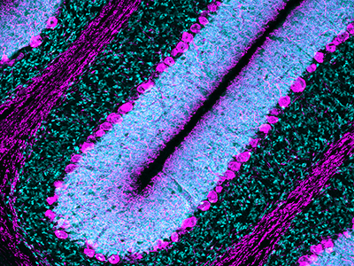
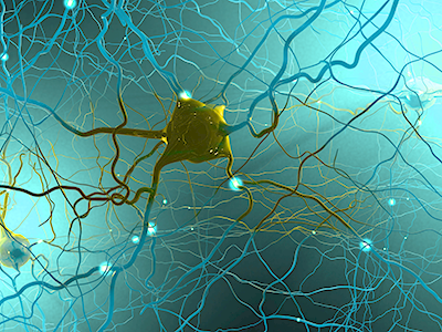
Primary cilia safeguard cortical neurons from environmental stress-induced dendritic degeneration

neuron on teal background

Fetus and neonates are under the risk of exposure to various external agents, such as alcohol and anesthetics taken by the mother. However, primary cilia can protect neurons by activating cilia-localized molecular signaling that inhibits degeneration of neuronal processes, according to the study’s findings.

A new study led by Kazue Hashimoto-Torii, Ph.D. and Masaaki Torii, Ph.D., both principal investigators for the Center for Neuroscience Research at Children’s National Hospital, found that primary cilia – tiny hair-like protrusions from the body of neuronal cells – protect neurons in the developing brain from adverse impacts of prenatal exposure.

Fetus and neonates are under the risk of exposure to various external agents, such as alcohol and anesthetics taken by the mother. However, primary cilia can protect neurons by activating cilia-localized molecular signaling that inhibits degeneration of neuronal processes, according to the study’s findings.

“Remarkably, the developing brain is equipped with intrinsic cell protection that helps to minimize the adverse impacts of to various external agents,” said Dr. Hashimoto-Torii. “However, the mechanisms of such protection have been unclear. Our study provides the first evidence that the tiny hair-like organelle protects neurons in the perinatal brain from adverse impacts of such external agents taken by the mother.”

The findings suggest that subtle alterations in primary cilia due to genetic conditions may lead to various neurodevelopmental disorders if combined with exposure to external agents from the environment. The findings also suggest that ciliopathy patients who have abnormal ciliary function due to genetic causes may have increased risk of abnormal brain development upon exposure to external agents.

“Clarifying diverse roles of cilia provides essential information for clinicians and patients with potential deficits in primary cilia to take extra precautions to avoid the risks for long-term negative impacts of external factors,” Dr. Torii explained. “We hope that further studies will define the whole picture of cilia-mediated neuroprotection and help us to advance our understanding of its importance in the pathogenesis of neurodevelopmental disorders.

This may ultimately lead to the development of treatment for various neurodevelopmental disorders,” he added.

The uniqueness of the study stems from the investigation of the role of cilia in brain development at the risk of exposure to various external factors that occur in the real world. Little is known about how the normal and abnormal brain development progresses in an environment where many external factors interact with intrinsic cellular mechanisms.

The study is a collaboration with researchers at Yale University and Keio University, Japan. Other Children’s National researchers who contributed to this study include Seiji Ishii, Ph.D.; Nobuyuki Ishibashi, M.D.; Toru Sasaki, M.D., Ph.D.; Shahid Mohammad, Ph.D.; Hye Hwang; Edwin Tomy; and Fahad Somaa.

Vittorio Gallo

Special issue of “Neurochemical Research” honors Vittorio Gallo, Ph.D.

Vittorio Gallo

Investigators from around the world penned manuscripts that were assembled in a special issue of “Neurochemical Research” that honors Vittorio Gallo, Ph.D., for his leadership in the field of neural development and regeneration.

At a pivotal moment early in his career, Vittorio Gallo, Ph.D., was accepted to work with Professor Giulio Levi at the Institute for Cell Biology in Rome, a position that leveraged courses Gallo had taken in neurobiology and neurochemistry, and allowed him to work in the top research institute in Italy directed by the Nobel laureate, Professor Rita Levi-Montalcini.

For four years as a student and later as Levi’s collaborator, Gallo focused on amino acid neurotransmitters in the brain and mechanisms of glutamate and GABA release from nerve terminals. Those early years cemented a research focus on glutamate neurotransmission that would lead to a number of pivotal publications and research collaborations that have spanned decades.

Now, investigators from around the world who have worked most closely with Gallo penned tributes in the form of manuscripts that were assembled in a special issue of “Neurochemical Research” that honors Gallo “for his contributions to our understanding of glutamatergic and GABAergic transmission during brain development and to his leadership in the field of neural development and regeneration,” writes guest editor Arne Schousboe, of the University of Copenhagen in Denmark.

Dr. Gallo as a grad student

Vittorio Gallo, Ph.D. as a 21-year-old mustachioed graduate student.

“In spite of news headlines about competition in research and many of the negative things we hear about the research world, this shows that research is also able to create a community around us,” says Gallo, chief research officer at Children’s National Hospital and scientific director for the Children’s National Research Institute.

As just one example, he first met Schousboe 44 years ago when Gallo was a 21-year-old mustachioed graduate student.

“Research can really create a sense of community that we carry on from the time we are in training, nurture as we meet our colleagues at periodic conferences, and continue up to the present. Creating community is bi-directional: influencing people and being influenced by people. People were willing to contribute these 17 articles because they value me,” Gallo says. “This is a lot of work for the editor and the people who prepared papers for this special issue.”

In addition to Gallo publishing more than 140 peer-reviewed papers, 30 review articles and book chapters, Schousboe notes a number of Gallo’s accomplishments, including:

  • He helped to develop the cerebellar granule cell cultures as a model system to study how electrical activity and voltage-dependent calcium channels modulate granule neuron development and glutamate release.
  • He developed a biochemical/neuropharmacological assay to monitor the effects of GABA receptor modulators on the activity of GABA chloride channels in living neurons.
  • He and Maria Usowicz used patch-clamp recording and single channel analysis to demonstrate for the first time that astrocytes express glutamate-activated channels that display functional properties similar to neuronal counterparts.
  • He characterized one of the spliced isoforms of the AMPA receptor subunit gene Gria4 and demonstrated that this isoform was highly expressed in the cerebellum.
  • He and his Children’s National colleagues demonstrated that glutamate and GABA regulate oligodendrocyte progenitor cell proliferation and differentiation.
Purkinje cells

Purkinje cells are large neurons located in the cerebellum that are elaborately branched like interlocking tree limbs and represent the only source of output for the entire cerebellar cortex.

Even the image selected to grace the special issue’s cover continues the theme of continuity and leaving behind a legacy. That image of Purkinje cells was created by a young scientist who works in Gallo’s lab, Aaron Sathyanesan, Ph.D. Gallo began his career working on the cerebellum – a region of the brain important for motor control – and now studies with a team of scientists and clinician-scientists Purkinje cells’ role in locomotor adaptive behavior and how that is disrupted after neonatal brain injury.

“These cells are the main players in cerebellar circuitry,” Gallo says. “It’s a meaningful image because goes back to my roots as a graduate student and is also an image that someone produced in my lab early in his career. It’s very meaningful to me that Aaron agreed to provide this image for the cover of the special issue.”

mitochondria

Molecular gatekeepers that regulate calcium ions key to muscle function

mitochondria

Controlled entry of calcium ions into the mitochondria, the cell’s energy powerhouses, makes the difference between whether muscles grow strong or easily tire and perish from injury, according to research published in Cell Reports.

Calcium ions are essential to how muscles work effectively, playing a starring role in how and when muscles contract, tap energy stores to keep working and self-repair damage. Not only are calcium ions vital for the repair of injured muscle fibers, their controlled entry into the mitochondria, the cell’s energy powerhouses, spells the difference between whether muscles will be healthy or if they will easily tire and perish following an injury, according to research published Oct. 29, 2019, in Cell Reports.

“Lack of the protein mitochondrial calcium uptake1 (MICU1) lowers the activation threshold for calcium uptake mediated by the mitochondrial calcium uniporter in both, muscle fibers from an experimental model and fibroblast of  a patient lacking MICU1,” says Jyoti K. Jaiswal, MSc, Ph.D., a principal investigator in the Center for Genetic Medicine Research at Children’s National Hospital and one of the paper’s corresponding authors. “Missing MICU1 also tips the calcium ion balance in the mitochondria when muscles contract or are injured, leading to more pronounced muscle weakness and myofiber death.”

Five years ago, patients with a very rare disease linked to mutations in the mitochondrial gene MICU1 were described to suffer from a neuromuscular disease with signs of muscle weakness and damage that could not be fully explained.

To determine what was going awry, the multi-institutional research team used a comprehensive approach that included fibroblasts donated by a patient lacking MICU1 and an experimental model whose MICU1 gene was deleted in the muscles.

Loss of MICU1 in skeletal muscle fibers leads to less contractile force, increased fatigue and diminished capacity to repair damage to their cell membrane, called the sarcolemma. Just like human patients, the experimental model suffers more pronounced muscle weakness, increased numbers of dead myofibers, with greater loss of muscle mass in certain muscles, like the quadriceps and triceps, the research team writes.

“What was happening to the patient’s muscles was a big riddle that our research addressed,” Jaiswal adds. “Lacking this protein is not supposed to make the muscle fiber die, like we see in patients with this rare disease. The missing protein is just supposed to cause atrophy and weakness.”

Patients with this rare disease show early muscle weakness, fluctuating levels of fatigue and lethargy, muscle aches after exercise, and elevated creatine kinase in their bloodstream, an indication of cell damage due to physical stress.

“One by one, we investigated these specific features in experimental models that look normal and have normal body weight, but also show lost muscle mass in the quadriceps and triceps,” explains Adam Horn, Ph.D., the lead researcher in Jaiswal’s lab who conducted this study. “Our experimental model lacking MICU1 only in skeletal muscles responded to muscle deficits so similar to humans that it suggests that some of the symptoms we see in patients can be attributed to MICU1 loss in skeletal muscles.”

Future research will aim to explore the details of how the impact of MICU1 deficit in muscles may be addressed therapeutically and possible implications of lacking MICU1 or its paralog in other organs.

In addition to Jaiswal and Horn, Children’s National Hospital Center for Genetic Medicine Research co-authors include Marshall W. Hogarth and Davi A. Mazala. Additional co-authors include Lead Author Valentina Debattisti, Raghavendra Singh, Erin L. Seifert, Kai Ting Huang, and Senior Author György Hajnóczky, all from Thomas Jefferson University; and Rita Horvath, from Newcastle University.

Financial support for research described in this post was provided by the National Institutes of Health under award numbers R01AR55686, U54HD090257 and RO1 GM102724; National Institute of Arthritis and Musculoskeletal and Skin Diseases under award number T32AR056993; and Foundation Leducq.