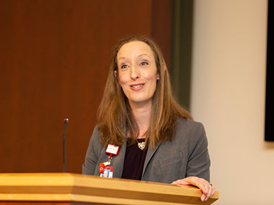
Dr. Gropman serves as Chief of the Division of Neurogenetics and Developmental Pediatrics at Children’s National Hospital. She is also a Professor of Pediatrics and Professor of Neurology at George Washington School of Medicine and Health Sciences.
About the award
Dr. Gropman joins a distinguished group of Children’s National physicians and scientists who hold an endowed chair. The Margaret O’Malley Professor of Genetic Medicine is one of 47 endowed chairs at Children’s National.
Professorships support groundbreaking work on behalf of children and their families and foster new discoveries and innovations in pediatric medicine. These appointments carry prestige and honor that reflect the recipient’s achievements and donor’s forethought to advance and sustain knowledge.
Dr. Gropman’s research focuses on neuroimaging, inborn errors of metabolism such as urea cycle disorders and mitochondrial disorders, and neurogenetics. She is the principal investigator of the Urea Cycle Disorders Consortium (UCDC) and the UCDC imaging consortium. She is the deputy clinical director of the Mito EpiGen Program.
Thomas and Mary Alice O’Malley, through their vision and generosity, are ensuring that Dr. Gropman and future holders of this professorship will launch bold, new initiatives to rapidly advance the field of pediatric genetic medicine, elevate our leadership and improve the lifetimes of children with genetic diseases.
About the donors
Tom and Mary Alice O’Malley have partnered with Children’s National to improve the lives of patients with urea cycles disorders for more than two decades. In 2003, their transformational philanthropy helped launch the Urea Cycle Disorders Consortium. This pioneering network grew to include 16-sites worldwide. It garnered 20 years of funding from the NIH’s Rare Diseases Clinical Research Network — the only center to sustain continuous funding over this period. This consortium’s research has yielded multiple effective treatment strategies, including government approval of three lifesaving therapies.
“The O’Malley family’s steadfast generosity helped us grow into the robust community of investigators and families we are today,” says Dr. Gropman. “They transformed care for UCD patients everywhere.”
https://innovationdistrict.childrensnational.org/wp-content/uploads/2023/09/Andrea-Gropman-feature.png300400Innovation Districthttps://innovationdistrict.childrensnational.org/wp-content/uploads/2023/12/innovationdistrict_logo-1-1030x165.pngInnovation District2023-09-25 13:53:152023-09-25 13:54:41Andrea L. Gropman, M.D., FAAP, FACMG, FANA, named as the Margaret O’Malley Professor of Genetic Medicine
About three years ago, Zika virus emerged as a newly recognized congenital infection, and a growing body of research indicates the damage it causes differs from other infections that occur in utero.
Seventy-one of 110 Brazilian infants at the highest risk for experiencing problems due to exposure to the Zika virus in the womb experienced a wide spectrum of brain abnormalities, including calcifications and malformations in cortical development, according to a study published July 31, 2019 in JAMA Network Open.
The infants were born at the height of Brazil’s Zika epidemic, a few months after the nation declared a national public health emergency. Already, many of the infants had been classified as having the severe form of congenital Zika syndrome, and many had microcephaly, fetal brain disruption sequence, arthrogryposis and abnormal neurologic exams at birth.
These 110 infants “represented a group of ZIKV-exposed infants who would be expected to have a high burden of neuroimaging abnormalities, which is a difference from other reported cohorts,” Sarah B. Mulkey, M.D., Ph.D., writes in an invited commentary published in JAMA Network Open that accompanies the Rio de Janeiro study. “Fortunately, many ZIKV-exposed infants do not have abnormal brain findings or a clinical phenotype associated with congenital Zika syndrome,” adds Dr. Mulkey, a fetal–neonatalneurologist in the Division of Fetal and Transitional Medicine at Children’s National in Washington, D.C.
Indeed, a retrospective cohort of 82 women exposed to Zika during their pregnancies led by a research team at Children’s National found only three pregnancies were complicated by severe fetal brain abnormalities. Compared with the 65% abnormal computed tomography (CT) or magnetic resonance imaging (MRI) findings in the new Brazilian study, about 1 in 10 (10%) of babies born to women living in the continental U.S. with confirmed Zika infections during pregnancy had Zika-associated birth defects, according to the Centers for Disease Control and Prevention.
“There appears to be a spectrum of brain imaging abnormalities in ZIKV-exposed infants, including mild, nonspecific changes seen at cranial US [ultrasound], such as lenticulostriate vasculopathy and germinolytic cysts, to more significant brain abnormalities, such as subcortical calcifications, ventriculomegaly and, in its most severe form, thin cortical mantle and fetal brain disruption sequence,” Dr. Mulkey writes.
About three years ago, Zika virus emerged as a newly recognized congenital infection, and a growing body of research indicates the damage it causes differs from other infections that occur in utero. Unlike congenital cytomegalovirus infection, cerebral calcifications associated with Zika are typically subcortical, Dr. Mulkey indicates. What’s more, fetal brain disruption sequence seen in Zika-exposed infants is unusual for other infections that can cause microcephaly.
“Centered on the findings of Pool, et al, and others, early neuroimaging remains one of the most valuable investigations of the Zika-exposed infant,” Dr. Mulkey writes, including infants who are not diagnosed with congenital Zika syndrome. She recommends:
Cranial ultrasound as the first-line imaging option for infants, if available, combined with neurologic and ophthalmologic exams, and brainstem auditory evoked potentials
Zika-exposed infants with normal cranial ultrasounds do not need additional imaging unless they experience a developmental disturbance
Zika-exposed infants with abnormal cranial ultrasounds should undergo further neuroimaging with low-dose cranial CT or brain MRI.
https://innovationdistrict.childrensnational.org/wp-content/uploads/2017/06/zika-virus.jpg300400Innovation Districthttps://innovationdistrict.childrensnational.org/wp-content/uploads/2023/12/innovationdistrict_logo-1-1030x165.pngInnovation District2019-07-31 14:32:302021-08-20 10:45:19Neuroimaging essential for Zika cases
A mutation of the gene PAC1R may be linked to the severity of social deficits experienced by kids with autism spectrum disorder (ASD), finds a study from a multi-institutional research team led by Children’s National faculty. If the pilot findings are corroborated in larger, multi-center studies, the research published online Dec. 17, 2018, in Autism Research represents the first step toward identifying a potential novel biomarker to guide interventions and better predict outcomes for children with autism.
As many as 1 in 40 children are affected by ASD. Symptoms of the disorder – such as not making eye contact, not responding to one’s name when called, an inability to follow a conversation of more than one speaker or incessantly repeating certain words or phrases – usually crop up by the time a child turns 3.
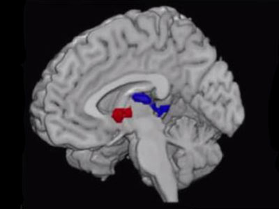
The developmental disorder is believed to be linked, in part, to disrupted circuitry within the amygdala, a brain structure integral for processing social-emotional information. This study reveals that PAC1R is expressed during key periods of brain development when the amygdala – an almond-shaped cluster of neurons – develops and matures. A properly functioning amygdala, along with brain structures like the prefrontal cortex and cerebellum, are crucial to neurotypical social-emotional processing.
“Our study suggests that an individual with autism who is carrying a mutation in PAC1R may have a greater chance of more severe social problems and disrupted functional brain connectivity with the amygdala,” says Joshua G. Corbin, Ph.D., interim director of the Center for Neuroscience Research at Children’s National Health System and the study’s co-senior author. “Our study is one important step along the pathway to developing new biomarkers for autism spectrum disorder and, hopefully, predicting patients’ outcomes.”
The research team’s insights came through investigating multiple lines of evidence:
They looked at gene expression in the brains of an experimental model at days 13.5 and 18.5 of fetal development and day 7 of life, dates that correspond with early, mid and late amygdala development. They confirmed that Pac1r is expressed in the experimental model at a critical time frame for brain development that coincides with the timing for altered brain trajectories with ASD.
They looked at gene expression in the human brain by mining publicly available genome-wide transcriptome data, plotting median PAC1R expression values for key brain regions. They found high levels of PAC1R expression at multiple ages with higher PAC1R expression in male brains during the fetal period and higher PAC1R expression in female brains during childhood and early adulthood.
One hundred twenty-nine patients with ASD aged 6 to 14 were recruited for behavioral assessment. Of the 48 patients who also participated in neuroimaging, 20 were able to stay awake for five minutes without too much movement as the resting state functional magnetic resonance images were captured. Children who were carriers of the high-risk genotype had higher resting-state connectivity between the amygdala and right posterior temporal gyrus. Connectivity alterations in a region of the brain involved in processing visual motion may influence how kids with ASD perceive socially meaningful information, the authors write.
Each child also submitted a saliva sample for DNA genotyping. Previously published research finds that a G to C single nucleotide polymorphism, a single swap in the nucleotides that make up DNA, in PAC1R is associated with higher risk for post traumatic stress disorder in girls. In this behavioral assessment, the research team found children with autism who carried the homozygous CC genotype had higher scores as measured through a validated tool, meaning they had greater social deficits than kids with the heterozygous genotype.
All told, the project is the fruit of six years of painstaking research and data collection, say the researchers. That includes banking patients’ saliva samples collected during clinical visits for future retrospective analyses to determine which genetic mutations were correlated with behavioral and functional brain deficits, Corbin adds.
“Lauren Kenworthy, who directs our Center for Autism Spectrum Disorders, and I have been talking over the years about how we could bring our programs together. We homed in on this project to look at about a dozen genes to assess correlations and brought in experts from genetics and genomics at Children’s National to sequence genes of interest,” he adds. “Linking the bench to bedside is especially difficult in neuroscience. It takes a huge amount of effort and dozens of discussions, and it’s very rare. It’s an exemplar of what we strive for.”
Financial support for the research described in this report was provided by DC-IDDRC under awards HD040677-07 and 1U54HD090257, the Clinical and Translational Science Institute at Children’s National, The Isidore and Bertha Gudelsky Family Foundation and the National Institutes of Health under awards MH083053-01A2 and MH084961.
https://innovationdistrict.childrensnational.org/wp-content/uploads/2019/01/DNA.jpg300400Innovation Districthttps://innovationdistrict.childrensnational.org/wp-content/uploads/2023/12/innovationdistrict_logo-1-1030x165.pngInnovation District2019-01-08 09:40:512022-05-17 12:34:08PAC1R mutation may be linked to severity of social deficits in autism
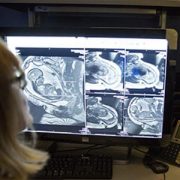
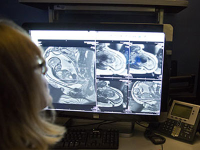
For the first time, a team of researchers led by Chandan Vaidya, Ph.D., chair of the Department of Psychology at Georgetown University, has used functional magnetic resonance imaging (fMRI) to capture the brain function of a small population of adolescents with obesity, both before and after bariatric surgery.
Obesity affects the whole body, from more obvious physical impacts on bones and joints to more subtle, internal impacts on organs like the brain.
For the first time, a team of researchers has used functional magnetic resonance imaging (fMRI) to capture the brain function of a small population of adolescents with obesity, both before and after bariatric surgery. The goal is to better understand the neural changes that occur when an adolescent is obese, and determine the effectiveness of interventions, such as vertical sleeve gastrectomy, at improving brain function as weight is lost.
The study, published as the November Editors’ Choice in the journal Obesity, found that executive and reward-related brain functions of study participants with obesity improved following the surgical procedure and initial weight loss.
“We’ve known for some time that severe obesity has negative consequences on some neurocognitive function areas for adults,” says Chandan Vaidya, Ph.D., chair of the Department of Psychology at Georgetown University and a senior author of the study. “But for the first time, we’ve captured fMRI evidence in young patients, and also shown that surgical intervention and the resulting weight loss can reverse some of those deficits.”
“For me, this early evidence makes a strong case that when kids are struggling with severe obesity, we need to consider surgical intervention as an option sooner in the process,” notes Evan Nadler, M.D., director of the Bariatric Surgery Program at Children’s National Health System, who also contributed to the study. “The question that remains is whether the neurocognitive function improves more if surgery, and thus weight loss, happens earlier – and is there a time factor that should help us determine when to perform a procedure that will maximize improvements?”
The preliminary study included 36 participants and was conducted using patients recruited from the Children’s National Bariatric Surgery program, one of the first children’s hospitals to achieve national accreditation by the Metabolic and Bariatric Surgery Accreditation and Quality Improvement Program.
“We asked these questions because we know that in the kids we see, their behavioral, brain, and physical health are all very closely related to one another and have an impact on each other,” adds Eleanor Mackey, Ph.D., study senior author and co-principal investigator on the National Institute of Diabetes and Digestive and Kidney Diseases grant that funded the project. “We expected that as physical health improves, we might see corresponding improvements in brain and behavior such as cognitive and school performance.”
The study also pointed out some technical and practical challenges to studying this particular young population. Anyone with a BMI greater than 50 was not able to fit within the MR bore used in the study, preventing fMRI participation by those patients.
“In addition to future studies with a larger sample size, we’d like to see if there are neuroimaging markers of plasticity differences in a population with BMI greater than 50,” says Dr. Vaidya. “Does the severity of the obesity change how quickly the brain can adapt following surgery and weight loss?”
The abstract was selected by the journal’s editors as one that provides insights into preventing and treating obesity. It was featured at the Obesity Journal Symposium during Obesity Week 2017 in Washington, D.C., as part of the Obesity Week recognition, and a digital video abstract was also released about the findings.
https://innovationdistrict.childrensnational.org/wp-content/uploads/2017/11/Adolescent-brain-scan-from-obesity-study.jpg300400Innovation Districthttps://innovationdistrict.childrensnational.org/wp-content/uploads/2023/12/innovationdistrict_logo-1-1030x165.pngInnovation District2017-11-03 10:00:432024-02-27 10:14:25Imaging captures obesity’s impact on the adolescent brain
Magnetic resonance imaging and ultrasound provide complementary data needed to assess ongoing changes to the brains of fetuses exposed to Zika in utero, says Sarah B. Mulkey, M.D., Ph.D.
For Zika-affected pregnancies, fetal magnetic resonance imaging (MRI) used in addition to standard ultrasound (US) imaging can better assess potential brain abnormalities in utero, according to research presented by Children’s National Health System during IDWeek 2017. In cases of abnormal brain structure, fetal MRI can reveal more extensive areas of damage to the developing brain than is seen with US.
“MRI and US provide complementary data needed to assess ongoing changes to the brains of fetuses exposed to Zika in utero,” says Sarah B. Mulkey, M.D., Ph.D., a fetal/neonatal neurologist at Children’s National Health System and lead author of the research paper. “In addition, our study found that relying on ultrasound alone would have given one mother the false assurance that her fetus’ brain was developing normally while the sharper MRI clearly pointed to brain abnormalities.”
As of Sept. 13, the Centers for Disease Control and Prevention (CDC) reported that 1,901 U.S. women were exposed to Zika at some point during their pregnancies but their infants appeared normal at birth. Another 98 U.S. women, however, gave birth to infants with Zika-related birth defects. And eight more women had pregnancy losses with Zika-related birth defects, according to CDC registries.
The longitudinal neuroimaging study led by Children’s National enrolled 48 pregnant women exposed to the Zika virus in the first or second trimester whose infection was confirmed by reverse transcription polymerase chain reaction, which detects Zika viral fragments shortly after exposure, and/or Immunoglobulin M testing, which reveals antibodies the body produces to neutralize the virus. Forty-six of the study volunteers live in Barranquilla, Colombia, where Zika infection is endemic. Two women live in the Washington region and were exposed to Zika during travel elsewhere.
All of the women underwent at least one diagnostic imaging session while pregnant, receiving an initial MRI or US at 25.1 weeks’ gestational age. Thirty-six women underwent a second MRI/US imaging pair at roughly 31 weeks’ gestation. Children’s National radiologists read every image.
Three of 48 pregnancies, or 6 percent, were marked by abnormal fetal MRIs:
One fetus had heterotopias (clumps of grey matter located at the wrong place) and abnormal cortical indent (a deformation at the outer layer of the cerebrum, a brain region involved in consciousness). The US taken at the same gestational age for this fetus showed its brain was developing normally.
Another fetus had parietal encephalocele (an uncommon skull defect) and Chiari malformation Type II (a life-threatening structural defect at the base of the skull and the cerebellum, the part of the brain that controls balance). The US for this fetus also detected these brain abnormalities.
The third fetus had a thin corpus callosum (bundle of nerves that connects the brain’s left and right hemispheres), an abnormally developed brain stem, temporal cysts, subependymal heterotopias and general cerebral/cerebellar atrophy. This fetal US showed significant ventriculomegaly (fluid-filled structures in the brain that are too large) and a fetal head circumference that decreased sharply from the 32nd to 36th gestational week, a hallmark of microcephaly.
After they were born, infants underwent a follow-up MRI without sedation and US. For nine infants, these ultrasounds revealed cysts in the choroid plexus (cells that produce cerebrospinal fluid) or germinal matrix (the source for neurons and glial cells that migrate during brain development). And one infant’s US after birth showed lenticulostriate vasculopathy (brain lesions).
“Because a number of factors can trigger brain abnormalities, further studies are needed to determine whether the cystic changes to these infants’ brains are attributable to Zika exposure in the womb or whether some other insult caused these troubling results,” Dr. Mulkey says.
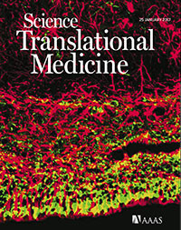
The cover of Science Translational Medicine features a new study of the cellular-level changes in the brain induced by congenital heart disease. Reprinted with permission from AAAS. Not for download
Disruptions in cerebral oxygen supply caused by congenital heart disease have significant impact on cortical growth, according to a research led by Children’s National Health System. The findings of the research team, which include co-authors from the National Institutes of Health, Boston Children’s Hospital and Johns Hopkins School of Medicine, appear on the cover of Science Translational Medicine. The subventricular zone (SVZ) in normal newborns’ brains is home to the largest stockpile of neural stem/progenitor cells, with newly generated neurons migrating from this zone to specific regions of the frontal cortex and differentiating into interneurons. When newborns experience disruptions in cerebral oxygen supply due to congenital heart disease, essential cellular processes go awry and this contributes to reduced cortical growth.
The preliminary findings point to the importance of restoring these cells’ neurogenic potential, possibly through therapeutics, to lessen children’s long-term neurological deficits.
“We know that congenital heart disease (CHD) reduces cerebral oxygen at a time when the developing fetal brain most needs oxygen. Now, we are beginning to understand the mechanisms of CHD-induced brain injuries at a cellular level, and we have identified a robust supply of cells that have the ability to travel directly to the site of injury and potentially provide help by replacing lost or damaged neurons,” says Nobuyuki Ishibashi, M.D., Director of the Cardiac Surgery Research Laboratory at Children’s National, and co-senior study author.
The third trimester of pregnancy is a time of dramatic growth for the fetal brain, which expands in volume and develops complex structures and network connections that growing children rely on throughout adulthood. According to the National Heart, Lung, and Blood Institute, congenital heart defects are the most common major birth defect, affecting 8 in 1,000 newborns. Infants born with CHD can experience myriad neurological deficits, including behavioral, cognitive, social, motor and attention disorders, the research team adds.
Cardiologists have tapped noninvasive imaging to monitor fetal hearts during gestation in high-risk pregnancies and can then perform corrective surgery in the first weeks of life to fix damaged hearts. Long term neurological deficits due to immature cortical development also have emerged as major challenges in pregnancies complicated by CHD.
“I think this is an enormously important paper for surgeons and for children and families who are affected by CHD. Surgeons have been worried for years that the things we do during corrective heart surgery have the potential to affect the development of the brain. And we’ve learned to improve how we do heart surgery so that the procedure causes minimal damage to the brain. But we still see some kids who have behavioral problems and learning delays,” says Richard A. Jonas, M.D., Chief of the Division of Cardiac Surgery at Children’s National, and co-senior study author. “We’re beginning to understand that there are things about CHD that affect the development of the brain before a baby is even born. What this paper shows is that the low oxygen level that sometimes results from a congenital heart problem might contribute to that and can slow down the growth of the brain. The good news is that it should be possible to reverse that problem using the cells that continue to develop in the neonate’s brain after birth.”
Among preclinical models, the spatiotemporal progression of brain growth in this particular model most closely parallels that of humans. Likewise, the SVZ cytoarchitecture of the neonatal preclinical model exposed to hypoxia mimics that of humans in utero and shortly after birth. The research team leveraged CellTracker Green to follow the path traveled by SVZ derived cells and to illuminate their fate, with postnatal SVZ supplying the developing cortex with newly generated neurons. SVZ derived cells were primarily neuroblasts. Superparamagnetic iron oxide nanoparticles supplied answers about long term SVZ migration, with SVZ derived cells making their way to the prefrontal cortex and the somatosensory cortex of the brain.
“We demonstrated that in the postnatal period, newly generated neurons migrate from the SVZ to specific cortices, with the majority migrating to the prefrontal cortex,” says Vittorio Gallo, Ph.D., Director of the Center for Neuroscience Research at Children’s National, and co-senior study author. “Of note, we revealed that the anterior SVZ is a critical source of newborn neurons destined to populate the upper layers of the cortex. We challenged this process through chronic hypoxia exposure, which severely impaired neurogenesis within the SVZ, depleting this critical source of interneurons.”
In the preclinical model of hypoxia as well as in humans, brains were smaller, weighed significantly less and had a significant reduction in cortical gray matter volume. In the prefrontal cortex, there was a significant reduction in white matter neuroblasts. Taken as a whole, according to the study authors, the findings suggest that impaired neurogenesis within the SVZ represents a cellular mechanism underlying hypoxia induced, region specific reduction in immature neurons in the cortex. The prefrontal cortex, the region of the brain that enables such functions as judgment, decision making and problem solving, is most impacted. Impairments in higher order cognitive functions involving the prefrontal cortex are common in patients with CHD.
https://innovationdistrict.childrensnational.org/wp-content/uploads/2017/01/CHD-Impaired-Cortical-Growth-HP.jpg248400Innovation Districthttps://innovationdistrict.childrensnational.org/wp-content/uploads/2023/12/innovationdistrict_logo-1-1030x165.pngInnovation District2017-02-07 14:08:072023-09-18 11:53:42Congenital heart disease and cortical growth
Although recent advances have greatly improved the survival of children with congenital heart disease, up to 55 percent will be left with injury to their brain’s white matter – an area that is critical for aiding connection and communication between various regions in the brain.
What’s known
Eight of every 1,000 children born each year have congenital heart disease (CHD). Although recent advances have greatly improved the survival of these children, up to 55 percent will be left with injury to their brain’s white matter – an area that is critical for aiding connection and communication between various regions in the brain. The resulting spectrum of neurological deficits can have significant costs for the individual, their family and society. Although studies have demonstrated that white matter injuries due to CHD have many contributing factors, including abnormal blood flow to the fetal brain, many questions remain about the mechanisms that cause these injuries and the best interventions.
What’s new
A Children’s National Health System research team combed existing literature, reviewing studies from Children’s as well as other research groups, to develop an article detailing the current state of knowledge on CHD and white matter injury. The scientists write that advances in neuroimaging – including magnetic resonance imaging, magnetic resonance spectroscopy, Doppler ultrasound and diffusion tensor imaging – have provided a wealth of knowledge about brain development in patients who have CHD. Unfortunately, these techniques alone are unable to provide pivotal insights into how CHD affects cells and molecules in the brain. However, by integrating animal models with findings in human subjects and in postmortem human tissue, the scientists believe that it will be possible to find novel therapeutic targets and new standards of care to prevent developmental delay associated with cardiac abnormalities.
For example, using a porcine model, the Children’s team was able to define a strategy for white matter protection in congenital heart surgery through cellular and developmental analysis of different white matter regions. Another study from Children’s combined rodent hypoxia with a brain slice model to replicate the unique brain conditions in neonates with severe and complex congenital heart disease. This innovative animal model provided novel insights into the possible additive effect of preoperative hypoxia on brain insults due to cardiopulmonary bypass and deep hypothermic circulatory arrest.
The Children’s research team also recently published an additional review article describing the key windows of development during which the immature brain is most vulnerable to CHD-related injury.
Questions for future research
Q: Can we create an animal model that recapitulates the morphogenic and developmental aspects of CHD without directly affecting other organs or developmental processes?
Q: What are the prenatal and neonatal cellular responses to CHD in the developing brain?
Q: What are the molecular mechanisms underlying white matter immaturity and vulnerability to CHD, and how can we intervene?
Q: How can we accurately assess the dynamic neurological outcomes of CHD and/or corrective surgery in animal models?
Q: Prenatal or postnatal insults to the developing brain: which is most devastating in regards to developmental and behavioral disabilities?
Q: How can we best extrapolate from, and integrate, neuroimaging findings/correlations in human patients with cellular/molecular approaches in animal models?
Source: Reprinted from Trends in Neurosciences, Vol. 38/Ed. 6, Paul D. Morton, Nobuyuki Ishibashi, Richard A. Jonas and Vittorio Gallo, “Congenital cardiac anomalies and white matter injury,” pp. 353-363, Copyright 2015, with permission from Elsevier.
https://innovationdistrict.childrensnational.org/wp-content/uploads/2017/02/CHD_White_Matter_article-1.jpg300400Innovation Districthttps://innovationdistrict.childrensnational.org/wp-content/uploads/2023/12/innovationdistrict_logo-1-1030x165.pngInnovation District2017-02-06 07:39:392017-06-07 14:12:46Congenital heart disease and white matter injury

 Children’s National Hospital named Andrea L. Gropman, M.D., FAAP, FACMG, FANA, as the Margaret O’Malley Professor of Genetic Medicine at Children’s National Hospital.