Serving patients with polycystic kidney disease
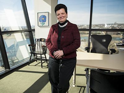
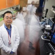
Lisa M. Guay-Woodford, M.D., is internationally recognized for her examination of the mechanisms that make certain inherited renal disorders particularly lethal, a research focus inspired by her patients.
When Children’s National pediatric nephrologist Lisa Guay-Woodford, M.D., was an intern at Boston Children’s Hospital, a baby with autosomal recessive polycystic kidney disease (ARPKD) was admitted to one of the hospital’s neonatal intensive care units (NICU). This disease, which causes cysts to form in the kidney and liver, kills about one-fifth of babies within the newborn period due to related problems that affect lung development.
But this baby seemed like a survivor, Dr. Guay-Woodford remembers. The child passed the newborn period and graduated from the NICU, although she went home with severe blood pressure issues. Along with a team of colleagues, Dr. Guay-Woodford helped to manage this patient’s care, juggling normal infant concerns with her ARPKD.
As far as Dr. Guay-Woodford knew at the time, this baby was beating the odds against her, growing and thriving. But one day near the end of her internship period, Dr. Guay-Woodford was called to the emergency department. Her patient was in a hypertensive crisis that ultimately killed her.
“It was absolutely devastating to all of us. This was supposed to be a good news kind of story, that she survived the newborn period and had gone home and was growing and developing,” Dr. Guay-Woodford says. “I realized then that a big part of the tragedy of this disease is how little we knew about it.”
Dr. Guay-Woodford vowed to change that. Since then, she’s devoted her career to studying ARPKD and other inherited kidney diseases.
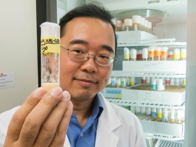
After finishing her residency and fellowship in Boston, Dr. Guay-Woodford was recruited to the University of Alabama, where she began caring for a cadre of 40 patients with inherited renal disorders. Fueled by the research questions that arose while working with these patients, she and her colleagues searched for PKD-related genes in the cpk mouse model, an animal that mimics many of the features of human ARPKD.
Dr. Guay-Woodford and her team cloned several of the key genes that caused recessive PKD in this mouse and other mouse models and eventually went on to identify the first major genetic modifier of PKD in these animals – a gene that wasn’t directly responsible for the disease but could sway its course. In time, her collaborative group became one of two that co-indentified the major gene responsible for human ARPKD. In 2005, Dr. Guay-Woodford led a team of investigators at the University of Alabama-Birmingham to establish one of just four PKD translational core centers funded by a National Institutes of Health P30 grant.
After moving to Children’s National in 2012, Dr. Guay-Woodford still co-directs this PKD translational core center while also caring for patients at her inherited renal disorders clinic. She and her colleagues here and beyond continue to work with mouse models of this disease, trying to ferret out the vast network of genes that interact in ARPKD and their specific roles.
“You can use a variety of strategies to compare these patients’ gene portfolios with those of healthy patients and pick out the disease genes. But at the end of the day, to me, that’s just the opening chapter,” she says. “To really make a story, you’ve got to understand what is it that gene does, what protein it makes, and how that protein works together with others involved in this disease.”
She and her team also are currently working with a pharmaceutical company to develop the first clinical trial to test a treatment for ARPKD. This effort has relied heavily on a clinical database that Dr. Guay-Woodford and colleagues worldwide maintain to track patients with this and related conditions. Through the extensive collection of clinical information in this database – including a variety of data on patients’ gestation and birth, growth, and kidney structure and function – the team has identified a core cohort of patients whose disease is rapidly progressing, a characteristic that makes them prime candidates to test this potential new treatment.
“Everything I do in the clinic informs the work I do in the lab, and everything I do in the lab is to help the patients I see in the clinic. It’s this constant dance back and forth between our human patients and animal models,” she says. “One day, this dance will help lessen the burden of this disease for these kids and their families.”


 Wilms tumor, also known as nephroblastoma, is the most common pediatric kidney cancer, typically seen in children ages three to four. Compared to patients with unilateral Wilms tumors, children with bilateral Wilms tumors (BWT) have poorer event-free survival (EFS) and are at higher risk for later effects such as renal failure. The treatment of BWT is challenging because it involves surgical removal of the cancer, while preserving as much healthy kidney tissue as possible to avoid the need for an organ transplant.