Marius George Linguraru, D.Phil., M.A., M.Sc., awarded Department of Defense grant for Neurofibromatosis application development
Marius George Linguraru, D.Phil., M.A., M.Sc., is a principal investigator in the Sheikh Zayed Institute for Pediatric Surgical Innovation at Children’s National, where he founded and directs the Precision Medical Imaging Laboratory. He’s an expert in quantitative imaging and artificial intelligence.
Marius George Linguraru, D.Phil., M.A., M.Sc., a principal investigator in the Sheikh Zayed Institute for Pediatric Surgical Innovation at Children’s National has been awarded a Congressionally Directed Medical Research Program (CDMRP) grant through the Department of Defense. This grant allows Dr. Linguraru to develop a novel quantitative MRI application that can inform treatment decisions by accurately identifying which children with Neurofibromatosis type 1 (NF1) and optic pathway glioma (OPG) are at risk of losing their vision.
This grant is part of the Neurofibromatosis Research Program of the CDMRP, which fills research gaps by funding high impact, high risk and high gain projects. Dr. Linguraru, who directs the Precision Medical Imaging Laboratory in the Sheikh Zayed Institute, is collaborating with the Gilbert Family Neurofibromatosis Institute and the Children’s Hospital of Philadelphia on this project.
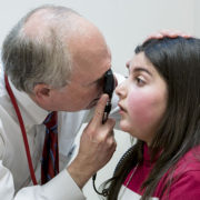
An expert in quantitative imaging and artificial intelligence, Dr. Linguraru has published several peer-reviewed studies on NF1 and OPG, a tumor that develops in 20 percent of children with NF1. The OPG tumor can cause irreversible vision loss, leading to permanent disability in about 50 percent of children with the tumor. This project, titled “MRI Volumetrics for Risk Stratification of Vision Loss in Optic Pathway Gliomas Secondary to NF1” will provide doctors certainty when identifying which children with NF1-OPG will lose vision and when the vision loss will occur.
Dr. Linguraru and his team will validate the quantitative MRI application that they’re developing by studying children at 25 NF1 clinics from around the world. Doctors using the application, which will perform comprehensive measurements of the OPG tumor’s volume, shape and texture, will upload their patient’s MRI into Dr. Linguraru’s application. Using recent advances in quantitative image analysis and machine learning, the application will then definitively determine whether the child’s NF1-OPG is going to cause vision loss and therefore requires treatment.
This diagnosis can occur before visual acuity starts to decline, which provides an opportunity for early treatment in children at risk for vision loss. Dr. Linguraru believes that early diagnosis and treatment can help to avoid lifelong visual impairment for these patients while preventing unnecessary MRIs and aggressive chemotherapy in pediatric patients who are not at risk of vision loss.
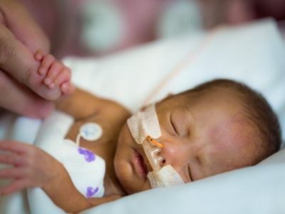
Occurring in one in 3,000 to 4,000 live births, NF1 is a genetic condition that manifests in early childhood and is characterized by changes in skin coloring and the growth of tumors along nerves in the skin, brain and other parts of the body. It is unknown why the OPG tumor caused by NF1 only results in vision loss for 50 percent of children. Some children will sustain lifelong disability from their vision loss, despite receiving treatment for their tumor, likely because treatment was started late. In other instances, doctors are unknowingly treating NF1-OPGs that would never cause vision loss.
Dr. Linguraru and his team have already proven that their computer-based, quantitative imaging measures are more objective and reliable than the current clinical measures, enabling doctors to make earlier and more accurate diagnoses and develop optimal treatment plans.