Tag Archive for: Children’s Research Institute

2019 pitch competition

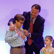
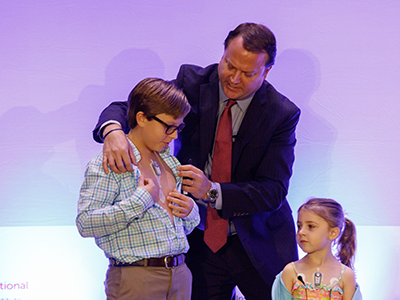
Pediatric medical device pitch competition deadline extended

2019 pitch competition

Pediatric innovators pitch for up to $250,000 in FDA-funded grant awards.

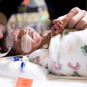
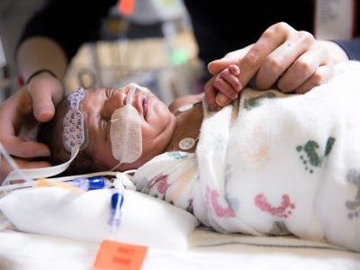
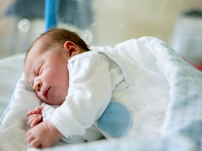
The National Capital Consortium for Pediatric Device Innovation (NCC-PDI) announced today that the application deadline for its annual “Make Your Medical Device Pitch for Kids!” competition is extended one week to Feb. 22 at midnight EST. Innovators and startup companies with devices in the pediatric cardiovascular, orthopedic and spine, or NICU sectors are invited to apply for a share of up to $250,000 in FDA-funded awards and access to a newly created NCC-PDI pediatric device accelerator program led by MedTech Innovator. Submissions are being accepted now.

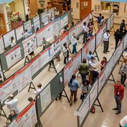
Up to 30 companies will be selected for the first round of competition scheduled for March 23, 2020 at the University of Maryland, College Park. Up to 10 finalists chosen from that event will compete for up to $250,000 in grant awards in Toronto, Canada on October 4. Finalists also receive a spot in the MedTech Innovator 2020 Accelerator – Pediatric Track, which provides a customized curriculum and in-depth mentorship.  Finalists will be announced in May, 2020.

This is the ninth competition in seven years hosted by NCC-PDI, one of five FDA Pediatric Device Consortia Grant Program members supporting the development and commercialization of pediatric medical devices. NCC-PDI is led by the Sheikh Zayed Institute for Pediatric Surgical Innovation at Children’s National Hospital and the A. James Clark School of Engineering at the University of Maryland. Additional consortium members include accelerators Medtech Innovator, BioHealth Innovation and design firm partner Archimedic.

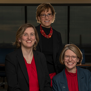
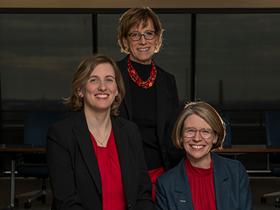
“This year’s competition focuses on three medical device areas of critical need for pediatric patients, so we want to give innovators as much time as possible to prepare their submissions,” said Kolaleh Eskandanian, Ph.D., MBA, PMP, vice president and chief innovation officer at Children’s National Hospital and principal investigator of NCC-PDI . “Our goal is to support devices that will improve care for children by helping them advance on the pathway to commercialization. We have seen how this competition can provide significant momentum for pediatric innovations, so we want to encourage as much participation as possible.”

To date, NCC-PDI has mentored over 100 medical device sponsors to help advance their pediatric innovations, notes Eskandanian, with six devices having received either their FDA market clearance or CE marking. Along with the positive exposure of presenting at this competition, she notes that the success of NCC-PDI’s portfolio companies is attributed to funding, mentorship, support from partners and facilitated interactions between device innovators and potential investors.

Eskandanian notes that enhancing access to resources for pediatric innovators is one aim of the Children’s National Research & Innovation Campus, a first-of-its-kind campus focused on pediatric healthcare innovation, currently under development on the former Walter Reed Army Medical Center campus in Washington, D.C. With its proximity to federal research institutions and agencies, universities, academic research centers, as well as on site accelerator Johnson & Johnson Innovation – JLABS, the campus will create a rich ecosystem of public and private partners which, like the NCC-PDI network, will help bolster pediatric innovation and commercialization. Opening is scheduled for December 2020.

Pediatric device competition

Premier annual pediatric medical device competition now accepting submissions

Pediatric device competition

Pediatric innovators pitch for grant awards and participation in a special accelerator program.

The official call for submissions is underway for the premiere annual pediatric medical device competition, sponsored by National Capital Consortium for Pediatric Device Innovation (NCC-PDI). The competition is led by Sheikh Zayed Institute for Pediatric Surgical Innovation at Children’s National Hospital, the A. James Clark School of Engineering at the University of Maryland and non-profit accelerator MedTech Innovator. The three organizations are all an integral part of the FDA-funded NCC-PDI, which aims to facilitate the development, production and distribution of pediatric medical devices. Additional NCC-PDI members include accelerator BioHealth Innovation and design firm Archimedic.

The competition focuses on pediatric devices in three areas of critical need: cardiovascular, orthopedic and spine, and neonatal intensive care (NICU) and is now accepting applications. Contestants will pitch for a share of up to $250K in grant awards and the opportunity to participate in the MedTech Innovator 2020 Accelerator – Pediatric Track.

The first stage of competition will be held on March 23 at the University of Maryland and will include up to 30 companies selected from all submissions received. Up to 10 finalists selected from that event will move on to the “Make Your Medical Device Pitch for Kids!” finals on October 4, 2020 in Toronto, Canada. Finalists from the March qualifying round will be notified in May, 2020.

“While there is a great need for pediatric devices in many specialty areas, the development and commercialization process is very challenging because of the small market size and dynamic characteristics of the patient population,” says Kolaleh Eskandanian, Ph.D., MBA, PMP, vice president and chief innovation officer at Children’s National Hospital and principal investigator of NCC-PDI. “To provide pediatric innovators with greater support in meeting these unique challenges, we must go beyond grant funding, which is why we are collaborating with MedTech Innovator to offer an accelerator program with a pediatric track.”

To date, NCC-PDI has mentored over 100 medical device sponsors to help advance their pediatric innovations, notes Eskandanian, with six devices having received either their FDA market clearance or CE marking. She says the success of NCC-PDI’s portfolio companies is attributed to funding, mentorship, support from partners, facilitated interactions between device innovators and potential investors, and being discovered during their presentations at the signature “Make Your Medical Device Pitch for Kids!” competitions.

While advancements have been made in some pediatric specialties, there is still a critical need for novel devices in cardiovascular, orthopedic and spine, and NICU areas. On average over the past decade, only 24 percent of life-saving medical devices approved by FDA – those that go through PMA and HDE regulatory pathways – have an indication for pediatric use. Of those, most are designated for children age 12 or older. “Devices designed specifically for the younger pediatric population are vitally needed and, at this early stage of the intervention, can significantly improve developmental outcomes for a child,” Eskandanian said.

Enhancing access to resources for pediatric innovators is also one of the aims of the Children’s National Research and Innovation Campus, a first-of-its-kind focused on pediatric healthcare innovation, currently under development on the former Walter Reed Army Medical Center campus in Washington, D.C. and opening in December, 2020. With its proximity to federal research institutions and agencies, universities, academic research centers, as well as on site accelerator Johnson and Johnson Innovation – JLABS, the campus will create a rich ecosystem of public and private partners which, like the NCC-PDI network, will help bolster pediatric innovation and commercialization.

NOTE: The deadline for submissions has been extended to February 22 at midnight EST.

Gilbert Vezina

Gilbert Vezina, M.D., recognized with American Society of Pediatric Neuroradiology Gold Medal Award

Gilbert Vezina

Gilbert Vezina, M.D., Director of Neuroradiology in the Division of Diagnostic Imaging and Radiology at Children’s National Hospital, is being recognized at the 2020 American Society of Pediatric Neuroradiology 2nd Annual Meeting with the society’s most distinguished honor, the Gold Medal Award.

The American Society of Pediatric Neuroradiology (ASPNR) Gold Medal is awarded for both professional and personal excellence, honoring individuals who are superb pediatric neuroradiologists, scientists, and/or physicians, and mentors and who also are truly outstanding people. Recipients have consistently extended themselves beyond self-interest to make contributions to the field of pediatric neuroradiology and as such, have elevated the subspecialty. This medal recognizes the exceptional service and achievements of these individuals.

Dr. Vezina completed his undergraduate degree at the Collège Jean-de-Brébeuf, Montréal, Canada and medical school at McGill Medical School, Montréal, Canada. He completed a mixed internship at Montreal General Hospital, Montreal, Canada; residency in Diagnostic Radiology, Massachusetts General Hospital, Boston, Massachusetts followed by a fellowship in Neuroradiology, Boston, Massachusetts.

He began his career at Children’s National Hospital in 1990. He is currently the Director of the Neuroradiology Program at Children’s National Hospital and Professor of Radiology and Pediatrics at George Washington University School of Medicine and Health Sciences, Washington DC. He created the Neuroradiology Fellowship Program in 1993 where he impacted medical students, residents and fellows from around the world. He served as president of ASPNR from 2001-2002 and past President from 2002-2005. He also served as the Interim Chief, Diagnostic Imaging and Radiology at Children’s National for a brief period in 2017.

Congratulations, Dr. Vezina!

Catherine Limperopoulos

Stressful pregnancies can leave fingerprint on fetal brain

Catherine Limperopoulos

“We were alarmed by the high percentage of pregnant women with a diagnosis of a major fetal heart problem who tested positive for stress, anxiety and depression,” says Catherine Limperopoulos, Ph.D., director of the Center for the Developing Brain at Children’s National and the study’s corresponding author.

When a diagnosis of fetal congenital heart disease causes pregnant moms to test positive for stress, anxiety and depression, powerful imaging can detect impaired development in key fetal brain regions, according to Children’s National Hospital research published online Jan. 13, 2020, in JAMA Pediatrics.

While additional research is needed, the Children’s National study authors say their unprecedented findings underscore the need for universal screening for psychological distress as a routine part of prenatal care and taking other steps to support stressed-out pregnant women and safeguard their newborns’ developing brains.

“We were alarmed by the high percentage of pregnant women with a diagnosis of a major fetal heart problem who tested positive for stress, anxiety and depression,” says Catherine Limperopoulos, Ph.D., director of the Center for the Developing Brain at Children’s National and the study’s corresponding author. “Equally concerning is how prevalent psychological distress is among pregnant women generally. We report for the first time that this challenging prenatal environment impairs regions of the fetal brain that play a major role in learning, memory, coordination, and social and behavioral development, making it all the more important for us to identify these women early during pregnancy to intervene,” Limperopoulos adds.

Congenital heart disease (CHD), structural problems with the heart, is the most common birth defect. Still, it remains unclear how exposure to maternal stress impacts brain development in fetuses with CHD.

The multidisciplinary study team enrolled 48 women whose unborn fetuses had been diagnosed with CHD and 92 healthy women with uncomplicated pregnancies. Using validated screening tools, they found:

  • 65% of pregnant women expecting a baby with CHD tested positive for stress
  • 27% of women with uncomplicated pregnancies tested positive for stress
  • 44% of pregnant women expecting a baby with CHD tested positive for anxiety
  • 26% of women with uncomplicated pregnancies tested positive for anxiety
  • 29% of pregnant women expecting a baby with CHD tested positive for depression and
  • 9% women with uncomplicated pregnancies tested positive for depression

All told, they performed 223 fetal magnetic resonance imaging sessions for these 140 fetuses between 21 and 40 weeks of gestation. They measured brain volume in cubic centimeters for the total brain as well as volumetric measurements for key regions such as the cerebrum, cerebellum, brainstem, and left and right hippocampus.

Maternal stress and anxiety in the second trimester were associated with smaller left hippocampi and smaller cerebellums only in pregnancies affected by fetal CHD. What’s more, specific regions — the hippocampus head and body and the left cerebellar lobe – were more susceptible to stunted growth. The hippocampus is key to memory and learning, while the cerebellum controls motor coordination and plays a role in social and behavioral development.

The hippocampus is a brain structure that is known to be very sensitive to stress. The timing of the CHD diagnosis may have occurred at a particularly vulnerable time for the developing fetal cerebellum, which grows faster than any other brain structure in the second half of gestation, particularly in the third trimester.

“None of these women had been screened for prenatal depression or anxiety. None of them were taking medications. And none of them had received mental health interventions. In the group of women contending with fetal CHD, 81% had attended college and 75% had professional educations, so this does not appear to be an issue of insufficient resources,” Limperopoulos adds. “It’s critical that we routinely to do these screenings and provide pregnant women with access to interventions to lower their stress levels. Working with our community partners, Children’s National is doing just that to help reduce toxic prenatal stress for both the health of the mother and for the future newborns. We hope this becomes standard practice elsewhere.”

Adds Yao Wu, Ph.D., a research associate working with Limperopoulos at Children’s National and the study’s lead author: “Our next goal is exploring effective prenatal cognitive behavioral interventions to reduce psychological distress felt by pregnant women and improve neurodevelopment in babies with CHD.”

In addition to Limperopoulos and Wu , Children’s National study co-authors include Kushal Kapse, MS, staff engineer; Marni Jacobs, Ph.D., biostatistician; Nickie Niforatos-Andescavage, M.D., neonatologist; Mary T. Donofrio, M.D., director, Fetal Heart Program; Anita Krishnan, M.D., associate director, echocardiography; Gilbert Vezina, M.D., director, Neuroradiology Program; David Wessel, M.D., Executive Vice President and Chief Medical Officer; and Adré  J. du Plessis, M.B.Ch.B., director, Fetal Medicine Institute. Jessica Lynn Quistorff, MPH, Catherine Lopez, MS, and Kathryn Lee Bannantine, BSN, assisted with subject recruitment and study coordination.

Financial support for the research described in this post was provided by the National Institutes of Health under grant No. R01 HL116585-01 and the Thrasher Research Fund under Early Career award No. 14764.

Drs. Tarini, Steinhorn, and Beers

Children’s National Hospital: Starting the new year with strong leadership

Drs. Tarini, Steinhorn, and Beers

Drs. Tarini, Steinhorn and Beers are also in leadership roles within professional societies, elected by their peers, further highlighting the strength of the leadership at Children’s National and professional respect within the health care community.

Three Children’s National Hospital executives are also in leadership roles within professional societies, elected by their peers, further highlighting the strength of the leadership at Children’s National and professional respect within the health care community.

Lee Savio Beers, M.D., FAAP, medical director of Community Health and Advocacy at the Child Health Advocacy Institute (CHAI) at Children’s National, was elected by her peers to become president-elect of the American Academy of Pediatrics (AAP) beginning Jan. 1, 2020. Dr. Beers will then serve as AAP president in 2021 for a one-year term.

“I am humbled and honored to have the support of my peers in taking on this newest leadership role,” says Dr. Beers. “AAP has been a part of my life since I first became a pediatrician, and my many leadership roles in the DC chapter and national AAP have given me a glimpse of the collective good we pediatricians can accomplish by working together toward common strategic goals.”

Dr. Beers is looking forward to continuing her work bringing together the diverse voices of pediatricians, children and families as well as other organizations to support improving the health of all children.

Robin Steinhorn, M.D., senior vice president of the Center for Hospital-Based Specialties at Children’s National was elected by her peers to become Vice President and President-elect of the American Pediatric Society (APS) in May 2018 and she is currently serving her role as the Society’s president, which began in May 2019.

“This is a tremendous honor. I look forward to leveraging the collective leadership and research accomplishments by our members to improve the health of infants and children throughout the U.S.,” said Dr. Steinhorn.

Dr. Steinhorn is particularly passionate about mentoring faculty and supporting the growth and career development of young neonatologists and scientists, with several having developed their own research laboratories and assumed division and department leadership positions. She was selected as a ‘Top Doctor’ by Northern Virginia Magazine in 2019.

Beth A. Tarini, M.D., MS, associate director, Center for Translational Research at The Children’s Research Institute, became vice president of the Society for Pediatric Research (SPR) in May 2019. Dr. Tarini will transition to President-Elect in May 2020 and become President in May 2021.

Dr. Tarini’s personal mission during this tenure will be to ensure that more pediatric researchers get to know SPR and are so excited about the organization that they become active members.

Dr. Tarini says she looks forward to working with other SPR leaders to find ways to build more productive, collaborative professional networks among faculty, especially emerging junior faculty. “Facilitating ways to network for research and professional reasons across pediatric research is vital – albeit easier said than done. I have been told I’m a connector, so I hope to leverage that skill in this new role,” says Dr. Tarini.

READ MORE:


Joseph Letzelter

Offering more options for children with hand and upper extremity ailments

Joseph Letzelter

The Children’s National Hand and Upper Extremities Program is one of few dedicated pediatric programs in the country, and it just added another hand surgeon to the team of extensively trained and experienced specialists.

Joseph Letzelter III, M.D., joined Children’s National Hospital and Pediatric Specialists of Virginia in September 2019 following comprehensive medical training in orthopaedic surgery with a focus on pediatric hand and upper extremity surgery. Dr. Letzelter received his undergraduate and medical degrees from Georgetown University, and also completed his internship and orthopaedic surgery residency there. He then completed a fellowship in hand and upper extremity surgery at New York University before continuing his studies with Dr. Michael Boland at the prestigious Hand Institute of New Zealand in Auckland. To round out his fellowship training, Dr. Letzelter focused purely on pediatric hand and upper extremity surgery at Shriner’s Hospital in Sacramento.

With the addition of Dr. Letzelter, the combined Hand and Upper Extremities Program expands its capacity to treat children with pediatric hand disorders. The joint orthopaedic surgery and plastic surgery program, led by Emily Hattwick, M.D., and Gary Rogers, M.D., uses pediatric-specific surgical methods to treat children without affecting their growth. Partnering with the world-class researchers at the Sheikh Zayed Institute for Surgical Innovation at Children’s National, the team is part of new discoveries that change the way we care for rare conditions. Learn more about the Hand and Upper Extremities Program here.

brain network illustration

$2.5M to protect the brain from metabolic insult

brain network illustration

The brain comprises only 2% of the body’s volume, but it uses more than 20% of its energy, which makes this organ particularly vulnerable to changes in metabolism.

More than 30 million Americans have diabetes, with the vast majority having Type 2 disease. Characterized by insulin resistance and persistently high blood sugar levels, poorly controlled Type 2 diabetes has a host of well-recognized complications: compared with the general population, a greatly increased risk of kidney disease, vision loss, heart attacks and strokes and lower limb amputations.

But more recently, says Nathan A. Smith, MS, Ph.D., a principal investigator in Children’s National Research Institute’s Center for Neuroscience Research, another consequence has become increasingly apparent. With increasing insulin resistance comes cognitive damage, a factor that contributes significantly to dementia diagnoses as patients age.

The brain comprises only 2% of the body’s volume, but it uses more than 20% of its energy, Smith explains – which makes this organ particularly vulnerable to changes in metabolism. Type 2 diabetes and even prediabetic changes in glucose metabolism inflict damage upon this organ in mechanisms with dangerous synergy, he adds. Insulin resistance itself stresses brain cells, slowly depriving them of fuel. As blood sugar rises, it also increases inflammation and blocks nitric oxide, which together narrow the brain’s blood vessels while also increasing blood viscosity.

When the brain’s neurons slowly starve, they become increasingly inefficient at doing their job, eventually succumbing to this deprivation. These hits don’t just affect individual cells, Smith adds. They also affect connectivity that spans across the brain, neural networks that are a major focus of his research.

While it’s well established that Type 2 diabetes significantly boosts the risk of cognitive decline, Smith says, it’s been unclear whether this process might be halted or even reversed. It’s this question that forms the basis of a collaborative Frontiers grant, $2.5 million from the National Science Foundation split between his laboratory; the lead institution, Stony Brook University; and Massachusetts General Hospital/Harvard Medical School.

Smith and colleagues at the three institutions are testing whether changing the brain’s fuel source from glucose to ketones – byproducts from fat metabolism – could potentially save neurons and neural networks over time. Ketones already have shown promise for decades in treating some types of epilepsy, a disease that sometimes stems from an imbalance in neuronal excitation and inhibition. When some patients start on a ketogenic diet – an extreme version of a popular fat-based diet – many can significantly decrease or even stop their seizures, bringing their misfiring brain cells back to health.

Principal Investigator Smith and his laboratory at the Children’s National Research Institute are using experimental models to test whether ketones could protect the brain against the ravages of insulin resistance. They’re looking specifically at interneurons, the inhibitory cells of the brain and the most energy demanding. The team is using a technique known as patch clamping to determine how either insulin resistance or insulin resistance in the presence of ketones affect these cells’ ability to fire.

They’re also looking at how calcium ions migrate in and out of the cells’ membranes, a necessary prerequisite for neurons’ electrical activity. Finally, they’re evaluating whether these potential changes to the cells’ electrophysiological properties in turn change how different parts of the brain communicate with each other, potentially restructuring the networks that are vital to every action this organ performs.

Colleagues at Athinoula A. Martinos Center for Biomedical Imaging at Massachusetts General Hospital and Harvard Medical School, led by Principal Investigator Eva-Maria Ratai, Ph.D.,  will perform parallel work in human subjects. They will use imaging to determine how these two fuel types, glucose or ketones, affect how the brain uses energy and produces the communication molecules known as neurotransmitters. They’re also investigating how these factors might affect the stability of neural networks using techniques that investigate the performance of these networks both while study subjects are at rest and performing a task.

Finally, colleagues at the Laufer Center for Physical and Quantitative Biology at Stony Brook University, led by Principal Investigator Lilianne R. Mujica-Parodi, Ph.D., will use results generated at the other two institutions to construct computational models that can accurately predict how the brain will behave under metabolic stress: how it copes when deprived of fuel and whether it might be able to retain healthy function when its cells receive ketones instead of glucose.

Collectively, Smith says, these results could help retain brain function even under glucose restraints. (For this, the research team owes a special thanks to Mujica-Parodi, who assembled the group to answer this important question, thus underscoring the importance of team science, he adds.)

“By supplying an alternate fuel source, we may eventually be able to preserve the brain even in the face of insulin resistance,” Smith says.

Xanxin Pei

Dr. Yanxin Pei receives prestigious grant from V Foundation for Cancer Research

Xanxin Pei

When asked about this award, Dr. Pei noted “I am so deeply grateful to receive this support from the V Foundation for Cancer Research…I will use these resources to aid our goal of discovering new therapies to treat medulloblastoma.”

Yanxin Pei, Ph.D., assistant professor in the Brain Tumor Institute and the Children’s Research Institute at Children’s National Hospital in Washington, D.C., has recently been awarded a prestigious grant by the V Foundation for Cancer Research to support her groundbreaking work in finding new treatments for childhood medulloblastoma.

Dr. Pei, who joined Children’s National in 2014 after training in the Wechsler-Reya lab at the Sanford-Burnham Institute in La Jolla, CA, has focused her work on the biology of medulloblastoma, the most common malignant brain tumor in children, with a major emphasis on the study of the medulloblastoma subtype most resistant to treatment. Children with this form of medulloblastoma have less than a 30% chance of survival five years from their diagnosis.

Having already developed one of the most important mouse models of this disease, Dr. Pei’s present V Foundation for Cancer Research Award, which includes becoming a V scholar, will explore the role of metabolism in the development of metastasis in MYC-amplified medulloblastomas (the most virulent form of medulloblastoma).

The V Foundation for Cancer Research Award is one of a series of prestigious awards Dr. Pei has received over the past 18 months for her work, including an NIH-sponsored 5-year award (ROI) evaluating other aspects of medulloblastoma development and resistance to therapy, and grants from the Rally Foundation, the Meghan Rose Bradley Foundation and the Children’s Cancer Foundation.

When asked about this award, Dr. Pei noted “I am so deeply grateful to receive this support from the V Foundation for Cancer Research…I will use these resources to aid our goal of discovering new therapies to treat medulloblastoma.”

Her cutting-edge work is generating national and international attention and firmly places Dr. Pei as an international leader in medulloblastoma research.

Nikki Gillum Posnack

Research team develops new and improved method for studying cardiac function

Nikki Gillum Posnack

While researching how plastic affects heart function in sensitive populations, such as children born with congenital heart defects, Children’s National researcher Nikki Posnack, Ph.D., led a team that developed a new and improved, replicable method of performing simultaneous dual optical mapping to examine electrical activity and calcium for the study of cardiac function.

Since arriving at the Sheikh Zayed Institute for Pediatric Surgical Innovation, researcher Nikki Gillum Posnack, Ph.D., a principal investigator with the institute and assistant professor of pediatrics at the George Washington University School of Medicine and Health Sciences, has been focused on examining how exposure to plastic affects heart function in sensitive populations, such as children born with congenital heart defects. She performs optical mapping to conduct this research, but the industry standard approaches of either using dual cameras or sequential single cameras were cost prohibitive and technically challenging while also diminishing the quality of the imaging results.

Fast forward to July 2019 when Dr. Posnack and her team published “Plasticizer Interaction With the Heart” in the journal Arrhythmia and Electrophysiology, which used imaging techniques to reveal the impact of plastic chemicals on the electrical activity of the heart. Dr. Posnack’s laboratory has since expanded this technique and revealed a new replicable method of performing simultaneous dual optical mapping to examine electrical activity and calcium handling in the heart.

Sharing a new method for studying cardiac function

This groundbreaking method is itself the focus of a new BMC Biomedical Engineering journal article titled “Lights, camera, path splitter: a new approach for truly simultaneous dual optical mapping of the heart with a single camera.”

The article compares and contrasts the current standard for dual camera simultaneous configurations and single camera sequential configurations to Dr. Posnack’s new single camera simultaneous configuration.

Simultaneous dual mapping systems use two probes and dual dyes – one for electrical voltage and the other for calcium. While dual-dye combinations like Di-4-ANEPPS with Indo-1, Di-2-ANEPEQ and calcium green have been developed to separate fluorescence signals by emission, these dye combinations can have spectral overlap, creating the need for non-ideal emission bandpass to negate spectral overlap and/or the inclusion of a calcium probe with an inferior dissociation constant. Additionally, dual-sensor systems require proper alignment to ensure that fluorescence signals are being analyzed from the same tissue region on each individual detector, which could lead to erroneous results. The dual-camera optical setup is expensive, technically challenging and requires a large physical footprint that is often not feasible for basic science and teaching laboratories conducting critical research.

As an alternative, some researchers use a single camera configuration to sequentially image the voltage and calcium probes using excitation light patterning. This approach also has limitations. These single-sensor designs use dual-dye combinations that require two or more excitation light sources, but share a single emission band. Like the dual camera system, this platform design is also technically challenging since the different excitation light wavelengths require light source triggering, camera synchronization and frame interleaving. Due to timing coordination, decreased frame rates, excitation light ramp up/down times and shutter open/close times, single system setups require shorter exposure times compared to dual sensor setups, diminishing the signal-to-noise quality without offering the same temporal fidelity. There is a cost advantage to the single camera system, however, because the additional camera is often one of the most expensive components.

This new single camera, simultaneous dual optical mapping approach is the first multiparametric mapping system that simultaneously acquires calcium and voltage signals from cardiac preparations, using a commercially available optical path splitter, single camera and single excitation light. Using a large field of view sCMOS sensor that is faster and more sensitive, this configuration separates the two emission bands for voltage and calcium probes and simultaneously directs them to either sides of the single, large camera sensor. This protocol employs a commonly used dual-dye combination (RH237 and Rhod2-AM). In contrast, other protocols may require genetically-encoded indicators or fluorescent probes that are not yet commercially available.

The team validated the utility of the approach by performing high-speed simultaneous dual imaging with sufficient signal-to-noise ratio for calcium and voltage signals and specificity of emission signals with negligible cross-talk. Demonstrating the need for simultaneous electrical and calcium sensors, they found that when ventricular tachycardia is induced, there is spatially discordant calcium alternans present in different regions of the heart even when the electrical alternans remain concordant.

Having eliminated the second camera as well as the need for multiple excitation light sources, light pattering and frame interleaving, this system is more cost effective, simpler, and can be easily setup by various types of researchers, not just those with engineering backgrounds.

With a limited research budget and a background in physiology, Dr. Posnack worked collaboratively with her post-doctoral fellow Rafael Jaimes III, an engineer in the Sheikh Zayed Institute for Pediatric Surgical Innovation, to develop a cost-effective system that would enable her to truly study the effects of plastics on the heart.

Multidisciplinary approach

“We’re fortunate to have a multidisciplinary team in the Sheikh Zayed Institute so that I could work with an engineer to develop the technology and system we needed to propel our research,” said Dr. Posnack. “There are so many researchers who have the science background, but not necessarily the technical aptitude, and they get stymied in their research, so we’re proud that this paper will help other researchers replicate the system to study cardiac function.”

The research paper was funded by a grant from the National Institutes of Health as well as support from the Children’s Research Institute, Children’s National Heart Institute and the Sheikh Zayed Institute for Pediatric Surgical Innovation.

The applications for this optical mapping system are significant and Dr. Posnack has been consulted by other research teams looking to implement it in their labs. Additionally, Dr. Posnack has collaborated with several neuroscience teams at Children’s National Hospital, including one that is investigating the effects of hypoxia on brain and heart development, and another that is interested in using image modalities and data processing to analyze calcium as an indicator of neuron firing.

Dr. Posnack continues to use this new dual optical mapping system to further her research as she anticipates the publication of a new article about age-dependent changes in cardiac electrophysiology and calcium handling.

t-cells

Tailored T-cell therapies neutralize viruses that threaten kids with PID

t-cells

Tailored T-cells specially designed to combat a half dozen viruses are safe and may be effective in preventing and treating multiple viral infections, according to research led by Children’s National Hospital faculty.

Catherine Bollard, M.B.Ch.B., M.D., director of the Center for Cancer and Immunology Research at Children’s National and the study’s senior author, presented the teams’ findings Nov. 8, 2019, during a second-annual symposium jointly held by Children’s National and the National Institute of Allergy and Infectious Diseases (NIAID), part of the National Institutes of Health (NIH). Children’s National and NIAID formed a research partnership in 2017 to develop and conduct collaborative clinical research studies focused on young children with allergic, immunologic, infectious and inflammatory diseases. Each year, they co-host a symposium to exchange their latest research findings.

According to the NIH, more than 200 forms of primary immune deficiency diseases impact about 500,000 people in the U.S. These rare, genetic diseases so impair the person’s immune system that they experience repeated and sometimes rare infections that can be life threatening. After a hematopoietic stem cell transplantation, brand new stem cells can rebuild the person’s missing or impaired immune system. However, during the window in which the immune system rebuilds, patients can be vulnerable to a host of viral infections.

Because viral infections can be controlled by T-cells, the body’s infection-fighting white blood cells, the Children’s National first-in-humans Phase 1 dose escalation trial aimed to determine the safety of T-cells with antiviral activity against a half dozen opportunistic viruses: adenovirus, BK virus, cytomegalovirus (CMV), Epstein-Barr virus (EBV), Human Herpesvirus 6 and human parainfluenza-3 (HPIV3).

Eight patients received the hexa-valent, virus-specific T-cells after their stem cell transplants:

  • Three patients were treated for active CMV, and the T-cells resolved their viremia.
  • Two patients treated for active BK virus had complete symptom resolution, while one had hemorrhagic cystitis resolved but had fluctuating viral loads in their blood and urine.
  • Of two patients treated prophylactically, one developed EBV viremia that was treated with rituximab.

Two additional patients received the T-cell treatments under expanded access for emergency treatment, one for disseminated adenoviremia and the other for HPIV3 pneumonia. While these critically ill patients had partial clinical improvement, they were being treated with steroids which may have dampened their antiviral responses.

“These preliminary results show that hexaviral-specific, virus-specific T-cells are safe and may be effective in preventing and treating multiple viral infections,” says Michael Keller, M.D., a pediatric immunologist at Children’s National and the lead study author. “Of note, enzyme-linked immune absorbent spot assays showed evidence of antiviral T-cell activity by three months post infusion in three of four patients who could be evaluated and expansion was detectable in two patients.”

In addition to Drs. Bollard and Keller, additional study authors include Katherine Harris M.D.; Patrick J. Hanley Ph.D., assistant research professor in the Center for Cancer and Immunology; Allistair Abraham, M.D., a blood and marrow transplantation specialist; Blachy J. Dávila Saldaña, M.D., Division of Blood and Marrow Transplantation; Nan Zhang Ph.D.; Gelina Sani BS; Haili Lang MS; Richard Childs M.D.; and Richard Jones M.D.

###

Children’s National-NIAID 2019 symposium presentations

“Welcome and introduction”
H. Clifford Lane, M.D., director of NIAID’s Division of Clinical Research

“Lessons and benefits from collaboration between the NIH and a free-standing children’s hospital”
Marshall L. Summar, M.D., director, Rare Disease Institute, Children’s National

“The hereditary disorders of PropionylCoA and Cobalamin Metabolism – past, present and future”
Charles P. Venditti, M.D., Ph.D., National Human Genome Research Institute Collaboration

“The road(s) to genetic precision therapeutics in pediatric neuromuscular disease: opportunities and challenges”
Carsten G. Bönnemann, M.D., National Institute of Neurological Disorders and Stroke

“Genomic diagnostics in immunologic diseases”
Helen Su, M.D., Ph.D., National Institute of Allergy and Infectious Diseases

“Update on outcomes of gene therapy clinical trials for X-SCID and X-CGD and plans for future trials”
Harry Malech, M.D., National Institute of Allergy and Infectious Diseases

“Virus-specific T-cell therapies: broadening applicability for PID patients”
Catherine Bollard, M.D., Children’s National 

“Using genetic testing to guide therapeutic decisions in Primary Immune Deficiency Disease”
Vanessa Bundy, M.D., Ph.D., Children’s National 

Panel discussion moderated by Lisa M. Guay-Woodford, M.D.
Drs. Su, Malech, Bollard and Bundy
Morgan Similuk, S.C.M., NIAID
Maren Chamorro, Parent Advocate

“Underlying mechanisms of pediatric food allergy: focus on B cells
Adora Lin, M.D., Ph.D., Children’s National 

“Pediatric Lyme outcomes study – interim update”
Roberta L. DeBiasi, M.D., MS, Children’s National 

“Molecular drivers and opportunities in neuroimmune conditions of pediatric onset”
Elizabeth Wells, M.D., Children’s National 


baby with tubes

BPA analogues may be less likely to disrupt heart rhythm

Some chemical alternatives to plastic bisphenol-a (BPA), which is still commonly used in medical settings such as operating rooms and intensive care units, may be less disruptive to heart electrical function than BPA.

A poster at the AHA Scientific Sessions suggests bisphenol-s (BPS) and bisphenol-f (BPF) may have less impact on heart function than bisphenol-a (BPA).

Some chemical alternatives to plastic bisphenol-a (BPA), which is still commonly used in medical settings such as operating rooms and intensive care units, may be less disruptive to heart electrical function than BPA, according to a pre-clinical study that explored how the structural analogues bisphenol-s (BPS) and bisphenol-f (BPF) interact with the chemical and electrical functions of heart cells.

The findings suggest that in terms of toxicity for heart function, these chemicals that are similar in structure to BPA may actually be safer for medically fragile heart cells, such as those in children with congenital heart disease. Previous research has found a high likelihood that BPA exposure may impact the heart’s electrical conductivity and disrupt heart rhythm, and patients are often exposed to the plastic via clinical equipment found in intensive care and in the operating room.

“There are still many questions that need to be answered about the safety and efficacy of using chemicals that look and act like BPA in medical settings, especially in terms of their potential contribution to endocrine disruption,” says Nikki Gillum Posnack, Ph.D., the poster’s senior author and a principal investigator in the Sheikh Zayed Institute for Pediatric Surgical Innovation at Children’s National Hospital. “What we can say is that, in this initial pre-clinical investigation, it appears that these structural analogues have less of an impact on the electrical activity within the heart and therefore, may be less likely to contribute to dysrhythmias.”

Future studies will seek to quantify the risk that these alternative chemicals pose in vulnerable populations, including pediatric cardiology and cardiac surgery patients. Since pediatric patients’ hearts are still growing and developing, the interactions may be different than what was seen in this pilot study.

Learn more the impacts of exposure to plastics such as bisphenol-A and plasticizers such as DEHP and MEHP that are commonly used in medical devices:

###

Bisphenol-a Analogues May Be Safer Alternatives For Plastic Medical Products
Rafael Jaimes, Damon McCullough, Luther M Swift, Marissa Reilly, Morgan Burke, Jiansong Sheng, Javier Saiz, Nikki G Posnack
Poster Presentation by senior author Nikki G Posnack
CH.APS.01 – Translational Research in Congenital Heart Disease
AHA Scientific Sessions
November 16, 2019
1:30 p.m. – 2:00 p.m.

Newborn baby laying in crib

Can cells collected from bone marrow stimulate generation of new neurons in babies with CHD?

Newborn baby laying in crib

The goal of the study will be to optimize brain development in babies with congenital heart disease (CHD) who sometimes demonstrate delay in the development of cognitive and motor skills.

An upcoming clinical trial at Children’s National Hospital will harness cardiopulmonary bypass as a delivery mechanism for a novel intervention designed to stimulate brain growth and repair in children who undergo cardiac surgery for congenital heart disease (CHD).

The NIH has awarded Children’s National $2.5 million to test the hypothesis that mesenchymal stromal cells (MSCs), which have been shown to possess regenerative properties and the ability to modulate immune responses in a variety of diseases, collected from allogeneic bone marrow, may promote regeneration of damaged neuronal and glial cells in the early postnatal brain. If successful, the trial will determine the safety of the proposed treatment in humans and set the stage for a Phase 2 efficacy trial of what could potentially be the first treatment for delays in brain development that happen before birth as a consequence of congenital heart disease. The study is a single-center collaboration between three Children’s National physician-researchers: Richard Jonas, M.D.Catherine Bollard, M.B.Ch.B., M.D. and Nobuyuki Ishibashi, M.D.

Dr. Jonas, chief of cardiac surgery at Children’s National, will outline the trial and its aims on Monday, November 18, 2019, at the American Heart Association’s Scientific Sessions 2019. Dr. Jonas was recently recognized by the Cardiac Neurodevelopmental Outcome Collaborative for his lifelong research of how cardiac surgery impacts brain growth and development in children with CHD.

Read more about the study: Researchers receive $2.5M grant to optimize brain development in babies with CHD.

###

Regenerative Cell Therapy in Congenital Heart Disease – Protecting the Immature Brain
Presented by Richard Jonas, M.D.
AHA Scientific Sessions
Session CH.CVS.608 Congenital Heart Disease and Pediatric Cardiology Seminar: A Personalized Approach to Heart Disease in Children
9:50 a.m. to 10:05 a.m.
November 18, 2019

pastel colored DNA strands

Germline microsatellite genotypes differentiate children with medulloblastoma

pastel colored DNA strands

A new study suggests that medulloblastoma-specific germline microsatellite variations mark those at-risk for medulloblastoma development.

Brian Rood, M.D., oncologist and medical director at the Brain Tumor Institute, and Harold “Skip” Garner, Ph.D., associate vice provost for research development at Edward Via College of Osteopathic Medicine, published a report in the Society for Neuro-Oncology’s Neuro-Oncology Journal about using a novel approach to identify specific markers in germline (non-tumor) DNA called microsatellites that can differentiate children who have the brain tumor medulloblastoma (MB) from those who don’t.

“Ultimately, the best way to save children from brain tumors and prevent them from bearing long-term side effects from treatment is to prevent those tumors from occurring in the first place,” says Dr. Rood. “New advancements hold the potential to finally realize the dream of cancer prevention, but we must first identify those children at-risk.”

While analyzing germline sequencing data from a training set of 120 MB subjects and 425 controls, the doctors identified 139 individual microsatellites whose genotypes differ significantly between the groups. Using a genetic algorithm, they were able to construct a subset of 43 microsatellites that distinguish MB subjects from controls with a sensitivity and specificity of 92% and 88% respectively.

“We made discoveries in an untapped part of the human genome, enabled by unique bioinformatics data mining approaches combined with clinical insight,” said Dr. Garner. “Our findings establish new genomic directions that can lead to high accuracy diagnostics for predicting susceptibility to medulloblastoma.”

What the doctors discovered and demonstrated in the study was that MB-specific germline microsatellite variations mark those at risk for MB development and suggest that other mechanisms of cancer predisposition beyond heritable mutations exist for MB.

“This work is the first to demonstrate the ability of specific DNA sequences to differentiate children with cancer from their healthy counterparts,” added Dr. Rood.

Contributing Authors to this research study included:  Brian R. Rood, M.D., Harold R. Garner, Ph.D., Samuel Rivero-Hinojosa, Ph.D., and Nicholas Kinney, Ph.D.

mitochondria

Molecular gatekeepers that regulate calcium ions key to muscle function

mitochondria

Controlled entry of calcium ions into the mitochondria, the cell’s energy powerhouses, makes the difference between whether muscles grow strong or easily tire and perish from injury, according to research published in Cell Reports.

Calcium ions are essential to how muscles work effectively, playing a starring role in how and when muscles contract, tap energy stores to keep working and self-repair damage. Not only are calcium ions vital for the repair of injured muscle fibers, their controlled entry into the mitochondria, the cell’s energy powerhouses, spells the difference between whether muscles will be healthy or if they will easily tire and perish following an injury, according to research published Oct. 29, 2019, in Cell Reports.

“Lack of the protein mitochondrial calcium uptake1 (MICU1) lowers the activation threshold for calcium uptake mediated by the mitochondrial calcium uniporter in both, muscle fibers from an experimental model and fibroblast of  a patient lacking MICU1,” says Jyoti K. Jaiswal, MSc, Ph.D., a principal investigator in the Center for Genetic Medicine Research at Children’s National Hospital and one of the paper’s corresponding authors. “Missing MICU1 also tips the calcium ion balance in the mitochondria when muscles contract or are injured, leading to more pronounced muscle weakness and myofiber death.”

Five years ago, patients with a very rare disease linked to mutations in the mitochondrial gene MICU1 were described to suffer from a neuromuscular disease with signs of muscle weakness and damage that could not be fully explained.

To determine what was going awry, the multi-institutional research team used a comprehensive approach that included fibroblasts donated by a patient lacking MICU1 and an experimental model whose MICU1 gene was deleted in the muscles.

Loss of MICU1 in skeletal muscle fibers leads to less contractile force, increased fatigue and diminished capacity to repair damage to their cell membrane, called the sarcolemma. Just like human patients, the experimental model suffers more pronounced muscle weakness, increased numbers of dead myofibers, with greater loss of muscle mass in certain muscles, like the quadriceps and triceps, the research team writes.

“What was happening to the patient’s muscles was a big riddle that our research addressed,” Jaiswal adds. “Lacking this protein is not supposed to make the muscle fiber die, like we see in patients with this rare disease. The missing protein is just supposed to cause atrophy and weakness.”

Patients with this rare disease show early muscle weakness, fluctuating levels of fatigue and lethargy, muscle aches after exercise, and elevated creatine kinase in their bloodstream, an indication of cell damage due to physical stress.

“One by one, we investigated these specific features in experimental models that look normal and have normal body weight, but also show lost muscle mass in the quadriceps and triceps,” explains Adam Horn, Ph.D., the lead researcher in Jaiswal’s lab who conducted this study. “Our experimental model lacking MICU1 only in skeletal muscles responded to muscle deficits so similar to humans that it suggests that some of the symptoms we see in patients can be attributed to MICU1 loss in skeletal muscles.”

Future research will aim to explore the details of how the impact of MICU1 deficit in muscles may be addressed therapeutically and possible implications of lacking MICU1 or its paralog in other organs.

In addition to Jaiswal and Horn, Children’s National Hospital Center for Genetic Medicine Research co-authors include Marshall W. Hogarth and Davi A. Mazala. Additional co-authors include Lead Author Valentina Debattisti, Raghavendra Singh, Erin L. Seifert, Kai Ting Huang, and Senior Author György Hajnóczky, all from Thomas Jefferson University; and Rita Horvath, from Newcastle University.

Financial support for research described in this post was provided by the National Institutes of Health under award numbers R01AR55686, U54HD090257 and RO1 GM102724; National Institute of Arthritis and Musculoskeletal and Skin Diseases under award number T32AR056993; and Foundation Leducq.

doctor checking pregnant woman's belly

Novel approach to detect fetal growth restriction

doctor checking pregnant woman's belly

Morphometric and textural analyses of magnetic resonance imaging can point out subtle architectural deviations associated with fetal growth restriction during the second half of pregnancy, a first-time finding that has the promise to lead to earlier intervention.

Morphometric and textural analyses of magnetic resonance imaging (MRI) can point out subtle architectural deviations that are associated with fetal growth restriction (FGR) during the second half of pregnancy. The first-time finding hints at the potential to spot otherwise hidden placental woes earlier and intervene in a more timely fashion, a research team led by Children’s National Hospital faculty reports in Pediatric Research.

“We found reduced placental size, as expected, but also determined that the textural metrics are accelerated in FGR when factoring in gestational age, suggesting premature placental aging in FGR,” says Nickie Andescavage, M.D., a neonatologist at Children’s National and the study’s lead author. “While morphometric and textural features can discriminate placental differences between FGR cases with and without Doppler abnormalities, the pattern of affected features differs between these sub-groups. Of note, placental insufficiency with abnormal Doppler findings have significant differences in the signal-intensity metrics, perhaps related to differences of water content within the placenta.”

The placenta, an organ shared by the pregnant woman and the developing fetus, delivers oxygen and nutrients to the developing fetus and ferries away waste products. Placental insufficiency is characterized by a placenta that develops poorly or is damaged, impairing blood flow, and can result in still birth or death shortly after birth. Surviving infants may be born preterm or suffer early brain injury; later in life, they may experience cardiovascular, metabolic or neuropsychiatric problems.

Because there are no available tools to help clinicians identify small but critical changes in placental architecture during pregnancy, placental insufficiency often is found after some damage is already done. Typically, it is discovered when FGR is diagnosed, when a fetus weighs less than 9 of 10 fetuses of the same gestational age.

“There is a growing appreciation for the prenatal origin of some neuropsychiatric disorders that manifest years to decades later. Those nine months of gestation very much define the breath of who we later become as adults,” says Catherine Limperopoulos, Ph.D., director of MRI Research of the Developing Brain at Children’s National and the study’s senior author. “By identifying better biomarkers of fetal distress at an earlier stage in pregnancy and refining our imaging toolkit to detect them, we set the stage to be able to intervene earlier and improve children’s overall outcomes.”

The research team studied 32 healthy pregnancies and compared them with 34 pregnancies complicated by FGR. These women underwent up to two MRIs between 20 weeks to 40 weeks gestation. They also had abdominal circumference, fetal head circumference and fetal femur length measured as well as fetal weight estimated.

In pregnancies complicated by FGR, placentas were smaller, thinner and shorter than uncomplicated pregnancies and had decreased placental volume. Ten of 13 textural and morphometric features that differed between the two groups were associated with absolute birth weight.

“Interestingly, when FGR is diagnosed in the second trimester, placental volume, elongation and thickness are significantly reduced compared with healthy pregnancies, whereas the late-onset of FGR only affects placental volume,” Limperopoulos adds. “We believe with early-onset FGR there is a more significant reduction in the developing placental units that is detected by gross measures of size and shape. By the third trimester, the overall shape of the placenta seems to have been well defined so that primarily volume is affected in late-onset FGR.”

In addition to Dr. Andescavage and Limperopoulos, study co-authors include Sonia Dahdouh, Sayali Yewale, Dorothy Bulas, M.D., chief of the Division of Diagnostic Imaging and Radiology, and Biostatistician, Marni Jacobs, Ph.D., MPH, all of Children’s National; Sara Iqbal, of MedStar Washington Hospital Center; and Ahmet Baschat, of Johns Hopkins Center for Fetal Therapy.

Financial support for research described in this post was provided by the National Institutes of Health under award number 1U54HD090257, R01-HL116585, UL1TR000075 and KL2TR000076, and the Clinical-Translational Science Institute-Children’s National.

illustration of brain showing cerebellum

Focusing on the “little brain” to rescue cognition

illustration of brain showing cerebellum

Research faculty at Children’s National in Washington, D.C., with colleagues recently published a review article in Nature Reviews Neuroscience that covers the latest research about how abnormal development of the cerebellum leads to a variety of neurodevelopmental disorders.

Cerebellum translates as “little brain” in Latin. This piece of anatomy – that appears almost separate from the rest of the brain, tucked under the two cerebral hemispheres – long has been known to play a pivotal role in voluntary motor functions, such as walking or reaching for objects, as well as involuntary ones, such as maintaining posture.

But more recently, says Aaron Sathyanesan, Ph.D., a postdoctoral research fellow at the Children’s Research Institute, the research arm of Children’s National  in Washington, D.C., researchers have discovered that the cerebellum is also critically important for a variety of non-motor functions, including cognition and emotion.

Sathyanesan, who studies this brain region in the laboratory of Vittorio Gallo, Ph.D., Chief Research Officer at Children’s National and scientific director of the Children’s Research Institute, recently published a review article with colleagues in Nature Reviews Neuroscience covering the latest research about how altered development of the cerebellum contributes to a variety of neurodevelopmental disorders.

These disorders, he explains, are marked by problems in the nervous system that arise while it’s maturing, leading to effects on emotion, learning ability, self-control, or memory, or any combination of these. They include diagnoses as diverse as intellectual disability, autism spectrum disorder (ASD), attention-deficit/hyperactivity disorder and Down syndrome.

“One reason why the cerebellum might be critically involved in each of these disorders,” Sathyanesan says, “is because its developmental trajectory takes so long.”

Unlike other brain structures, which have relatively short windows of development spanning weeks or months, the principal cells of the cerebellum – known as Purkinje cells – start to differentiate from stem cell precursors at the beginning of the seventh gestational week, with new cells continuing to appear until babies are nearly one year old.  In contrast, cells in the neocortex, a part of the brain involved in higher-order brain functions such as cognition, sensory perception and language is mostly finished forming while fetuses are still gestating in the womb.

This long window for maturation allows the cerebellum to make connections with other regions throughout the brain, such as extensive connections with the cerebral cortex, the outer layer of the cerebrum that plays a key role in perception, attention, awareness, thought, memory, language and consciousness. It also allows ample time for things to go wrong.

“Together,” Sathyanesan says, “these two characteristics are at the root of the cerebellum’s involvement in a host of neurodevelopmental disorders.”

For example, the review article notes, researchers have discovered both structural and functional abnormalities in the cerebellums of patients with ASD. Functional magnetic resonance imaging (MRI), an imaging technique that measures activity in different parts of the brain, suggests that significant differences exist between connectivity between the cerebellum and cortex in people with ASD compared with neurotypical individuals. Differences in cerebellar connectivity are also evident in resting-state functional connectivity MRI, an imaging technique that measures brain activity in subjects when they are not performing a specific task. Some of these differences appear to involve patterns of overconnectivity to different brain regions, explains Sathyanesan; other differences suggest that the cerebellums of patients with ASD don’t have enough connections to other brain regions.

These findings could clarify research from Children’s National and elsewhere that has shown that babies born prematurely often sustain cerebellar injuries due to multiple hits, including a lack of oxygen supplied by infants’ immature lungs, he adds. Besides having a sibling with ASD, premature birth is the most prevalent risk factor for an ASD diagnosis.

The review also notes that researchers have discovered structural changes in the cerebellums of patients with Down syndrome, who tend to have smaller cerebellar volumes than neurotypical individuals. Experimental models of this trisomy recapitulate this difference, along with abnormal connectivity to the cerebral cortex and other brain regions.

Although the cerebellum is a pivotal contributor toward these conditions, Sathyanesan says, learning more about this brain region helps make it an important target for treating these neurodevelopmental disorders. For example, he says, researchers are investigating whether problems with the cerebellum and abnormal connectivity could be lessened through a non-invasive form of brain stimulation called transcranial direct current stimulation or an invasive one known as deep brain stimulation. Similarly, a variety of existing pharmaceuticals or new ones in development could modify the cerebellum’s biochemistry and, consequently, its function.

“If we can rescue the cerebellum’s normal activity in these disorders, we may be able to alleviate the problems with cognition that pervade them all,” he says.

In addition to Sathyanesan and Senior Author Gallo, Children’s National study co-authors include Joseph Scafidi, D.O., neonatal neurologist; Joy Zhou and Roy V. Sillitoe, Baylor College of Medicine; and Detlef H. Heck, of University of Tennessee Health Science Center.

Financial support for research described in this post was provided by the National Institute of Neurological Disorders and Stroke under grant numbers 5R01NS099461, R01NS089664, R01NS100874, R01NS105138 and R37NS109478; the Hamill Foundation; the Baylor College of Medicine Intellectual and Developmental Disabilities Research Center under grant number U54HD083092; the University of Tennessee Health Science Center (UTHSC) Neuroscience Institute; the UTHSC Cornet Award; the National Institute of Mental Health under grant number R01MH112143; and the District of Columbia Intellectual and Developmental Disabilities Research Center under grant number U54 HD090257.

Eugene Hwang

Unexpected heterogeneity in CNS-PNET patients treated as a single entity

Eugene Hwang

“We found that some patients diagnosed with standard tools underwent much more treatment than necessary or intended,” said Eugene Hwang, M.D.

Eugene I. Hwang, M.D., a neuro-oncologist in the Center for Cancer and Blood Disorders, and other researchers at Children’s National Health System, Seattle Children’s Hospital and Research Institute, the Fred Hutchinson Cancer Research Center and the Hopp-Children’s Cancer Center at the NCT Heidelberg recently published the results of a clinical trial focusing on children with histologically diagnosed supratentorial primitive neuroectodermal tumors (CNS-PNET) and pineblastomas (PBLs).

The clinical trial, published online October 17, 2018 in the Journal of Clinical Oncology, included children and adolescents aged 3-22 with these brain cancers who were randomly assigned to receive carboplatin during radiation and/or isotretinoin after the standard intensive therapy (high-dose craniospinal radiation and months of inpatient chemotherapy).  Importantly, because each patient was treated prospectively according to the clinical trial design, the conclusions related to tumor biology were felt to be less affected by varied treatment plans.

“This trial really highlighted the importance of new molecular testing methods in accurately diagnosing some of the brain cancers included in the trial. We found that some patients diagnosed with standard tools underwent much more treatment than necessary or intended.” says Dr. Hwang. “Kids who aren’t receiving the right form of cancer treatment may not get better despite months and months of intensive treatment.”

During this clinical trial, 85 participants with institutionally-diagnosed CNS-PNETs/PBLs were enrolled. Out of the 60 patients with sufficient tissue, 31 were non-pineal in location, 22 of which represented tumors that did not fit in the diagnoses intended for trial inclusion.

The researchers discovered that patient outcomes across each molecularly-diagnosed tumor type were strikingly different. Patients with molecularly-confirmed supratentorial embryonal tumors/PBLs exhibited a five-year event free survival (EFS) and an overall survival rate of 62 percent and 78.5 percent, respectively. However, patients with molecularly-classified high-grade gliomas (HGGs) had a five-year EFS of 5.6 percent and OS of 12 percent, showing no benefit even with the chemotherapy and craniospinal radiation.

Researchers determined that for patients with CNS-PNETs/PBLs, prognosis is considerably better than previously assumed when molecularly-confirmed HGG are removed. Dr. Hwang and co-authors concluded that molecular diagnosis can greatly aid standard pathological diagnostic tools, preventing unnecessary intensive therapy for some patients while enabling more rational treatment for others.

“The findings from our clinical trial have highlighted the immense challenges of histology-based diagnosis for some types of pediatric brain tumors, and the enormous importance this has for children with brain cancer,” Dr. Hwang says. “We hope that ultimately our study will pave the way for molecular profiling to become a standard component of initial diagnosis.”

Yanxin Pei awarded St. Baldrick’s Foundation Research Grant for Childhood Cancer

Yanxin Pei, Ph.D., assistant professor at the Children’s Research Institute, was a recipient of a $100,000 grant that is being named the “Benicio Martinez Fund for Pediatric Cancer Research Grant” from the St. Baldrick’s Foundation, the largest private funder of childhood cancer research grants in the United States.

Dr. Pei studies medulloblastoma – one of the most common malignant brain tumors in children – and has identified a subpopulation of tumor cells that contribute to metastasis after radiotherapy. Her lab is now determining whether targeting these cells can eliminate or prevent the spread of medulloblastoma, thereby improving the outcome of patients with this disease.

In their latest round of funding, the St. Baldrick’s Foundation awarded 76 grants totaling more than $19.1 million to support physician-scientists studying innovative treatment options in the pediatric cancer space. The grants from St. Baldrick’s deliver on its commitment to support the most promising childhood cancer research and work to provide the best solutions possible for kids. The next St. Baldrick’s grant cycle will be announced in fall 2018.

“At St. Baldrick’s, we focus on funding research that has the best potential of giving kids the healthy childhoods they deserve,” said Kathleen Ruddy, CEO of the St. Baldrick’s Foundation.  “I’m proud to say that we have now funded more than a quarter billion dollars since 2005 to support lifesaving childhood cancer research.

Research and Education Week awardees embody the diverse power of innovation

cnmc-research-education-week

“Diversity powers innovation” was brought to life at Children’s National April 16 to 20, 2018, during the eighth annual Research and Education Week. Children’s faculty were honored as President’s Award winners and for exhibiting outstanding mentorship, while more than 360 scientific poster presentations were displayed throughout the Main Atrium.

Two clinical researchers received Mentorship Awards for excellence in fostering the development of junior faculty. Lauren Kenworthy, Ph.D received the award for Translational Science and Murray M. Pollack, M.D., M.B.A., was recognized in the Clinical Science category as part of Children’s National Health System’s Research and Education Week 2018.

Dr. Kenworthy has devoted her career to improving the lives of people on the autism spectrum and was cited by former mentees as an inspirational and tireless counselor. Her mentorship led to promising new lines of research investigating methods for engaging culturally diverse families in autism studies, as well as the impact of dual language exposure on cognition in autism.

Meanwhile, Dr. Pollack was honored for his enduring focus on motivating early-career professionals to investigate outcomes in pediatric critical care, emergency medicine and neonatology. Dr. Pollack is one of the founders of the Collaborative Pediatric Critical Care Research Network. He developed PRISM 1 and 2, which has revolutionized pediatric intensive care by providing a methodology to predict mortality and outcome using standardly collected clinical data. Mentees credit Dr. Pollack with helping them develop critical thinking skills and encouraging them to address creativity and focus in their research agenda.

In addition to the Mentorship and President’s Awards, 34 other Children’s National faculty, residents, interns and research staff were among the winners of Poster Presentation awards. The event is a celebration of the commitment to improving pediatric health in the form of education, research, scholarship and innovation that occurs every day at Children’s National.

Children’s Research Institute (CRI) served as host for the week’s events to showcase the breadth of research and education programs occurring within the entire health system, along with the rich demographic and cultural origins of the teams that make up Children’s National. The lineup of events included scientific poster presentations, as well as a full slate of guest lectures, educational workshops and panel discussions.

“It’s critical that we provide pathways for young people of all backgrounds to pursue careers in science and medicine,” says Vittorio Gallo, Ph.D., Children’s chief research officer and CRI’s scientific director. “In an accelerated global research and health care environment, internationalization of innovation requires an understanding of cultural diversity and inclusion of different mindsets and broader spectrums of perspectives and expertise from a wide range of networks,” Gallo adds.

“Here at Children’s National we want our current and future clinician-researchers to reflect the patients we serve, which is why our emphasis this year was on harnessing diversity and inclusion as tools to power innovation,” says Mark L. Batshaw, M.D., physician-in-chief and chief academic officer of Children’s National.

“Research and Education Week 2018 presented a perfect opportunity to celebrate the work of our diverse research, education and care teams, who have come together to find innovative solutions by working with local, national and international partners. This event highlights the ingenuity and inspiration that our researchers contribute to our mission of healing children,” Dr. Batshaw concludes.

Awards for the best posters were distributed according to the following categories:

  • Basic and translational science
  • Quality and performance improvement
  • Clinical research
  • Community-based research and
  • Education, training and program development.

Each winner illustrated promising advances in the development of new therapies, diagnostics and medical devices.

Diversity powers innovation: Denice Cora-Bramble, M.D., MBA
Diversity powers innovation: Vittorio Gallo, Ph.D.
Diversity powers innovation: Mark L. Batshaw, M.D.