Tag Archive for: Children’s National Research Institute

Research & Innovation Campus

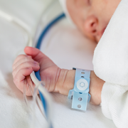
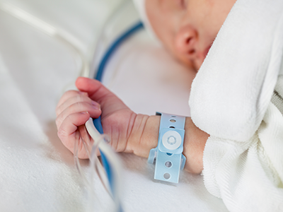
Virginia Tech, Children’s National Hospital award $100,000 to fund collaborative cancer research pilot projects

Research & Innovation Campus

This pilot research program represents a growing academic research partnership between Children’s National and Virginia Tech. Last year, the two institutions announced that Virginia Tech will establish a biomedical research facility on the Children’s National Research & Innovation Campus.

Children’s National Hospital and Virginia Tech have awarded two $50,000 one-year pilot grants to multi-institutional teams of scientists for pediatric brain cancer research.

The inter-institutional program, which launched in December, promotes cross-disciplinary collaborations among researchers at both institutions. At Virginia Tech, the program is part of the Virginia Tech Cancer Research Alliance. Financial support for the program was provided by the Offices of the Physician-in-Chief and Chief Academic Officer at Children’s National, and by Virginia Tech’s Office of the Vice President for Health Sciences and Technology.

“We were delighted to see so many innovative and competitive research proposals for our first round of pilot grants in the area of brain cancer. By forging new research collaborations with our partners at Children’s National, we hope to make major strides in addressing one of the most common and devastating groups of cancers in children,” said Michael Friedlander, Virginia Tech’s vice president for health sciences and technology, and the executive director of the Fralin Biomedical Research Institute at VTC. “The pilot funding will bootstrap several programs to be able to acquire ongoing sustainable funding by providing the opportunity to test novel high impact ideas for new strategies for treating these disorders. There are simply too few good options for children in this space now and this partnership can change that for the better.”

The collaborative research initiative began through an agreement between the Fralin Biomedical Research Institute and the Children’s National Research Institute. The collaborative teams formed through a series of interactive discussions among Virginia Tech’s Cancer Research Alliance faculty members from the university’s Blacksburg and Roanoke campuses, and Children’s National’s neuro-oncology researchers.

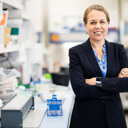
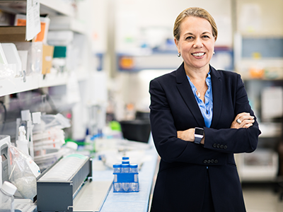
“I am extremely excited by this collaboration between VT and CNH that is focused on pediatric brain tumors which is such an area of unmet need,” said Catherine Bollard, M.D., M.B.Ch.B.,, director of Children’s National’s Center for Cancer and Immunology Research. “I am confident that the funded proposals will soon advance our understanding of pediatric brain tumors and, more importantly, facilitate more joint efforts between two world-class institutions which is especially timely with the development of the Children’s National Research & Innovation Campus.”

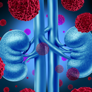
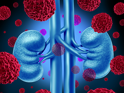
Yanxin Pei, Ph.D., an assistant professor in the Center for Cancer Immunology Research at Children’s National, and Liwu Li, Ph.D., a professor of biological sciences in Virginia Tech’s College of Science, were awarded one of the pilot research grants to study how white blood cells called neutrophils are involved in metastatic MYC-driven medulloblastoma, an aggressive type of brain tumor in children that often resists conventional radiation and chemotherapies.

Yuan Zhu, Ph.D., the Gilbert Family Professor of Neurofibromatosis Research at Children’s National, and Susan Campbell, Ph.D., an assistant professor of animal and poultry sciences in Virginia Tech’s College of Agriculture and Life Sciences, were awarded funds to study glioma-induced seizures in mice with a genetic mutation that inhibits the production of P53, a key protein involved in suppressing cancer cell growth and division.

The successful applicants will receive funding starting this month and are expected to deliver preliminary data to support an extramural research application by 2024.

This pilot research program represents a growing academic research partnership between Children’s National and Virginia Tech. Last year, the two institutions announced that Virginia Tech will establish a biomedical research facility on the Children’s National Research & Innovation Campus. It will be the first research and innovation campus in the nation focused on pediatrics when it opens later this year and will house newly recruited teams of pediatric brain cancer researchers.

Liwu Li, Yanxin Pei, Susan Campbell, and Yuan Zhu

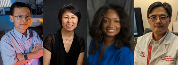
Liwu Li, Ph.D., Yanxin Pei, Ph.D., Susan Campbell, Ph.D., and Yuan Zhu, Ph.D., were awarded funding through the new pilot research program.

neuron on teal background

Primary cilia safeguard cortical neurons from environmental stress-induced dendritic degeneration

neuron on teal background

Fetus and neonates are under the risk of exposure to various external agents, such as alcohol and anesthetics taken by the mother. However, primary cilia can protect neurons by activating cilia-localized molecular signaling that inhibits degeneration of neuronal processes, according to the study’s findings.

A new study led by Kazue Hashimoto-Torii, Ph.D. and Masaaki Torii, Ph.D., both principal investigators for the Center for Neuroscience Research at Children’s National Hospital, found that primary cilia – tiny hair-like protrusions from the body of neuronal cells – protect neurons in the developing brain from adverse impacts of prenatal exposure.

Fetus and neonates are under the risk of exposure to various external agents, such as alcohol and anesthetics taken by the mother. However, primary cilia can protect neurons by activating cilia-localized molecular signaling that inhibits degeneration of neuronal processes, according to the study’s findings.

“Remarkably, the developing brain is equipped with intrinsic cell protection that helps to minimize the adverse impacts of to various external agents,” said Dr. Hashimoto-Torii. “However, the mechanisms of such protection have been unclear. Our study provides the first evidence that the tiny hair-like organelle protects neurons in the perinatal brain from adverse impacts of such external agents taken by the mother.”

The findings suggest that subtle alterations in primary cilia due to genetic conditions may lead to various neurodevelopmental disorders if combined with exposure to external agents from the environment. The findings also suggest that ciliopathy patients who have abnormal ciliary function due to genetic causes may have increased risk of abnormal brain development upon exposure to external agents.

“Clarifying diverse roles of cilia provides essential information for clinicians and patients with potential deficits in primary cilia to take extra precautions to avoid the risks for long-term negative impacts of external factors,” Dr. Torii explained. “We hope that further studies will define the whole picture of cilia-mediated neuroprotection and help us to advance our understanding of its importance in the pathogenesis of neurodevelopmental disorders.

This may ultimately lead to the development of treatment for various neurodevelopmental disorders,” he added.

The uniqueness of the study stems from the investigation of the role of cilia in brain development at the risk of exposure to various external factors that occur in the real world. Little is known about how the normal and abnormal brain development progresses in an environment where many external factors interact with intrinsic cellular mechanisms.

The study is a collaboration with researchers at Yale University and Keio University, Japan. Other Children’s National researchers who contributed to this study include Seiji Ishii, Ph.D.; Nobuyuki Ishibashi, M.D.; Toru Sasaki, M.D., Ph.D.; Shahid Mohammad, Ph.D.; Hye Hwang; Edwin Tomy; and Fahad Somaa.

Research & Innovation Campus

Children’s National pain expert and innovator shares global summit spotlight

Research & Innovation Campus

As a Johnson & Johnson Innovation Quickfire Children’s Challenge awardee, Dr. Finkel and AlgometRx will be among the first group of startups taking up residence at the new JLABS @ Washington, DC, located on the Children’s National Research & Innovation Campus, when it opens in 2021 at the historic former Walter Reed Army Medical Center site.

Medical technology innovator Julia Finkel, M.D., principal investigator for the Pain Medicine Initiative of the Sheikh Zayed Institute for Pediatric Surgical Innovation at Children’s National Hospital, recently participated in Galen Growth’s 2020 Global Healthtech Summit on a virtual panel featuring resident companies from Johnson & Johnson Innovation – JLABS who are utilizing artificial intelligence (AI) with the aim to create advanced solutions for diagnostics, treatment and clinical trials. The summit, hosted in Singapore, brought the innovators together to discuss their views on their progress, the challenges and opportunities for bringing medtech innovations to market in the current climate, as well as the tools needed to succeed.

Dr. Finkel’s innovation, AlgometRx, is a real-time pain measurement technology that captures a digital image of a patient’s pupillary response to a non-invasive stimulus and applies proprietary algorithms to measure pain type and intensity. AlgometRx, a spin-off of Children’s National, recently received a JLABS @Washington DC Quickfire Children’s Challenge award.

Joining Dr. Finkel on the panel were JLABS resident company leaders Don Crawford, CEO, Analytics 4 Life; Jim Havelka, CEO, Inform AI; and Kim Walpole, CEO, Trials.ai, which leverages AI to help research teams design more effective clinical trials. The 50-minute program, moderated by Kara Bortone, senior director, Portfolio and Sourcing Management, Johnson & Johnson Innovation – JLABS, focused on topics such as how these startups approached the market and regulatory processes as well as the up-and-coming trends in health technology.

A pediatric anesthesiologist, Dr. Finkel explained the significance of achieving real-time, objective pain measurement. “Pain is one word that represents a myriad of conditions,” she says. “Pain from acute post-operative conditions is very different from peripheral neuropathic pain and different from the type of inflammatory pain seen in lupus and rheumatoid arthritis. Being able to discern the drivers of pain, the etiology, is essential to treating it well and to developing better therapeutics in the future.”

Dr. Finkel points out that AlgometRx measures nociception, which is pain fiber activation, and that is also what medications are addressing. “We’re not discounting a patient’s perception of pain, as we recognize that one’s experience of pain is very complex,” she says. “What we aim to measure is the activity being transmitted by the pain nerve and the type of nerve fiber that is doing the transmitting.”

Aiming to identify pain phenotypes is an important part of current AlgometRx development work, says Dr. Finkel, as it could significantly aid clinical decision-making in treating and monitoring patients’ pain. The company’s current regulatory focus is to seek FDA clearance related to its potential use for patients with peripheral neuropathy, which is pain and numbness resulting from damage to the nerves outside of the brain and spinal cord. The company has also identified fibromyalgia cases as a place where the technology could potentially benefit a large number of patients as it considers regulatory clearance targets.

As the COVID-19 pandemic presented many unique challenges to healthcare startups this year, panel participants were asked to discuss the hurdles they faced and how it impacted device development.

Dr. Finkel notes that the pandemic slowed patient enrollment in AlgometRx clinical studies, but also presented some upside. “At first, that had a negative impact, but it wound up being a good thing,” she says. “It gave us a moment to pause, regroup and examine the data we’d already generated. That break gave us improved information and a new, more powerful approach. It changed our trajectory by altering our regulatory path in terms of the order of things in our pipeline, so we’ve been enormously productive.”

As a Johnson & Johnson Innovation Quickfire Children’s Challenge awardee, Dr. Finkel and AlgometRx will be among the first group of startups taking up residence at the new JLABS @ Washington, DC, located on the Children’s National Research & Innovation Campus, when it opens in 2021 at the historic former Walter Reed Army Medical Center site. Along with a one-year residency at the new JLABS @ Washington DC facility,* AlgometRx will receive mentorship from experts at the Johnson & Johnson Family of Companies and grant funding to help support its continued advancement to commercialization.

*Residency at JLABS @ Washington subject to acceptance and execution of a License Agreement with Children’s National.

feeding tubes

NIH grant funds development of pediatric feeding tube placement device

feeding tubes

A new grant will help to finalize development of the Pediatric PUMA-G System, the world’s first and only ultrasound-based procedure for placing feeding tubes into the stomach.

Researchers at Children’s National Hospital have received grant funding from the National Institute of Diabetes and Digestive and Kidney Diseases, within the National Institutes of Health (NIH), to finalize development of the Pediatric PUMA-G System, the world’s first and only ultrasound-based procedure for placing feeding tubes into the stomach. The funding will also support the first clinical trial of this technology in pediatric patients.

“Children’s National was chosen because we have a strong record of innovating pediatric devices and surgical procedures through the Sheikh Zayed Institute and we have a busy clinical interventional radiology service,” says Karun Sharma, M.D., Ph.D., director of Interventional Radiology and associate director of clinical translation at the Sheikh Zayed Institute for Pediatric Surgical Innovation (SZI) at Children’s National. “We are proud to be a part of this collaboration that will potentially help improve care of pediatric patients who cannot tolerate feeding by mouth.”

The feeding tubes are vital for children who cannot eat or swallow and require liquid nutrition (known as enteral feeding). Common feeding tube placement procedures for children may expose them to risks from invasive surgical tools or from ionizing radiation, which may lead to cancer in young patients at elevated rates. The PUMA-G System is less invasive and uses ultrasound to help physicians image the body during the procedure.

The grant, totaling $1.6M, will clinically evaluate the Pediatric PUMA-G System in collaboration with CoapTech, a biotechnology medical device company and two other premier pediatric medical centers — New York-Presbyterian Morgan Stanley Children’s Hospital and Children’s Hospital of Philadelphia.

Associations Between Resting State Functional Connectivity and Behavior in the Fetal Brain

Maternal anxiety affects the fetal brain

Associations Between Resting State Functional Connectivity and Behavior in the Fetal Brain

Anxiety in gestating mothers appears to affect the course of brain development in their fetuses, changing neural connectivity in the womb, a new study suggests.

Anxiety in gestating mothers appears to affect the course of brain development in their fetuses, changing neural connectivity in the womb, a new study by Children’s National Hospital researchers suggests. The findings, published Dec. 7, 2020, in JAMA Network Open, could help explain longstanding links between maternal anxiety and neurodevelopmental disorders in their children and suggests an urgent need for interventions to diagnose and decrease maternal stress.

Researchers have shown that stress, anxiety or depression in pregnant mothers is associated not only with poor obstetric outcomes but also social, emotional and behavioral problems in their children. Although the care environment after birth complicates the search for causes, postnatal imaging showing significant differences in brain anatomy has suggested that these problems may originate during gestation. However, direct evidence for this phenomenon has been lacking, says Catherine Limperopoulos, Ph.D., director of the Developing Brain Institute at Children’s National.

To help determine where these neurological changes might get their start, Dr. Limperopoulos, along with staff scientist Josepheen De Asis-Cruz, M.D., Ph.D., and their Children’s National colleagues used a technique called resting-state functional magnetic resonance imaging (rs-fMRI) to probe developing neural circuitry in fetuses at different stages of development in the late second and third trimester.

The researchers recruited 50 healthy pregnant volunteers from low-risk prenatal clinics in the Washington, D.C. area who were serving as healthy “control” volunteers in a larger study on fetal brain development in complex congenital heart disease. These study participants, spanning between 24 and 39 weeks in their pregnancies, each filled out widely used and validated questionnaires to screen for stress, anxiety and depression. Then, each underwent brain scans of their fetuses that showed connections between discrete areas that form circuits.

After analyzing rs-fMRI results for their fetuses, the researchers found that those with higher scores for either form of anxiety were more likely to carry fetuses with stronger connections between the brainstem and sensorimotor areas, areas important for arousal and sensorimotor skills, than with lower anxiety scores. At the same time, fetuses of pregnant women with higher anxiety were more likely to have weaker connections between the parieto-frontal and occipital association cortices, areas involved in executive and higher cognitive functions.

“These findings are pretty much in keeping with previous studies that show disturbances in connections reported in the years and decades after birth of children born to women with anxiety,” says Dr. De Asis-Cruz. “That suggests a form of altered fetal programming, where brain networks are changed by this elevated anxiety even before babies are born.”

Whether these effects during gestation themselves linger or are influenced by postnatal care is still unclear, adds Dr. Limperopoulos. Further studies will be necessary to follow children with these fetal differences in neural connectivity to determine whether these variations in neural circuitry development can predict future problems. In addition, it’s unknown whether easing maternal stress and anxiety can avoid or reverse these brain differences. Dr. Limperopoulos and her colleagues are currently studying whether interventions that reduce stress could alter the trajectory of fetal neural development.

In the meantime, she says, these findings emphasize the importance of making sure pregnant women have support for mental health issues, which helps ensure current and future health for both mothers and babies.

“Mental health problems remain taboo, especially in the peripartum period where the expectation is that this is a wonderful time in a woman’s life. Many pregnant mothers aren’t getting the support they need,” Dr. Limperopoulos says. “Changes at the systems level will be necessary to chip away at this critical public health problem and make sure that both mothers and babies thrive in the short and long term.”

Other Children’s National researchers who contributed to this study include Dhineshvikram Krishnamurthy, M.S., software engineer; Li Zhao, Ph.D., research faculty; Kushal Kapse, M.S., staff engineer; Gilbert Vezina, M.D., neuroradiologist; Nickie Andescavage, M.D., neonatologist; Jessica Quistorff, M.P.H., clinical research program lead; and Catherine Lopez, M.S., clinical research program coordinator.

This study was funded by R01 HL116585-01 from the National Heart, Lung, and Blood Institute and U54HD090257 from the Intellectual and Developmental Disabilities Research Center.

Roger Packer

Roger Packer, M.D., receives Lifetime Achievement Award

Roger Packer

“I am very honored and humbled to receive this recognition from the International Symposium on Pediatric Neuro-Oncology,” says Roger Packer, M.D. “I am proud of the contributions my team and I have made in this field and we look forward to continue to lead research focused on the advancement of the crucial areas neuro-oncology.”

Roger Packer, M.D., senior vice president of the Center for Neurosciences and Behavioral Medicine at Children’s National Hospital, will receive the 2020 Lifetime Achievement Award from the International Symposium on Pediatric Neuro-Oncology. Dr. Packer was selected as a recipient for the prestigious award for his substantial contributions to pediatric oncology and scientific achievements.

“I am very honored and humbled to receive this recognition from the International Symposium on Pediatric Neuro-Oncology,” says Dr. Packer. “I am proud of the contributions my team and I have made in this field and we look forward to continue to lead research focused on the advancement of the crucial areas neuro-oncology.”

Dr. Packer is also a Gilbert Distinguished Professor of Neurofibromatosis and is Director of both the Gilbert Neurofibromatosis Institute and the Brain Tumor Institute of Children’s National Hospital. Most of the current studies Dr. Packer coordinates are studies evaluating innovative agents aimed at the molecular underpinnings of neurologic disease. He has published over 400 original articles and 350 reviews and chapters.

The award will be presented at ISPNO 2020, the 19th International Symposium on Pediatric Neuro-Oncology, December 13-16, 2020, in Karuizawa, Japan.

Children’s National Hospital is incredibly proud of the work Dr. Packer has done in the neuro-oncology community.

graphic abstract for brain tumor paper

First large-scale proteogenomic analysis offers insights into pediatric brain tumor biology

graphic abstract for brain tumor paper

In the first large-scale, multicenter study of its kind, researchers conducted comprehensive analysis yielding a more complete understanding of pediatric brain tumors (PBT), which are the leading cause of cancer-related deaths in children. Researchers from the Clinical Proteomic Tumor Analysis Consortium (CPTAC) and Children’s Brian Tumor Network (CBTN) generated and analyzed proteomic data, which identifies common biological characteristics among different tumor types. The consortia consist of collaborators from the Icahn School of Medicine at Mount Sinai, National Cancer Institute, Fred Hutchinson Cancer Research Center, Children’s National Hospital and Children’s Hospital of Philadelphia. The study, published in Cell on November 25, 2020, provides a clearer understanding of the molecular basis of pediatric brain tumors and proposes new therapeutic avenues.

The molecular characterization of brain tumors has largely hinged upon the presence of unique alterations in the tumor genome ignoring the many layers of regulation that exist between DNA and the functional biology of the tumor cell that is actuated by proteins. The integration of proteomic data identifies common biological themes that span histologic boundaries, suggesting that treatments used for one histologic type may be applied effectively to other tumors sharing similar proteomic features.

Brian Rood, M.D., medical director of the Brain Tumor Institute and associate professor of pediatrics in the Center for Cancer and Blood Disorders at Children’s National Hospital, participated in this study and explains the importance of what the team discovered.

Q: Why was it important that researchers came together to do this work?

A: Comprehensive characterization of the fundamental biology of pediatric brain tumors, including the proteogenomic analysis done in this study, is essential to better understand and treat pediatric brain tumors.

Our study is based on the recognition that proteomics and phosphoproteomics needs to be integrated with other omics data to gain an improved systems biology view of the molecular features of brain tumors. In addition, characterizing biological themes that cross histologic boundaries and cells of origin can suggest extending treatments shown to be effective in one type of tumor to other histologically disparate tumors sharing the same proteomic features.

Proteomic data further reveal the functional impacts of somatic mutations and copy number variations (CNVs) not evident in transcriptomic data alone. Further, kinase-substrate network analyses identify activated biological mechanisms of tumor biology.

This work was only possible because of a unique collaboration between the CPTAC program of the NCI and the CBTN, of which Children’s National is a member.

Q: How will this work advance understanding and treatment of pediatric brain tumors?

A: Pediatric brain tumors have not benefitted from molecularly targeted drugs as much as other tumor types largely because they harbor relatively few gene mutations. Therefore, identifying key pathways to target in these patients’ tumors has been a challenge. The integration of proteomic and phosphoproteomic data with genomic data allows for the construction of a more comprehensive model of brain tumor biology and nominates specific key pathways to be targeted.

Q: What did you find that excites you?

A: Proteomic data revealed a number of findings that were not present in the genomic data. We found evidence to support a molecularly targeted approach to treating craniopharyngioma, a tumor that has previously been unresponsive to chemotherapy. We also found a prognostic marker for high grade gliomas that do not have a mutation in the H3 histone. We were able to identify specific kinases that may dictate the aggressive nature of certain ependymoma tumors. Importantly, we demonstrated the potential of proteomic studies to uncover unique tumor biology, paving the way for more extensive investigations using this approach.

You can find the full study published in Cell. Learn more about the Brain Tumor Institute at Children’s National.


Dr. Rood recently joined a live panel discussion with researchers from the Children’s Brain Tumor Network and the Clinical Proteomic Tumor Analysis Consortium to explore the impact of their landmark study.

pile of plastic bottles

The linkage between chemicals used in plastics and cardiovascular disease

pile of plastic bottles

For people across the globe, plastics are synonymous with modern life and it’s impossible to avoid exposure to them, including clinical environments where a variety of frequently used materials, such as tubing and blood storage bags, are made from plastics.

For people across the globe, plastics are synonymous with modern life and it’s impossible to avoid exposure to them, including clinical environments where a variety of frequently used materials, such as tubing and blood storage bags, are made from plastics. Led by Nikki Posnack, Ph.D, principal investigator at The Sheikh Zayed Institute for Pediatric Surgical Innovation at Children’s National Hospital, a team of Children’s National researchers has been studying the potential effects of chemicals found in plastics, such as BPA and DEHP, as possible contributors to cardiovascular disease.

Along with conducting proprietary studies of the potential effects, Posnack and her team recently reviewed available scientific studies to further identify and illuminate the potential links between exposure to the synthetic additives contained in plastics and cardiovascular mortality. The article was published this month in Nature Reviews Cardiology.

In the article Posnack cites a 10-year longitudinal study with the finding that high exposure to BPA was associated with a 46-49% higher hazard ratio for cardiovascular and all-cause mortality, compared with low exposure to BPA.

“Plastics may be indispensable materials, but their ubiquity does raise concerns about the effects of our continuous exposure to plasticizer additives like di(2-ethylhexyl) phthalate (DEHP) and synthetic chemicals used to create polymers like BPA,” said Posnack. “Although disease causation can be difficult to pinpoint in population and epidemiological studies, experimental work has clearly demonstrated a direct link to plastic chemicals and cardiac dysfunction. It is clear that future collaborative endeavors are necessary to bridge the gap between experimental, epidemiological and clinical investigations to resolve the impact of plastics on cardiovascular health.”

Nikki Gillum Posnack

Nikki Posnack, Ph.D, principal investigator at The Sheikh Zayed Institute for Pediatric Surgical Innovation at Children’s National Hospital.

Posnack added that, given the omnipresence of plastics and their related chemicals, biomonitoring studies have reported detectable levels of DEHP and BPA in 75-90% of the population. Occupational or clinical environments can also result in elevated exposures to these dangerous chemicals. Previous epidemiological studies have reported links between elevated urinary levels of phthalate or bisphenol, common additives in plastic, and an increased risk of coronary and peripheral artery disease, chronic inflammation, myocardial infarction, angina, suppressed heart rate variability and hypertension.

Additionally, available research has shown that incomplete polymerization or degradation of BPA-based plastic products can result in unsafe human exposure to BPA. Despite these links, the article points out, both BPA and DEHP are still manufactured in high volumes and are used to produce a wide variety of consumer and commercial products.

Further exploring implications for pediatrics, a June 2020 article published by Posnack in Birth Defects Research looks at the potential effects of plastic chemicals on the cardiovascular health of fetal, infant and pediatric groups. The article highlighted experimental work that suggests plasticizer chemicals such as bisphenols and phthalates may exert negative influence on pediatric cardiovascular health. The article systematically called out areas of concern supported by research findings. Also addressing current gaps in knowledge, Posnack outlined future research endeavors that would be needed to resolve the relationship between chemical exposures and the impact on pediatric cardiovascular physiology.

In related work, Posnack and her team are expanding their work on plastics used in blood bags to also investigate the role of blood storage duration on health outcomes. A recently published first study demonstrates that “older” blood products (stored 35 or more days) directly impact cardiac electrophysiology, using experimental models. Published October 22, 2020 in the Journal of the American Heart Association, the study concludes that the cardiac effects are likely caused by biochemical alterations in the supernatant from red blood cell units that occur over time, including but not limited to, hyperkalemia (elevated potassium levels).

newborn in ICU

Cardiac technology advances show promise for kids but only if right-sized

newborn in ICU

“Smaller patients, and those with congenital heart disease, can benefit from minimally-invasive methods of delivering pacemakers and defibrillators without the need for open-chest surgery,” says Charles Berul, M.D.

How to address the growing need for child-sized pacemakers and defibrillators, and finding better surgical techniques to place them, is the topic of an invited session called The Future is Now (or Coming Soon): Updates on New Technologies in Congenital Heart Care at the 2020 American Heart Association Scientific Sessions.

“Smaller patients, and those with congenital heart disease, can benefit from minimally-invasive methods of delivering pacemakers and defibrillators without the need for open-chest surgery,” says Charles Berul, M.D., co-director of the Children’s National Heart Institute and chief of Cardiology at Children’s National Hospital, who presented at the session.

“This unmet need can only be met by innovative pediatric research, geared towards miniaturization technologies for use in the smallest of children,” he says.

His presentation focused on the devices and approaches that have caught the attention of pediatric cardiology, such as pacemakers and subcutaneous defibrillators designed without lead wires, as well as less-invasive surgical approaches that may reduce recovery time for children with congenital heart disease who require these assist devices.

Using them in kids comes with added challenges, however. Often pediatric cardiologists have to be creative in how to make them work for smaller patients, Dr. Berul notes. This reiterates the important point that simply applying an adult technology to a child isn’t the right approach. The subcutaneous defibrillator, for example, is still pretty large for a child’s body. Some studies also show these devices may not be as accurate in children as in adults.

Investigators in the Sheikh Zayed Institute working together with the cardiologists at Children’s National Hospital are focused on product development and commercialization of tools and techniques to allow percutaneous minimally-invasive placement of devices, taking advantage of the newest devices and surgical techniques as they develop.

In his presentation, Dr. Berul stressed that as the technology for adults advances, it creates an opportunity for pediatric cardiology, but only if the devices, and the techniques to place them, are specifically redesigned for pediatric application.

American Heart Association Scientific Sessions 2020
The Future is Now (or Coming Soon): Updates on New Technologies in Congenital Heart Care – On Demand Session
CH.CVS.715
9:00am – 10:00am
Fri, Nov 13  (CST)

illustration of the brain

New research provides glimpse into landscape of the developing brain

illustration of the brain

Stem and progenitor cells exhibit diversity in early brain development that likely contributes to later neural complexity in the adult cerebral cortex, this according to a new study in Science Advances. This research expands on existing ideas about brain development, and could significantly impact the clinical care of neurodevelopmental diseases in the future.

Stem and progenitor cells exhibit diversity in early brain development that likely contributes to later neural complexity in the adult cerebral cortex, this according to a study published Nov. 6, 2020, in Science Advances. Researchers from the Center for Neuroscience Research (CNR) at Children’s National Hospital say this research expands on existing ideas about brain development, and could significantly impact the clinical care of neurodevelopmental diseases in the future. The study was done in collaboration with a research team at Yale University led by Nenad Sestan, M.D, Ph.D.

“Our study provides a new glimpse into the landscape of the developing brain. What we are seeing are new complex families of cells very early in development,” says Tarik Haydar, Ph.D., director of CNR at Children’s National, who led this study. “Understanding the role of these cells in forming the cerebral cortex is now possible in a way that wasn’t possible before.”

The cerebral cortex emerges early in development and is the seat of higher-order cognition, social behavior and motor control. While the rich neural diversity of the cerebral cortex and the brain in general is well-documented, how this variation arises is relatively poorly understood.

“We’ve shown in our previous work that neurons generated from different classes of cortical stem and progenitor cells have different functional properties,” says William Tyler, Ph.D., CNR research faculty member and co-first author of the study. “Part of the reason for doing this study was to go back and try to classify all the different progenitors that exist so that eventually we can figure out how each contributes to the diversity of neurons in the adult brain.”

Using a preclinical model, the researchers were able to identify numerous groups of cortical stem and precursor cells with distinct gene expression profiles. The team also found that these cells showed early signs of lineage diversification likely driven by transcriptional priming, a process by which a mother cell produces RNA for the sole purpose of passing it on to its daughter cells for later protein production.

Tarik Haydar

“Our study provides a new glimpse into the landscape of the developing brain. What we are seeing are new complex families of cells very early in development,” says Tarik Haydar, Ph.D., director of CNR at Children’s National, who led this study. “Understanding the role of these cells in forming the cerebral cortex is now possible in a way that wasn’t possible before.”

Using novel trajectory reconstruction methods, the team observed distinct developmental streams linking precursor cell types to particular excitatory neurons. After comparing the dataset of the preclinical model to a human cell database, notable similarities were found, such as the surprising cross-species presence of basal radial glial cells (bRGCs), an important type of progenitor cell previously thought to be found mainly in the primate brain.

“At a very high level, the study is important because we are directly testing a fundamental theory of brain development,” says Zhen Li, Ph.D., CNR research postdoctoral fellow and co-first author of the study. The results add support to the protomap theory, which posits that early stem and progenitor diversity paves the way for later neuronal diversity and cortical complexity. Furthermore, the results also hold exciting translational potential.

“There is evidence showing that neurodevelopmental diseases affect different populations of the neural stem cells differently,” says Dr. Li. “If we can have a better understanding of the complexity of these neural stem cells there is huge implication of disease prevention and treatment in the future.”

“If we can understand how this early landscape is affected in disorders, we can predict the resulting changes to the cortical architecture and then very narrowly define ways that groups of cells behave in these disorders,” adds Dr. Haydar. “If we can understand how the cortex normally achieves its complex architecture, then we have key entry points into improving the clinical course of a given disorder and improving quality of life.”

Future topics the researchers hope to study include the effects of developmental changes on brain function, the origin and operational importance of bRGCs, and the activity, connections and cognitive features enabled by different families of neurons.

Catherine Bollard

Catherine Bollard, M.D., M.B.Ch.B., named next editor-in-chief of Blood Advances

Catherine Bollard

“As editor-in-chief, I will aim to capture new developments in the hematology field, including immunology, immunotherapy, cell therapy, gene and cell therapy and transplant,” said Dr. Bollard.

The American Society of Hematology (ASH) has selected Catherine Bollard, M.D., M.B.Ch.B., director of the Center for Cancer and Immunology Research at the Children’s National Research Institute as the next editor-in-chief of its journal Blood Advances.

Blood Advances is a peer-reviewed online open access journal published by ASH that covers the latest developments in basic, translational and clinical hematology. Dr. Bollard’s term as editor-in-chief will begin in September 2021. She was selected by the ASH Executive Committee after a competitive international search.

Blood Advances has a broad presence, a large and growing number of manuscript submissions, and an engaged readership,” said Dr. Bollard. “I am so looking forward to continuing to build and strengthen the journal as its editor-in-chief.”

Since its launch in 2016, Blood Advances has taken advantage of its digital, open-access publication model to emphasize multimedia and a rapid, continuous publication format. Under the leadership of founding editor-in-chief Robert Negrin, M.D., of Stanford University, the peer-reviewed journal has pioneered new means of interactive, collaborative discussion and achieved an impact factor of 4.910.

“As editor-in-chief, I will aim to capture new developments in the hematology field, including immunology, immunotherapy, cell therapy, gene and cell therapy and transplant,” said Dr. Bollard. Her vision for the future of Blood Advances also includes further expansion of the journal’s global reach as well as continued efforts to recruit an editorial team representing geographic, ethnic and gender diversity.

“We have tremendous opportunities for growth, and I think in order to grow we must consider what readers want, how we can provide quality service for authors and reviewers, and how we can establish our own identity as a journal,” she said.

Dr. Bollard is a hematologist whose research interests include developing cell and gene therapies for patients with cancer and underlying immune deficiencies. Recognized as a national and international leader in the bone marrow transplant, immunology and immunotherapy space, Dr. Bollard has an expansive understanding of cancer, immune deficiencies and viral infections in pediatric and adult patients.

In her role as director of the Center for Cancer and Immunology Research at Children’s National Hospital, she works to establish clinical and research programs focused on developing and bringing novel cell therapies from bench to bedside.

Blood Advances is an important hub for hypothesis-generating papers, pilot studies and case reports, commentaries and other educational materials of interest to hematologists everywhere,” said ASH President Stephanie Lee, M.D., of Fred Hutchinson Cancer Research Center. “I am confident that Dr. Bollard will continue leveraging the journal’s unique digital platform to maintain its tradition of excellence as she executes her vision as editor-in-chief. I look forward to seeing how the journal evolves under her leadership.”

Artificial Intelligence concept image

Thrombectomy can be efficient and safe in childhood stroke, new study finds

Artificial Intelligence concept image

A recent study adds to the growing evidence that mechanical thrombectomy can be effective and safe not only in adults, but also in childhood stroke.

Previous randomized trials proved the effectiveness of thrombectomy for large intracranial vessel occlusions in adults only. However, a recent retrospective study led by Monica S. Pearl, M.D., Neurointerventional Radiology Program director at Children’s National Hospital, finds that thrombectomy can be safely performed in carefully selected cases of childhood stroke. The study further shows that treated children have good neurological outcomes.

In the findings, Dr. Pearl and other leading experts discussed specific circumstances and important considerations to take into account when managing a child with acute ischemic stroke due to a large vessel occlusion.

“We are raising the bar for the expected level of care for children with acute ischemic stroke,” said Dr. Pearl. “Care should be multidisciplinary and involve stroke neurology, neuroradiology, neurointerventional radiology, neurosurgery, cardiology, hematology and ICU teams.”

Prior to the study, clear guidelines for patient selection, thrombectomy technique and periprocedural care did not exist for the pediatric population despite the proven success of mechanical thrombectomy in adults.

Through a case-based approach encompassing a broad range of ages and clinical presentations, Dr. Pearl and other leading experts presented select cases of acute ischemic stroke in children and discussed the nuances, risks, benefits and management plan for each child.

Many of the clinical scenarios highlighted unanswered questions in the management and treatment of children with acute ischemic stroke due to large vessel occlusion. The study adds to the growing evidence that mechanical thrombectomy can be effective and safe not only in adults, but also in childhood stroke.

“It’s exciting to be shaping management for children with acute ischemic stroke,” said Dr. Pearl. “We are serving as the model for individualized, patient-centered care with multidisciplinary specialists and institutional collaboration caring for children with acute ischemic stroke.”

However, Dr. Pearl and experts encourage caution because etiology in childhood stroke differs substantially from that in acute ischemic stroke in adults, with potentially major impact on procedure success and safety.

The mission of the Neurointerventional Radiology Program, a new effort at Children’s National, is to provide exceptional family-centered care and cutting-edge diagnostic and endovascular treatment options for children with neurovascular disorders. Dr. Pearl serves as the program’s full time, dedicated neurointerventional radiologist, a specialized expertise found only in a handful of other pediatric hospitals in the country.

You can find the full study published in JAHA. Learn more about the Children’s National Research Institute Center for Neuroscience Research.

communication network concept image

Children’s National joins international AI COVID-19 initiative

communication network concept image

Children’s National Hospital is the first pediatric partner to join an international initiative led by leading technology firm NVIDIA and Massachusetts General Brigham Hospital, focused on creating solutions through machine and deep learning to benefit COVID-19 healthcare outcomes.

Children’s National Hospital is the first pediatric partner to join an international initiative led by leading technology firm NVIDIA and Massachusetts General Brigham Hospital, focused on creating solutions through machine and deep learning to benefit COVID-19 healthcare outcomes. The initiative, known as EXAM (EMR CXR AI Model) is the largest and most diverse federated learning enterprise, comprised of 20 leading hospitals from around the globe.

Marius George Linguraru, D.Phil., M.A., M.Sc., principal investigator at the Sheikh Zayed Institute for Pediatric Surgical Innovation at Children’s National Hospital, noted that one of the core goals of the initiative is to create a platform which brings resources together, from a variety of leading institutions, to advance the care of COVID-19 patients across the board, including children.

“Children’s National Hospital is proud to be the first pediatric partner joining the world’s leading healthcare institutions in this collaboration to advance global health,” says Linguraru. “We are currently living in a time where rapid access to this kind of global data has never been more important — we need solutions that work fast and are effective. That is not possible without this degree of collaboration and we look forward to continuing this important work with our partners to address one of the most significant healthcare challenges in our lifetime.”

A recent systematic review and meta-analysis from Children’s National Hospital became another core contribution to understanding how children are impacted by COVID-19. Led by Linguraru and accepted to be published in Pediatric Pulmonology, it offers the first comprehensive summary of the findings of various studies published thus far that describe COVID-19 lung imaging data across the pediatric population.

The review examined articles based on chest CT imaging in 1,026 pediatric patients diagnosed with COVID-19, and concluded that chest CT manifestations in those patients could potentially be used to prompt intervention across the pediatric population.

Marius George Linguraru

“Children’s National Hospital is proud to be the first pediatric partner joining the world’s leading healthcare institutions in this collaboration to advance global health,” says Marius George Linguraru, D.Phil., M.A., M.Sc.

“Until this point, pediatric COVID-19 studies have largely been restricted to case reports and small case series, which have prevented the identification of any specific pediatric lung disease patterns in COVID-19 patients,” says Linguraru. “Not only did this review help identify the common patterns in the lungs of pediatric patients presenting COVID-19 symptoms, which are distinct from the signs of other viral respiratory infections in children, it also provided insight into the differences between children and adults with COVID-19.”

Earlier this month, NVIDIA announced the EXAM initiative had – in just 20 days – developed an artificial intelligence (AI) model to determine whether a patient demonstrating COVID-19 symptoms in an emergency room would require supplemental oxygen hours – even days – after the initial exam. This data ultimately aids physicians in determining the proper level of care for patients, including potential ICU placement.

The EXAM initiative achieved a machine learning model offering precise prediction for the level of oxygen incoming patients would require.

In addition to Children’s National Hospital, other participants included Mass Gen Brigham and its affiliated hospitals in Boston; NIHR Cambridge Biomedical Research Centre; The Self-Defense Forces Central Hospital in Tokyo; National Taiwan University MeDA Lab and MAHC and Taiwan National Health Insurance Administration; Tri-Service General Hospital in Taiwan; Kyungpook National University Hospital in South Korea; Faculty of Medicine, Chulalongkorn University in Thailand; Diagnosticos da America SA in Brazil; University of California, San Francisco; VA San Diego; University of Toronto; National Institutes of Health in Bethesda, Maryland; University of Wisconsin-Madison School of Medicine and Public Health; Memorial Sloan Kettering Cancer Center in New York; and Mount Sinai Health System in New York.

illustration of cancer cells attacking kidneys

Renal cell carcinoma study shows excellent short-term outcomes without adjuvant therapy

illustration of cancer cells attacking kidneys

Researchers found that patients with localized pediatric renal cell carcinoma have excellent short-term outcomes without adjuvant therapy.

Although renal cell carcinoma (RCC) is the second most common kidney cancer diagnosed in children and adolescents, guidance regarding its clinical management has been confined to retrospective case series, which were limited by reporting bias, varied treatment approaches and a lack of central pathology review to confirm the diagnosis.

Research conducted by the Children’s Oncology Group (COG) and led by Jeffrey Dome, M.D., Ph.D., vice president of the Center for Cancer and Blood Disorders at Children’s National Hospital, found that patients with localized pediatric RCC have excellent short-term outcomes without adjuvant therapy with 4-year overall survival estimates of 96% for patients with stage I disease, 100% for patients with stage II disease and 88% for patients with stage III disease.

“The results of this study provide important practical insights into the management of pediatric RCC,” said Dr. Dome. “Oncologists now have validation that a surgery-only approach is appropriate management for the majority of children and adolescents with RCC.”

Jeffrey Dome

“The results of this study provide important practical insights into the management of pediatric RCC,” said Jeffrey Dome, M.D., Ph.D.

The excellent survival in patients with stage III disease held up even in those with tumor involvement of regional lymph nodes, a finding that differs from adult RCC. However, patients with metastatic disease (stage IV), had a 4-year overall survival estimate of only 29%, demonstrating the need to find active treatments for this group. Outcomes varied according to tumor histological subtype. Nearly all recurrences occurred in patients with the translocation histology and renal medullary carcinoma; recurrences were rare in other subtypes.

A follow-up study called AREN1721 is now open in the Children’s Oncology Group and adult cancer cooperative groups that participate in the National Clinical Trials Network. This study involves a comparison of two treatment regimens for metastatic or unresectable “translocation” renal cell carcinoma, the most common subtype of renal cell carcinoma in children, adolescents and young adults. The treatment regimens will include nivolumab, a PD1 immune checkpoint inhibitor, with or without axitinib, a tyrosine kinase inhibitor that targets vascular endothelial growth factor receptor (VEGFR).

illustration of lungs surrounded by virus

COVID-19: First comprehensive review of pediatric lung imaging features

illustration of lungs surrounded by virus

A systematic review and meta-analysis by Children’s National Hospital researchers, published in Pediatric Pulmonology, provides the first comprehensive review of the findings of published studies describing COVID-19 lung imaging data in children.

The number COVID-19 studies focused on children have been small and with limited data. This has prevented the identification of specific pediatric lung disease patterns in COVID-19. Although children make up around 9.5% of COVID-19 infections, less than 2% of the literature on the virus, its symptoms and effects, have focused on kids.

A systematic review and meta-analysis by Children’s National Hospital researchers, published in Pediatric Pulmonology, provides the first comprehensive review of the findings of published studies describing COVID-19 lung imaging data in children. The analysis concludes that chest CT manifestations in children with COVID‐19 could potentially prompt intervention in the pediatric population.

Marius George Linguraru, D.Phil., M.A., M.Sc., principal investigator in the Sheikh Zayed Institute for Pediatric Surgical Innovation at Children’s National, discusses the importance of this work.

Q: What findings stand out to you?

A: We found that more than a third of children with COVID-19 had normal imaging. The lung imaging findings in these children were overall less frequent and less severe than in adult patients, but they were also more heterogeneous than in adults. Importantly, children with COVID-19 were three times more likely to have a normal exam than adults.

Several common lung imaging findings reported in adults were extremely rare or not found in the pediatric studies. These discoveries, and other recent reports in this space, support the fact that children’s symptoms may be less obvious than adults or even absent, but they still carry the virus and may be at risk for serious and life-threatening illness.

Marius George Linguraru

Marius George Linguraru, D.Phil., M.A., M.Sc., principal investigator in the Sheikh Zayed Institute for Pediatric Surgical Innovation at Children’s National.

Q: How will the findings of this study benefit pediatric care?

A: In our study, we showed how the health of the lungs of these children is impacted. Our results from data from 1,026 children (from newborns to 18 year old) with COVID-19 present chest manifestations that could potentially prompt informed intervention and better recovery.

Another conclusion of our study is that the abnormalities reported on the chest scans of children infected with COVID-19 are distinct from the typical lung images seen during other viral respiratory infections in the pediatric population. This is important for preparing for the cold and flu season.

Q: Why was this review important to our understanding of how COVID-19 impacts children?

A: This is the first systematic review and meta-analysis focused on the manifestation of the COVID-19 infection in the lungs of children. Our study, and others from colleagues at Children’s National, helps lead the efforts on elucidating how the pandemic affects the health of children.

Though children were initially thought to be less susceptible to infection, the data has made it clear that many children are at high risk for hospitalization and severe health complications. Although there are similarities between how children and adults are affected by the pandemic, there are also critical differences.

Given the limited knowledge in the manifestation of COVID-19 in children, with children susceptible to infection and hospitalization, and with children returning to school, continued efforts to understand the impact of COVID-19 on young patients is critically important. Understanding how children fare through the pandemic is the foundation of discovering better ways to take care of young patients and their health.

You can find the full study published in Pediatric Pulmonology. Learn more about the Sheikh Zayed Institute for Pediatric Surgical Innovation at Children’s National.

Annual Pediatric Device Innovation Symposium panelists

Accelerating pediatric device innovation through legislative processes and industry changes

Annual Pediatric Device Innovation Symposium panelists
While the way we deliver healthcare is changing rapidly, far too often the tools we use to treat children are stuck in the past.

Over the last decade, pediatric medical device innovation, particularly for the youngest, most fragile children, has made dismal progress. Of the Class 3 (high risk/high benefit) medical devices approved by FDA for pediatrics in the last 10 years, less than 4% are for ages 0-2 years old; and even less for neo-natal patients. Simply put, as medical devices advance, children are not seeing the benefit of innovation.

The 8th Annual Pediatric Device Innovation Symposium presented by Children’s National Hospital in conjunction with the National Capital Consortium for Pediatric Medical Devices (NCC-PDI) featured a keynote panel, “Pediatric Device Innovation: What’s Next?”, to examine the legislative and industry changes needed to speed up device innovation for kids.

One of the keynote panelists, and leading voices on this issue, is Children’s National Hospital president and CEO Kurt Newman, M.D. Dr. Newman, a former pediatric surgeon, knows firsthand that every day in our nation’s pediatric hospitals, surgeons are manipulating adult medical devices to create creative solutions for children’s bodies because it’s the only available option.

“Children need and deserve devices that are conceived and designed with their biology and future in mind,” says Dr. Newman. “While children may only make up a small percentage of our population – maybe 20 or 25% – they are 100% of our future.”

Dr. Kurt Newman in front of the capitol building

“Children need and deserve devices that are conceived and designed with their biology and future in mind,” says Children’s National Hospital president and CEO Kurt Newman, M.D. “While children may only make up a small percentage of our population – maybe 20 or 25% – they are 100% of our future.”

Dr. Newman also addressed the current barriers to pediatric device innovation, which ranges from limited pediatric clinical trials to a market size that’s not financially appealing.

“The truth is, the frontiers of pediatric medicine are really in the innovative treatments, devices, therapies, and cures awaiting us on the other side of research and development.,” says Dr. Newman.

Former CNN correspondent, Jeanne Meserve, moderated the 45-minute keynote panel discussion, asking questions about the challenges to pediatric innovation, what policy changes need to take place to see improvement in the field of pediatric device innovation, and how federal funding can assist in creating change.

Michelle McMurry-Heath, new CEO of DC-based Biotechnology Innovation Organization (BIO), who joined Dr. Newman on this keynote panel, agreed that more needs to be done in the pediatric space. Dr. McMurry-Heath believes the Food and Drug Administration (FDA) is a public health advocate at heart and that the Pediatric Device Consortia (PDC), which Children’s National Hospital is part of, is starting to make new advancements in pediatric innovation by giving FDA clearance to more start-up companies than we’ve seen in the past.

“The FDA is interested in is what improves the health outcomes for the people and innovation is a huge piece. This is an important part of their mission and it is starting to yield benefits,” says Dr. Michelle McMurry-Heath. “Innovation is a team sport – it’s not easy. It takes a village of expertise and collaboration to progress and projects like the Pediatric Device Consortia is an important piece in this puzzle.”

NCC-PDI is one of five consortia in the FDA’s Pediatric Device Consortia (PDC) Grant Program created to support the development and commercialization of medical devices for children and is led by the Sheikh Zayed Institute for Pediatric Surgical Innovation at Children’s National Hospital and the A. James Clark School of Engineering at the University of Maryland, with support from partners MedTech Innovator, BioHealth Innovation and design firm Archimedic.

To date, NCC-PDI has mentored over 100 medical device sponsors to help advance their pediatric innovations, with seven devices having received either their FDA market clearance or CE marking.

Dr. McMurry-Heath also addressed the challenge of diseases that don’t exist in adults and posed the question, “How do you create a device for kids if it doesn’t exist in adults?” She cited the lack of market in pediatrics and the difficulty in bringing a device to market as problems that hinder innovation, which is why advocating for these devices is crucial to children’s healthcare everywhere.

“So much of our innovation comes from our small, innovative companies,” say Dr. McMurry-Heath. “For example, my company is working on a COVID-19 tracker now and 70% of the innovation is coming from our smallest biotech companies. It’s a race against time for these companies to bring their innovation to market in order to keep the lights on and pay their scientists; this dog-eat-dog world isn’t immediately obvious to outsiders.

Beyond advocating, Dr. Newman and Children’s National are developing the first-of-its-kind pediatric research and innovation campus, which is currently under construction at DC’s former Walter Reed Army Medical Center site.

“We secured 12 acres to create something that has never been done before and that’s a campus for innovation dedicated to children,” says Dr. Newman. “Our close proximity to federal research institutions and agencies enables the new Children’s National campus to leverage the rich ecosystem of public and private sectors to help bolster biohealth, medical device, and life science innovation.”

As Children’s National continues to champion ways to accelerate pediatric device development, one focus is the on-site incubator Johnson & Johnson Innovation – JLABS, which will help start-up companies strengthen their ideas by working with coaches, having access to mentors and learning how to interact with the FDA. This partnership also offers an audience for their device which could potentially lead to investments.

The Children’s National Research & Innovation Campus will create an ecosystem that can accelerate breakthroughs in pediatric healthcare discoveries and technologies: The new campus is currently under construction and expected to open in the first quarter of 2021.

Both panelists agreed they’d like to see more flexibility with regulators to work with innovators in order create more incentives for them to present their device, like the NCC-PDI “Make Your Medical Device Pitch for Kids!” Competition, which was recently held in September 2020. The six winners received up to $50,000 in FDA-funded grant awards in order to develop their device, eventually bring it to market in order to improve healthcare for kids.

sick child in palliative care hospital bed

Children’s National Research Institute receives NIH grant for palliative care study

sick child in palliative care hospital bed

A new NIH grant will support the first study that examines palliative care needs in pediatric rare disease community.

The National Institute of Health (NIH) has awarded $500,875 to the Children’s National Research Institute (CNRI), the academic arm of Children’s National Hospital, to support a new study examining the palliative care needs of children living with rare genetic diseases.

This is the first study of families of children with genetic and metabolic conditions, termed collectively as rare diseases, that is designed to intervene to support the well-being of family caregivers and create advance care plans for future medical decision making. In the United States, a rare disease is defined as a particular condition affecting fewer than 200,000 people. Pediatric patients with rare diseases experience high mortality rates, with 30 percent not living to see their fifth birthday.

“Children with ultra-rare or complex rare disorders are routinely excluded from research studies because of their conditions, creating a significant health disparity. Surveys show that families of children with rare diseases are adversely impacted by lack of easy access to peer and psychological support,” says Maureen Lyon, Ph.D., Clinical Health Psychologist and Professor of Pediatrics at the CNRI and principal investigator on the project. “This study will examine the palliative care needs of family caregivers of children with rare genetic disorders and advance care planning intervention, which will ultimately help facilitate discussions about future medical care choices that families are likely to be asked to make for their child.”

Although greatly needed, there are few empirically validated interventions to address these issues Currently, there is only one intervention described for families of children with rare diseases — a Swedish residential, competence program — which focuses on active coping. However, this intervention does not address pediatric advance care planning, a critical aspect of palliative care.

Lyon adds that the major benefit of this proposed project will be filling the gap in knowledge about what family caregivers of medically fragile children with rare diseases want with respect to palliative care. In the United States, these families are expected to provide a level of care that, until a few decades ago, was reserved for hospitals.

Maureen E Lyon

Maureen Lyon, Ph.D., Clinical Health Psychologist and Professor of Pediatrics at the CNRI and principal investigator on the project.

“Our hope is that this study will provide a structured model for facilitating family decisions about end-of-life care, for those families who do not have the good fortune to have children who have the capacity to share in decision-making,” Lyon says.

In addition to bridging the knowledge gap regarding palliative care in rare disease patients, the study will also help inform current clinical, ethical and policy discussions, as well as the legal issues in a variety of areas, such as the debate surrounding advocacy, particularly for those children with impairments in physical function.

“We look forward to the results of this study,” said Marshall Summar, M.D., director of the Rare Disease Institute and division chief, Genetics and Metabolism at Children’s National Hospital. “As a leader in rare disease care, we continually examine how we can improve care and support for our patient families at our clinic and want to share our findings with others engaged in caring for rare disease patients. Because rare diseases can be life limiting in some cases, we need to learn all we can about how best to care and support a patient and family as they prepare for a potential transition to palliative care.”

All research at Children’s National Hospital is conducted through the CNRI, including translational, clinical and community studies. The CNRI also oversees the educational activities and academic affairs of the hospital and the Department of Pediatrics at the George Washington University School of Medicine and Health Sciences, frequently partnering with many other research institutions regionally and nationally. CNRI conducts and promotes translational and clinical medical research and education programs within Children’s National Hospital that lead to improved understanding, prevention, treatment and care of childhood diseases.

Pediatric Device Innovation Symposium graphic

Real-world evidence and the impact on pediatric device innovation

Pediatric Device Innovation Symposium graphic

The 8th Annual Pediatric Device Innovation Symposium presented by @ChildrensNatl in conjunction with @Devices4kids took place Sept. 28-30.

The 8th Annual Pediatric Device Innovation Symposium presented by Children’s National Hospital in conjunction with the National Capital Consortium for Pediatric Medical Devices (NCC-PDI) kicked off on Monday, Sept. 28, 2020 with a panel featuring three fellow members of the FDA-funded Pediatric Device Consortia (PDC) Grants Program discussing real-world evidence and the vital role that innovation and technology play in advancing healthcare for the pediatric population.

As described by the FDA, real-world evidence (RWE) is the clinical evidence regarding the usage and potential benefits or risks of a medical product, derived from the analysis of patient data. RWE can be generated by different study designs or analyses, including but not limited to, randomized trials, including large simple trials, pragmatic trials and observational studies (prospective and/or retrospective).

The symposium panel, “Pediatric Device Consortia Update on the Use of Real-World Evidence (RWE) for Pediatric Device Innovation” examined real-world evidence (RWE) demonstration projects from Southwest Pediatric Device Consortium, UCSF-Stanford Pediatric Device Consortium and the West Coast Consortium for Technology and Innovation in Pediatrics (CTIP). The panel was moderated by Juan Espinoza, M.D., FAAP, director of CTIP.

“Real-world evidence projects are critical to the advancement of pediatric medical device innovation,” said Kolaleh Eskandanian, Ph.D., M.B.A., P.M.P., vice president and chief innovation officer at Children’s National Hospital, and principal investigator for NCC-PDI. “Bringing together our colleagues in pediatric healthcare through the symposium helps us together identify solutions that will bring medical device innovations to the market faster to benefit the children and families we all serve.”

Here are some of the key discussion points made by panelists regarding current RWE demonstration projects:

  • Emerging medical and consumer technologies are enabling the diabetes community to take great strides toward truly personalized, real-time, data-driven management.
  • “Connected” technologies such as smartphone apps, wearable devices and sensors create an ecosystem of data driven-tools that can link patients and care teams for precision management of conditions like diabetes, including predicting a hypoglycemic event.
  • RWE has an important future in treating rare diseases by using existing data and harnessing that to improve treatment among pediatric patients.
  • Through the rich data in academic healthcare systems, practitioners are better equipped to provide RWE to address important regulatory and research questions.
  • The creation of a pediatric device patient database, which provides real-time updates to clinical, device and patient-generated health data, offers several regulatory, safety and research advantages in advancing device innovation.
Kolaleh-Eskandanian

Kolaleh Eskandanian, PhD, MBA, PMP, vice president and chief innovation officer at Children’s National Hospital, and principal investigator for NCC-PDI.

The FDA currently supports RWE demonstration projects that are focused on understanding data quality, improving RWE tools and evaluating RWE approaches to study design and data analytics. Dr. Espinoza highlighted the importance of ongoing dialogue on the use of RWE as it pertains to innovations that advance pediatric healthcare across the board.

“Thank you to the NCC-PDI team for creating this opportunity for PDCs to talk about the impact of real-world evidence on pediatric medical device development and the projects we have to move that field forward,” said Dr. Espinoza, director of CTIP and principal investigator on the PDC’s RWE Demonstration Project. “These projects are intended to inform the FDA and the industry’s approach to RWE including study design, data standards, fitness for use and regulatory decision making and reproducibility. This is complicated work that involves research, IT infrastructure, clinical care and operations.”

NCC-PDI, which is led by the Sheikh Zayed Institute for Pediatric Surgical Innovation at Children’s National Hospital and the A. James Clark School of Engineering at the University of Maryland, is one of five members of the FDA’s Pediatric Device Consortia Grant Program. To date, NCC-PDI has mentored over 100 medical device sponsors to help advance their pediatric innovations, with seven devices having received either their FDA market clearance or CE marking.

illustration of brain showing cerebellum

NIH grant supports research on locomotor dysfunction in Down Syndrome

illustration of brain showing cerebellum

The National Institutes of Health (NIH) has granted the Children’s National Research Institute (CNRI) nearly $500,000 to better understand and identify specific alterations in the circuitry of the cerebellum that results in locomotor dysfunction in down syndrome.

Down syndrome (DS), the most commonly diagnosed chromosomal condition, affects a range of behavioral domains in children including motor and cognitive function. Cerebellar pathology has been consistently observed in DS, and is thought to contribute to dysfunction in locomotor and adaptive motor skills. However, the specific neural pathways underlying locomotor learning that are disrupted in DS remain poorly understood.

The National Institutes of Health (NIH) has granted the Children’s National Research Institute (CNRI) nearly $500,000 through their NIH-wide initiative INCLUDE – INvestigating Co-occurring conditions across the Lifespan to Understand Down syndrome – to better understand and identify specific alterations in the circuitry of the cerebellum that results in locomotor dysfunction in DS. The INCLUDE initiative aims to support the most promising high risk-high reward basic science.

“There is still a lot unknown about Down syndrome, in particular how fundamental cellular and physiological mechanisms of neural circuit function are altered in this syndrome,” says Vittorio Gallo, Ph.D., chief research officer at Children’s National Hospital and scientific director of CNRI. “Grant funding is particularly important to have the resources to develop and apply new cutting-edge methodology to study this neurodevelopmental disorder.”

The main goal of this research is to identify specific alterations in the circuitry of the cerebellum that result in locomotor dysfunction in DS. Defining specific abnormalities in motor behavior, and identifying the brain regions and neurons which are functionally involved will provide the basis for developing potential therapies for treating motor problems in individuals with DS.

“The last decade has brought rapid advances in neurotechnology to address questions at the ‘systems-level’ understanding of brain function,” says Aaron Sathyanesan, Ph.D., a Children’s National postdoctoral research fellow. “This technology has rarely been applied to preclinical models of neurodevelopmental disorders, and even more rarely to models of Down syndrome.”

An example is the use of fiber-optics to probe changes in neural circuitry during behavior. Using this technology, researchers can now directly correlate the changes in circuitry to deficits in behavior.

“Along with the other approaches in our proposal, this represents the synthesis of a new experimental paradigm that we hope will push the field forward,” says Dr. Sathyanesan.

In 1960, the average life expectancy of a baby with Down syndrome was around 10 years. Today, that life expectancy has increased to more than 47 years. That significant increase reflects critical advances in medicine, however, kids with DS still live with long-term challenges in motor and cognitive ability.

Children’s National strongly supports translation and innovation, and recently recruited internationally renowned DS researcher, Tarik Haydar, Ph.D., as its new director of the Center for Neuroscience Research.

“We’re building significant strength in this area of research. This grant helps open new avenues of investigation to define which cells and circuits are impacted by this common neurodevelopmental disorder,” says Dr. Gallo. “Our cutting-edge approach will help us answer questions that we could not answer before.”

NCC-PDI device competition

Medical device competition announces six winners to share in $250K

Judges award grants for pediatric medical devices that address cardiovascular, NICU, and orthopaedic and spine device innovations.