What’s Known
The placenta plays an essential role in the growth of a healthy fetus and, among other critical tasks, it ferries in oxygen and nutrients. During pregnancies complicated by fetal growth restriction (FGR), the failing placenta cannot support the developing fetus adequately. FGR is a major cause of stillbirth and death, and newborns who do survive face numerous risks for multiple types of ailments throughout their lives. In fact, studies have shown that nutrient depravation during gestation can have lasting consequences that may manifest themselves years or decades later in life. These risks can also cross generations, affecting future pregnancies.
What’s New
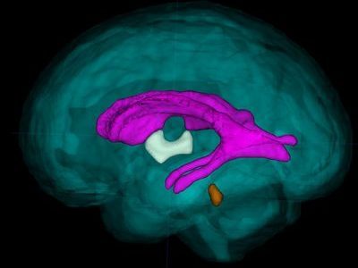
A team of researchers applied an advanced imaging technique, three-dimensional (3-D) MRI, to study brain development in these high-risk pregnancies. They are the first to report regional, tissue-specific volume delays for the developing fetal brain in FGR-affected pregnancies. The team compared overall fetal brain volume as well as regional brain volumes for a control group of healthy young pregnant women with a group of young women whose pregnancies were complicated by FGR. While fetuses in both groups grew exponentially as pregnancies progressed, the researchers began to see dramatic differences when they compared the volumes of specific regions of the brain, including the cerebellum, which coordinates balance and smooth movement; the deep gray matter, which also is involved in complex functions, such as memory and emotion; and the white matter, which is made up of millions of nerve fibers that connect to neurons in different regions. Because there are no biomarkers to spot early brain failure, 3-D MRI imaging may fill this knowledge gap.
Questions for Future Research
Q: Certain regions of the brains of FGR-affected infants show accelerated volume. Are these differences regional or global?
Q: Is accelerated brain volume in FGR-affected infants a result of heightened stress that these fetuses experience in the womb?
Q: How do differences in regional brain volume relate to later neurodevelopmental impairment that some FGR-affected infants experience?
Source: “Impaired Global and Tissue-Specific Brain Development in the Growth-Restricted Fetus.” N. Andescavage, J. Cruz, M. Metzler, A. du Plessis, and C. Limperopoulos. Presented during the 2016 Pediatric Academic Societies Annual Meeting, Baltimore, MD. May 2, 2016.


